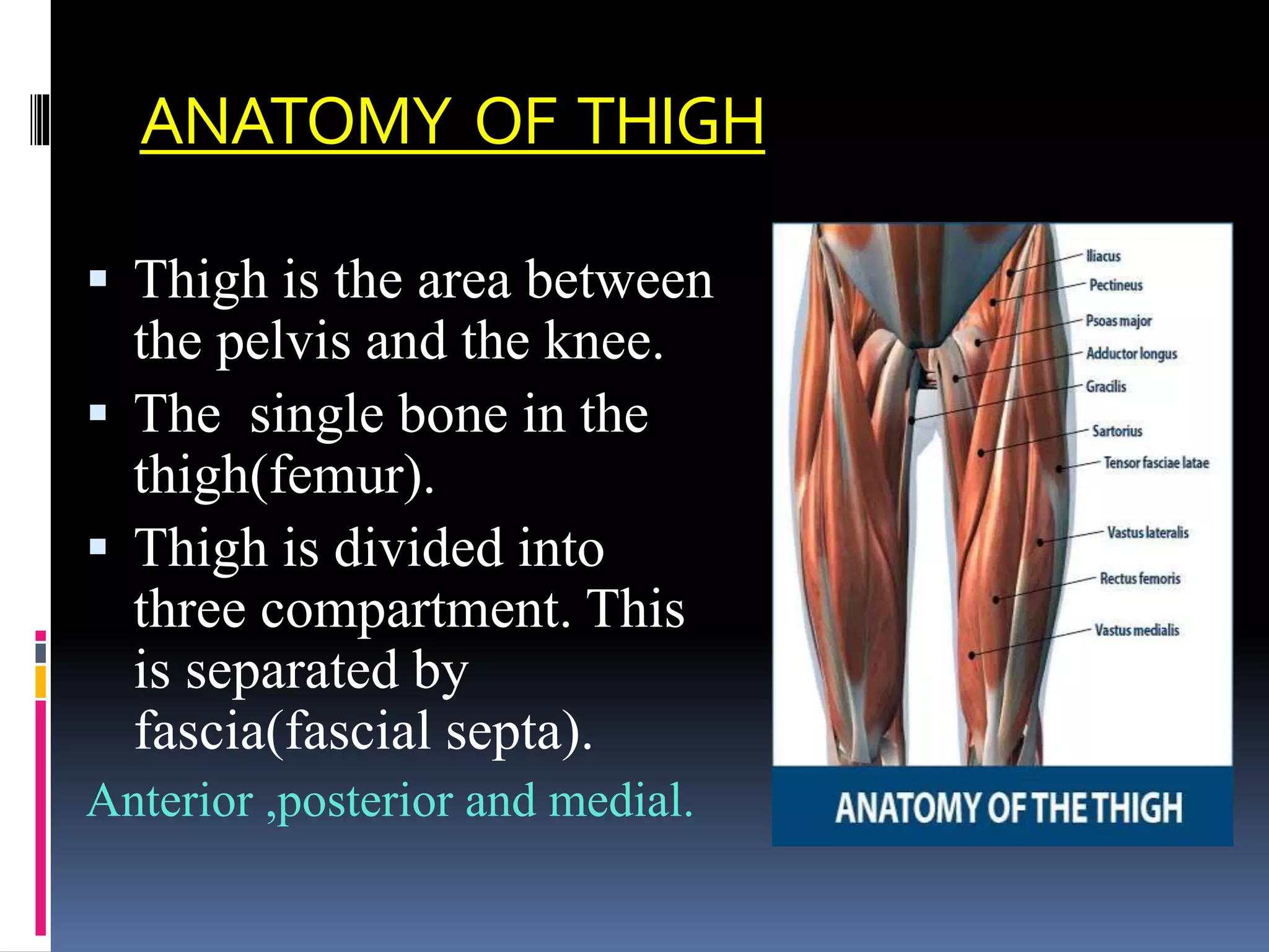
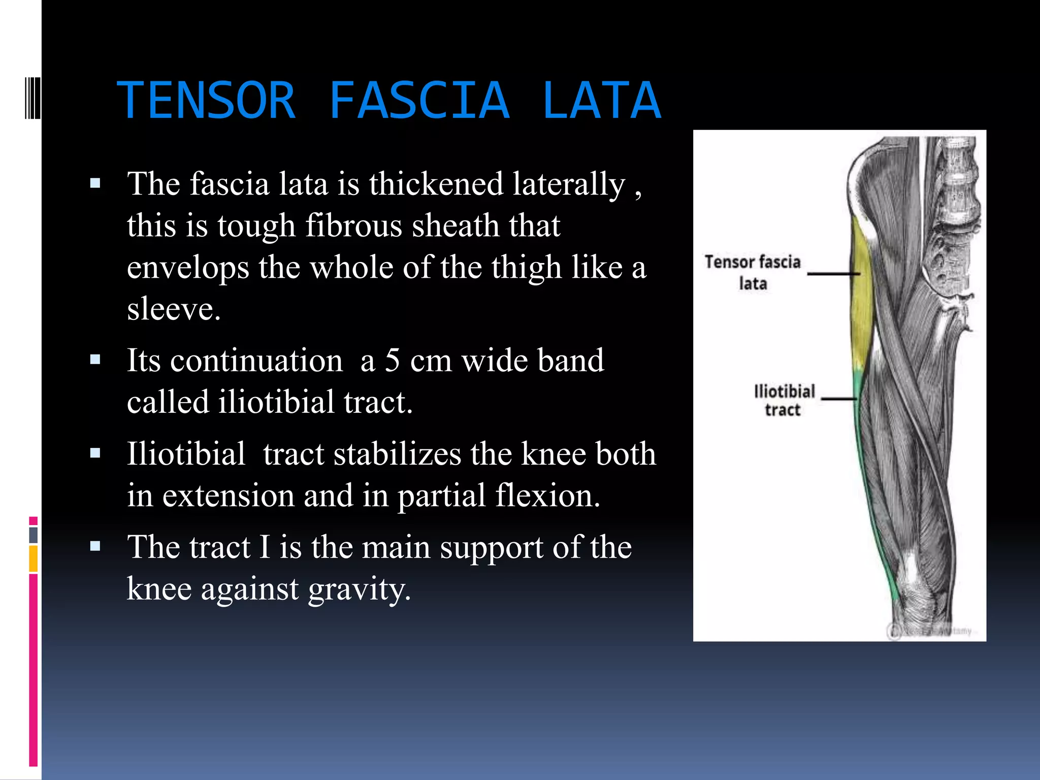
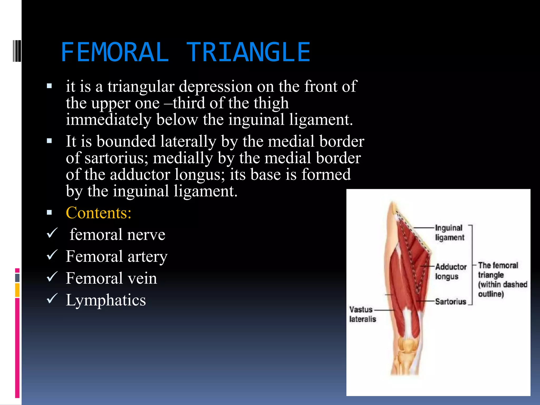
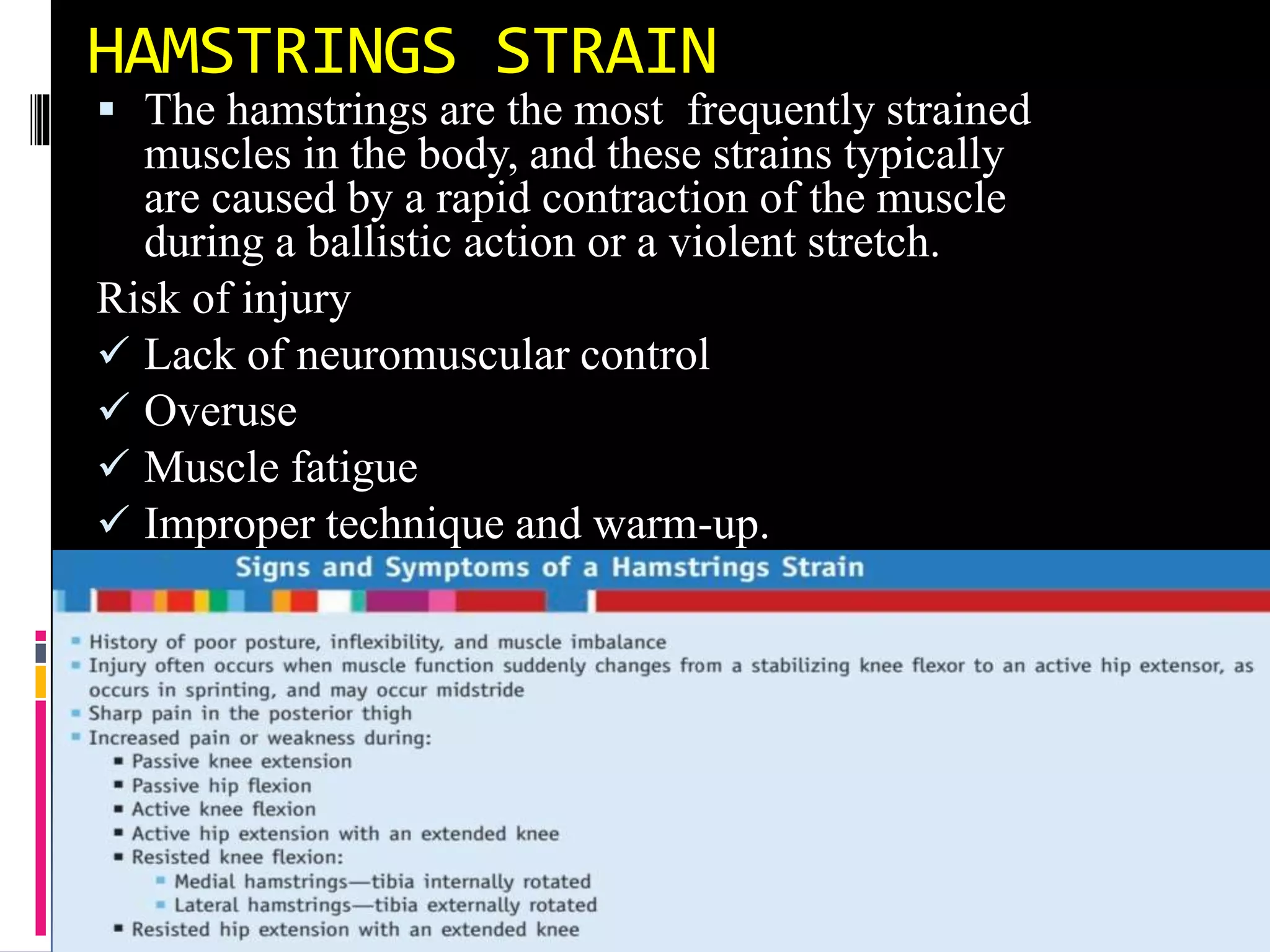
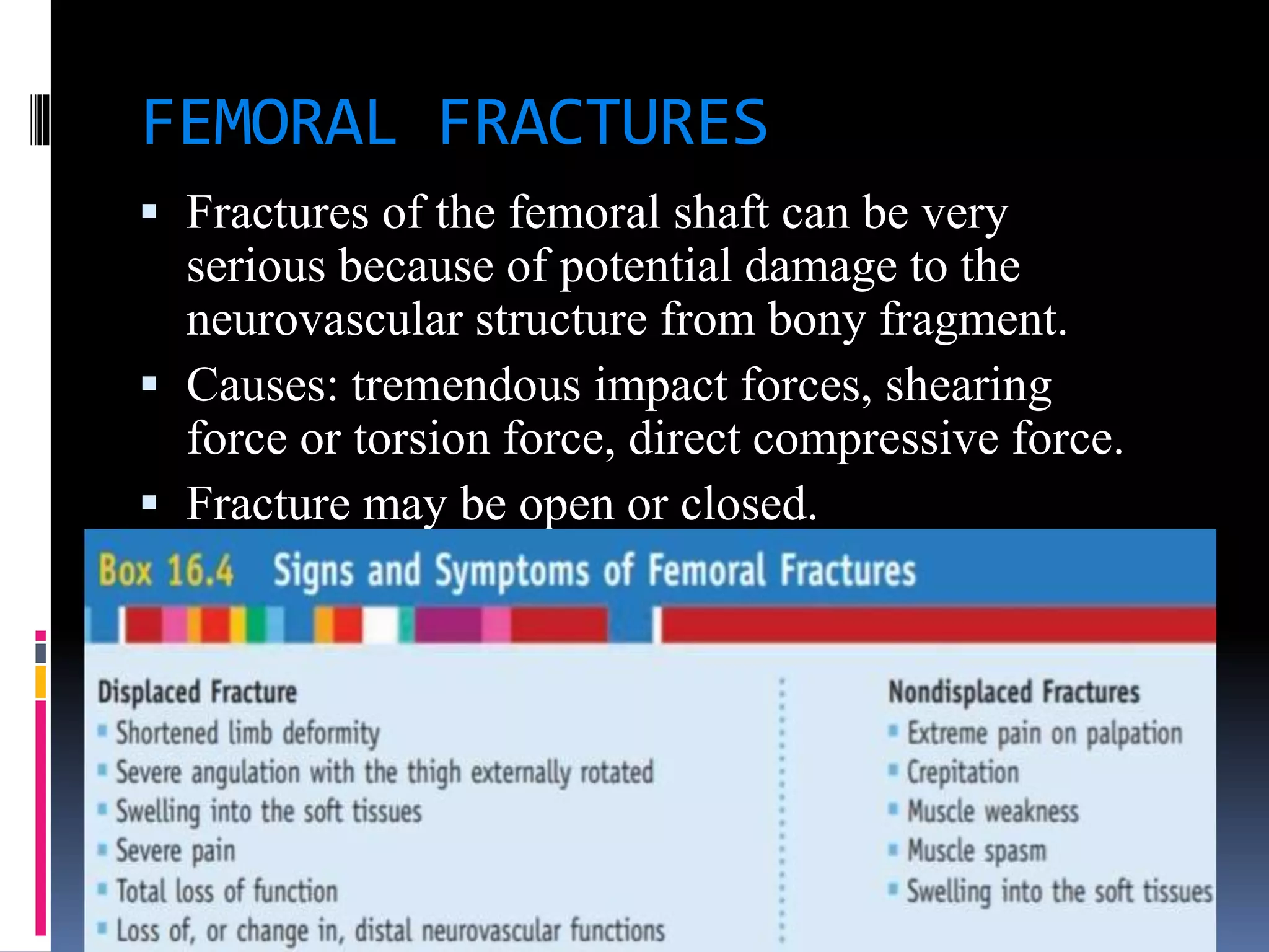
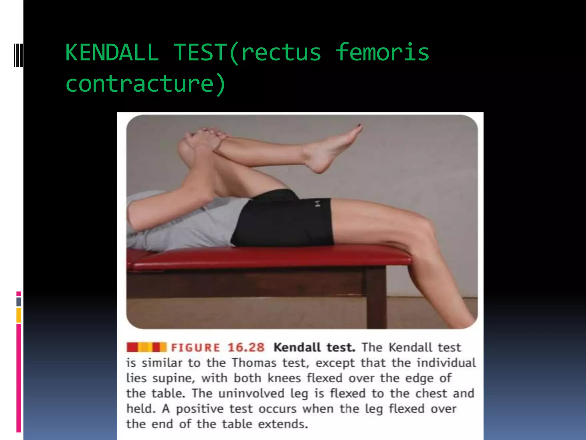
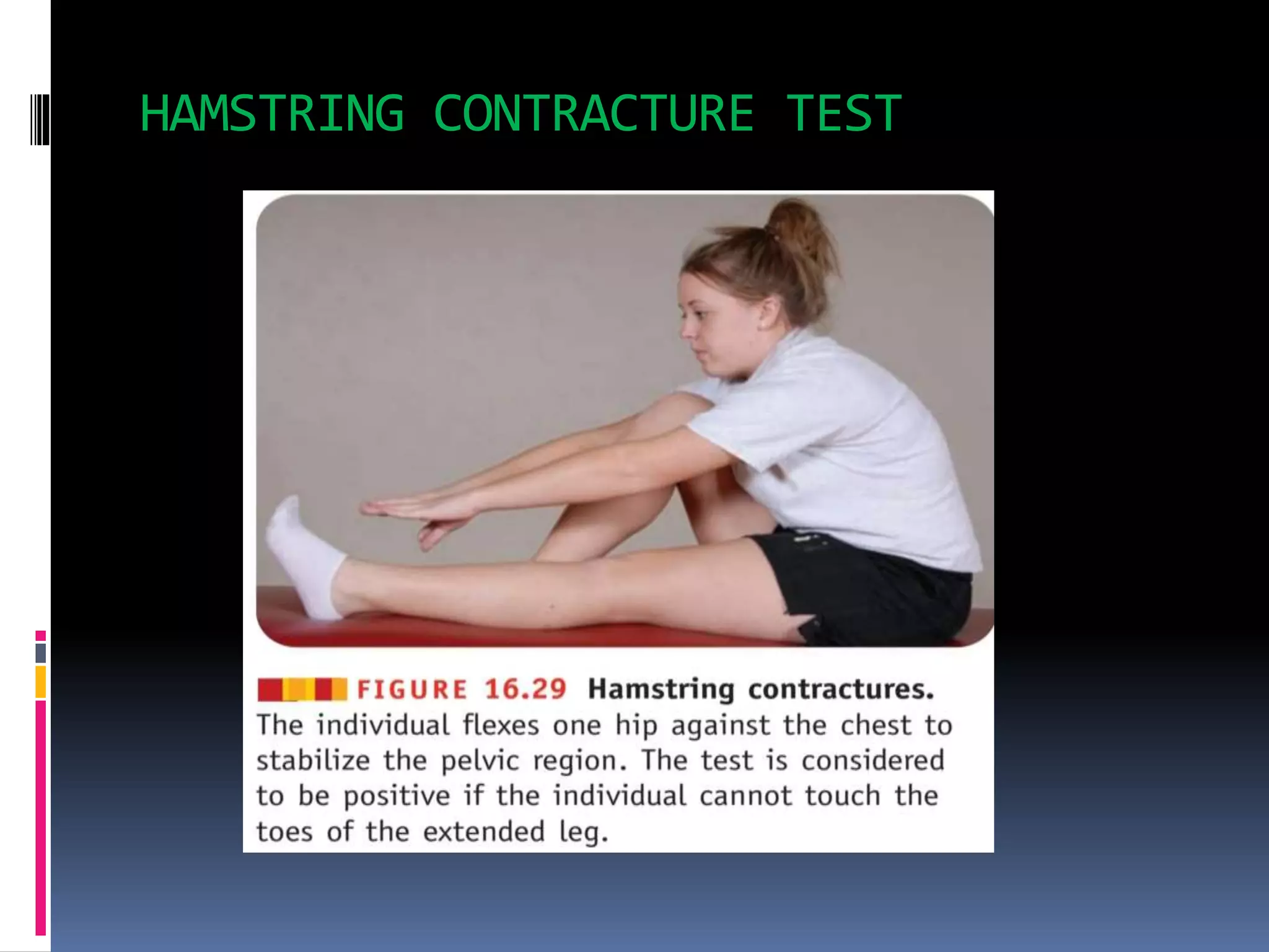
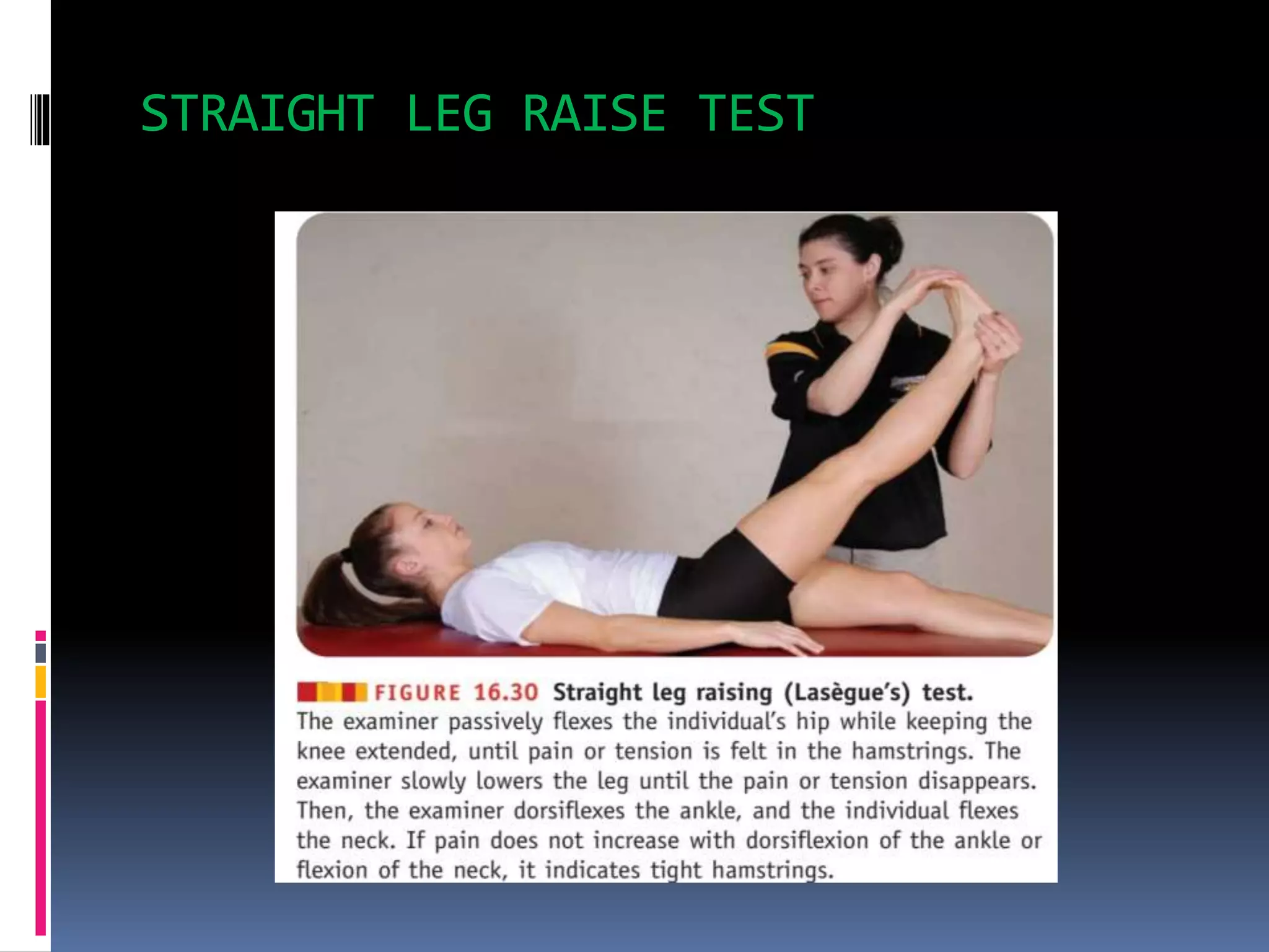
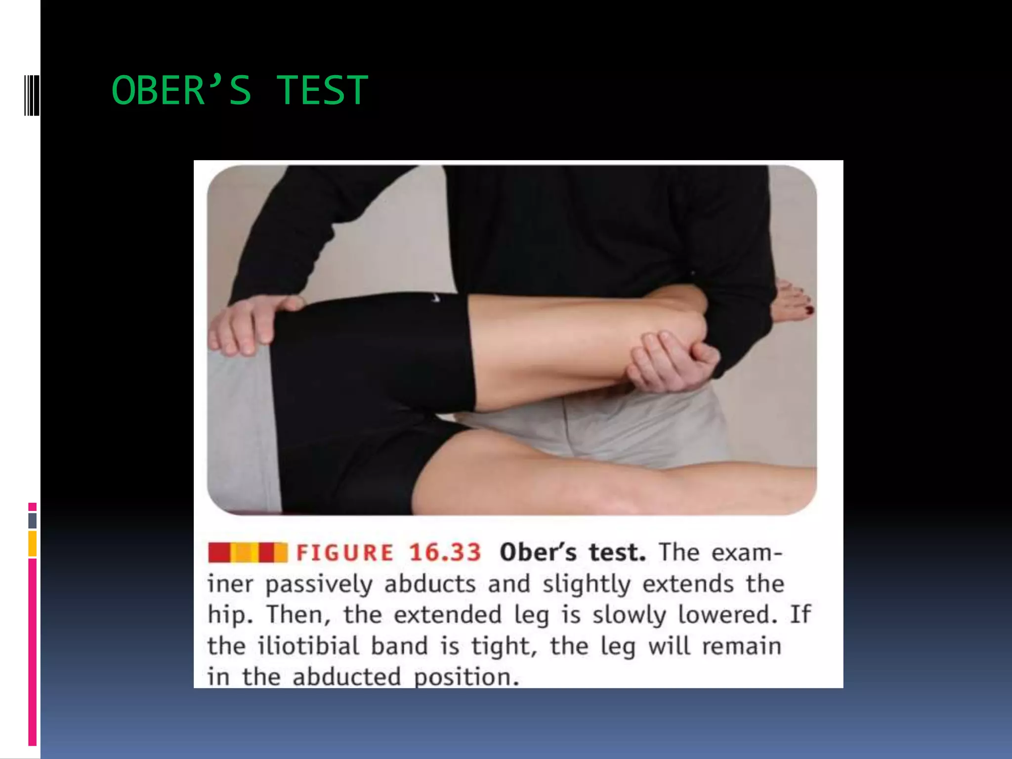
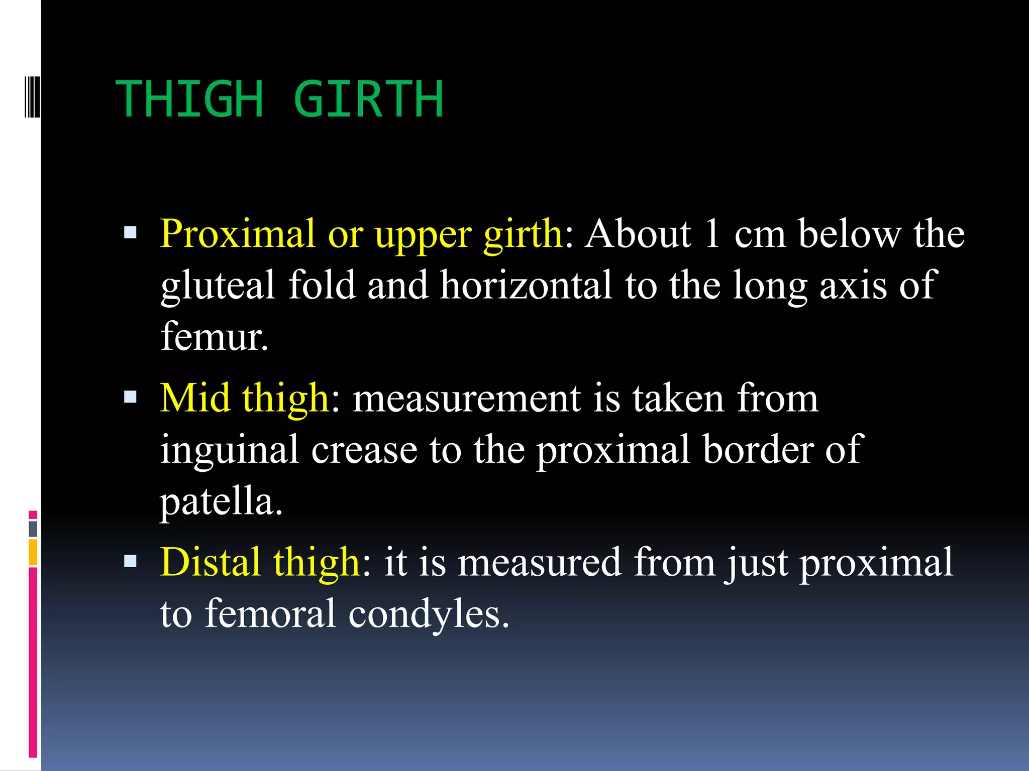
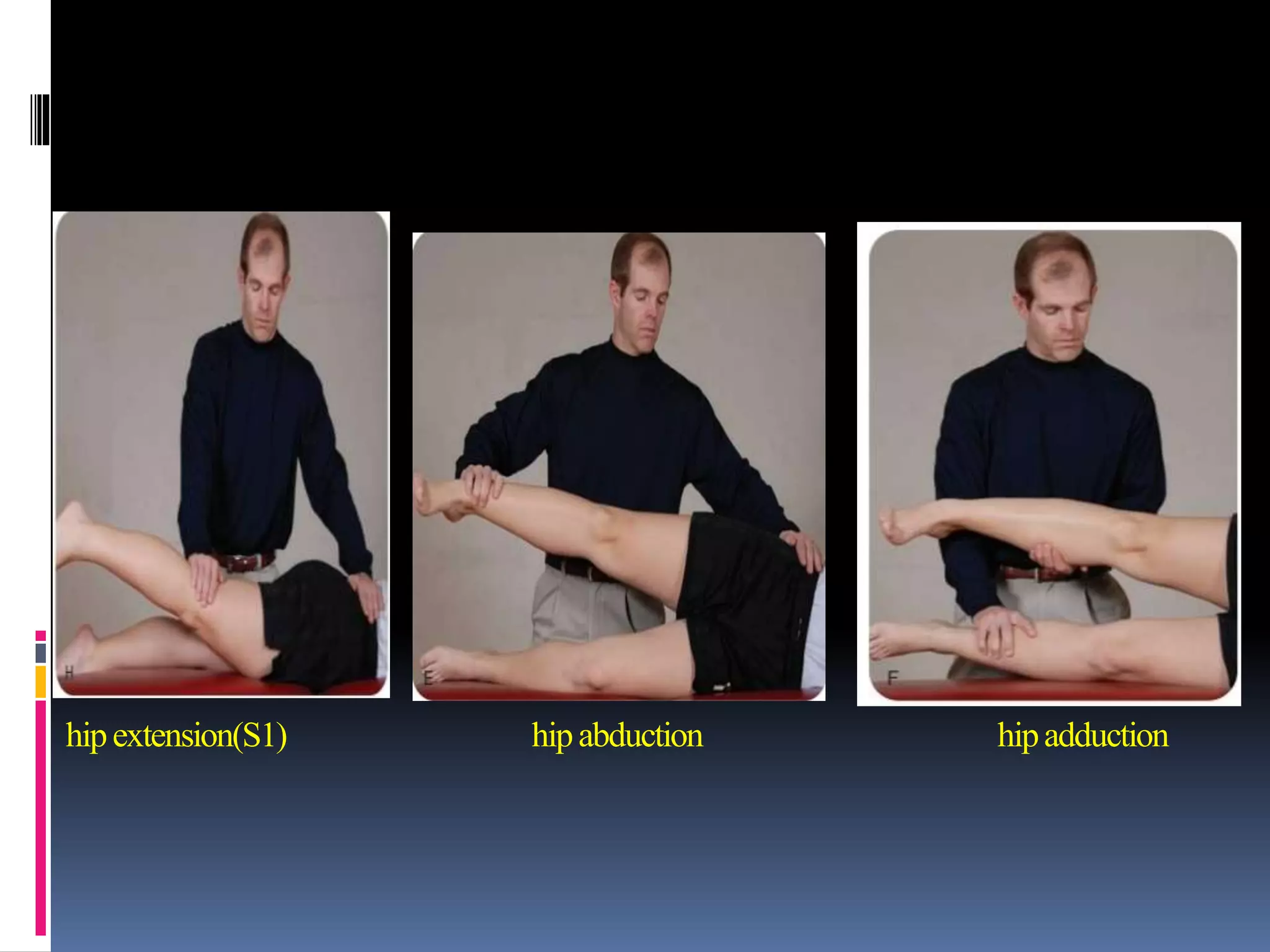
ASSESSMENT OF THIGH
Pravinraj v MPT 1st year
ANATOMY OF THIGH
 Thigh is the area between
the pelvis and the knee.
 The single bone in the
thigh(femur).
 Thigh is divided into
three compartment. This
is separated by
fascia(fascial septa).
Anterior ,posterior and medial.
ANTERIOR COMPARTMENT
MUSCLES
 Iliopsoas
 Sartorious
 Quadriceps
 Rectus femoris.
 Vastus lateralis.
 Vastus medialis.
 Vastus intermedius.
INNERVATION
Femoral nerve(L2,3,4).
ACTIONS
 Hip flexion
 Knee extension.
MEDIAL COMPARTMENT
MUSCLES
 Gracilis
 Adductor longus.
 Adductor brevis.
 Adductor magnus.
 Pectineus.(femoral nerve)
INNERVATION
Obturator nerve(L2,3,4).
ACTION
Adduction.
POSTERIOR COMPARTMENT
MUSCLES
 Hamstrings
 Biceps femoris.
long head.
Short head.
 Semimembranous.
 Semitendinosus.
 Adductor magnus.
INNERVATION
Common peroneal nerve(short head).
Sciatic nerve(L4,5,S1,2,3).
ACTION
 Hip extension.
 Knee flexion.
TENSOR FASCIA LATA
 The fascia lata is thickened laterally ,
this is tough fibrous sheath that
envelops the whole of the thigh like a
sleeve.
 Its continuation a 5 cm wide band
called iliotibial tract.
 Iliotibial tract stabilizes the knee both
in extension and in partial flexion.
 The tract I is the main support of the
knee against gravity.
FEMORAL TRIANGLE
 it is a triangular depression on the front of
the upper one –third of the thigh
immediately below the inguinal ligament.
 It is bounded laterally by the medial border
of sartorius; medially by the medial border
of the adductor longus; its base is formed
by the inguinal ligament.
 Contents:
 femoral nerve
 Femoral artery
 Femoral vein
 Lymphatics
INJURIES
CONTUSIONS
HIP POINTER
 A hip pointer generally refers to a contusion of the iliac
crest over the tensor fascia latae muscle belly with an
associated hematoma, but the term also may be used to
identify tearing of the external oblique muscle from the
iliac crest, periostitis of the crest, and trochondric
contusion.
Mechanism
A direct blow impacts the iliac crest.
QUADRICEPS CONTUSION
 The most common site for a this contusion is the anterolateral
thigh.
 If the contusion is located adjacent to the intermuscular septum,
pain and hemorrhage tend to resolve more rapidly.
 Associated with greater tearing, hemorrhage, and pain with great
tendency toward abnormal pathology.
Signs and symptoms
 Pain and swelling immediately after impact(grade1) able to walk
without a limp, passive flexion beyond 90 degree may be painful,
but resisted knee extension may cause less discomfort.
 Grade 2: flex the knee between 45 to 90 degree and walk, swelling
prevents the knee fully flexed.
 Grade 3: bruising seen initially, but within 24 hours, progressive
bleeding and swelling occur, firm hematoma, resulting in an
inability to contract the quadriceps or do a straight leg raise.
MYOSITIS OSSIFICATION
 abnormal ossification involving bone deposition within muscle tissue. It may
stem from a single traumatic blow, or from repeated blows, to the quadriceps.
 Risk factors:
 Innate predisposition to ectopic bone formation.
 Continuing to play after injury.
 Early massage, hydrotherapy, or thermotherapy during the acute stage.
 Passive, forceful stretching.
 Too rapid a progression in the rehabilitation program.
 Premature return to play.
 Re-injury of same area.
 Common sites are the anterior and lateral thigh. Although the precise
mechanism that triggers the bone formation has yet to be established, it is
thought that during resolution of the hematoma,within a week after injury, the
existing fibroblasts involved in the repair process begin to differentiate into
osteoblasts.
Signs and symptoms
 A warm, firm, swollen thigh nearly 2 to 4 cm larger than the unaffected side.
 Painful mass may limit passive knee flexion to 20 to 30 degree.
 Quadriceps contraction and straight leg raise may be impossible.
ACUTE COMPARTMENT SYNDROME
 This is advance stage of the quadriceps contusion.
 Defined as increased tissue pressure in a facial
compartment that compromises circulation to the
nerves and muscles within that compartment.
 A result of crushing injury or fracture of the
femur.
Signs and symptoms
 Severe pain occur with passive motion and
isometric contraction of the quadriceps.
 Motor weakness and decreased femoral sensation.
STRAINS
Quadriceps strain
 This strain is less common than hamstring strain.
 Tears more commonly occur in the mid-substance
muscle belly of the rectus femoris, because this is
most superficial.
 Vastus medialis and lateralis are more rarely injured.
HAMSTRINGS STRAIN
 The hamstrings are the most frequently strained
muscles in the body, and these strains typically
are caused by a rapid contraction of the muscle
during a ballistic action or a violent stretch.
Risk of injury
 Lack of neuromuscular control
 Overuse
 Muscle fatigue
 Improper technique and warm-up.
ADDUCTOR(groin)STRAIN
 Adductor strain are common in activities requires quick changes
of direction as well as explosive propulsion and acceleration.
 A strength imbalance between the hip abductors and adductors
may be a predisposing factor in many of these injuries.
 The more severe strains typically occur at proximal; attachment
of adductor longus.
Signs and symptoms
 Sharp pain
 Stiffness
 Weakness in adductors.
 Increased pain during
• Side-to-side movement
• Passive stretching
• Abducted position
• Externally rotated and with resisted hip adduction.
PIRIFORMIS SYNDROME
 The sciatic nerve passes through the sciatic notch beneath
the piriformis muscles to travel into the posterior thigh.
 The nerve to compression from trauma, hemorrhage, or
spasm of the piriformis muscle, most commonly peroneal
part of nerve is compressed.
Signs and symptoms
 Low back pain is not usual
 Difficulty in walking up-stairs
 Weakness or numbness present in back of the leg.
 pain and weakness present in hip external rotation.
 SLR may be limitted.
STRESS FRACTURE
 Stress fractures of the pubis, femoral neck, and proximal
third of the femur are seen in individuals who engage in
extensive jogging or aerobic dance activities to the point
of muscle fatigue. Several factor increase the risk of stress
fracture
 Improper footwear.
 Biomechanical abnormalities.
 Change in the running surface or terrain.
 Sudden increase in training.
Signs and symptoms
 Aching pain in the groin or thigh, during weight bearing.
 Night pain.
 Antalgic gait.
OSTEITIS PUBIS
 osteitis pubis is an inflammatory process involving
continued stress on the pubic symphysis from
repeated overload of the adductors muscles or
repetitive running activity.
Signs and symptoms
 Pain in the adductors
 Pain aggravating to the kicking, running and
pivoting on one leg.
 Pain radiating distally into medial thigh.
FEMORAL FRACTURES
 Fractures of the femoral shaft can be very
serious because of potential damage to the
neurovascular structure from bony fragment.
 Causes: tremendous impact forces, shearing
force or torsion force, direct compressive force.
 Fracture may be open or closed.
 Observation
 Muscle wasting
 Oedema
 Any bandages
 Palpation
 Tenderness
 Temperature
 Spasm
 Swelling
SPECIAL TESTS
THOMAS TEST
KENDALL TEST(rectus femoris
contracture)
HAMSTRING CONTRACTURE TEST
STRAIGHT LEG RAISE TEST
PIRIFORMIS TEST
OBER’S TEST
THIGH GIRTH
 Proximal or upper girth: About 1 cm below the
gluteal fold and horizontal to the long axis of
femur.
 Mid thigh: measurement is taken from
inguinal crease to the proximal border of
patella.
 Distal thigh: it is measured from just proximal
to femoral condyles.
RESISTED MANUAL MUSCLE TESTING
kneeextension(L3) kneeflexion(S2) hipflexion(L2)
hipextension(S1) hipabduction hipadduction
REFERENCE
 Foundation of athletic training-Marcia K.
Anderson, Gail P. Parr, Susan J. Hall.
 Human anatomy- BD chaurasia.
THANK YOU…...

Assessment of Thigh

  • 1.
  • 2.
    ANATOMY OF THIGH Thigh is the area between the pelvis and the knee.  The single bone in the thigh(femur).  Thigh is divided into three compartment. This is separated by fascia(fascial septa). Anterior ,posterior and medial.
  • 3.
    ANTERIOR COMPARTMENT MUSCLES  Iliopsoas Sartorious  Quadriceps  Rectus femoris.  Vastus lateralis.  Vastus medialis.  Vastus intermedius. INNERVATION Femoral nerve(L2,3,4). ACTIONS  Hip flexion  Knee extension.
  • 4.
    MEDIAL COMPARTMENT MUSCLES  Gracilis Adductor longus.  Adductor brevis.  Adductor magnus.  Pectineus.(femoral nerve) INNERVATION Obturator nerve(L2,3,4). ACTION Adduction.
  • 5.
    POSTERIOR COMPARTMENT MUSCLES  Hamstrings Biceps femoris. long head. Short head.  Semimembranous.  Semitendinosus.  Adductor magnus. INNERVATION Common peroneal nerve(short head). Sciatic nerve(L4,5,S1,2,3). ACTION  Hip extension.  Knee flexion.
  • 6.
    TENSOR FASCIA LATA The fascia lata is thickened laterally , this is tough fibrous sheath that envelops the whole of the thigh like a sleeve.  Its continuation a 5 cm wide band called iliotibial tract.  Iliotibial tract stabilizes the knee both in extension and in partial flexion.  The tract I is the main support of the knee against gravity.
  • 7.
    FEMORAL TRIANGLE  itis a triangular depression on the front of the upper one –third of the thigh immediately below the inguinal ligament.  It is bounded laterally by the medial border of sartorius; medially by the medial border of the adductor longus; its base is formed by the inguinal ligament.  Contents:  femoral nerve  Femoral artery  Femoral vein  Lymphatics
  • 8.
    INJURIES CONTUSIONS HIP POINTER  Ahip pointer generally refers to a contusion of the iliac crest over the tensor fascia latae muscle belly with an associated hematoma, but the term also may be used to identify tearing of the external oblique muscle from the iliac crest, periostitis of the crest, and trochondric contusion. Mechanism A direct blow impacts the iliac crest.
  • 10.
    QUADRICEPS CONTUSION  Themost common site for a this contusion is the anterolateral thigh.  If the contusion is located adjacent to the intermuscular septum, pain and hemorrhage tend to resolve more rapidly.  Associated with greater tearing, hemorrhage, and pain with great tendency toward abnormal pathology. Signs and symptoms  Pain and swelling immediately after impact(grade1) able to walk without a limp, passive flexion beyond 90 degree may be painful, but resisted knee extension may cause less discomfort.  Grade 2: flex the knee between 45 to 90 degree and walk, swelling prevents the knee fully flexed.  Grade 3: bruising seen initially, but within 24 hours, progressive bleeding and swelling occur, firm hematoma, resulting in an inability to contract the quadriceps or do a straight leg raise.
  • 11.
    MYOSITIS OSSIFICATION  abnormalossification involving bone deposition within muscle tissue. It may stem from a single traumatic blow, or from repeated blows, to the quadriceps.  Risk factors:  Innate predisposition to ectopic bone formation.  Continuing to play after injury.  Early massage, hydrotherapy, or thermotherapy during the acute stage.  Passive, forceful stretching.  Too rapid a progression in the rehabilitation program.  Premature return to play.  Re-injury of same area.  Common sites are the anterior and lateral thigh. Although the precise mechanism that triggers the bone formation has yet to be established, it is thought that during resolution of the hematoma,within a week after injury, the existing fibroblasts involved in the repair process begin to differentiate into osteoblasts. Signs and symptoms  A warm, firm, swollen thigh nearly 2 to 4 cm larger than the unaffected side.  Painful mass may limit passive knee flexion to 20 to 30 degree.  Quadriceps contraction and straight leg raise may be impossible.
  • 12.
    ACUTE COMPARTMENT SYNDROME This is advance stage of the quadriceps contusion.  Defined as increased tissue pressure in a facial compartment that compromises circulation to the nerves and muscles within that compartment.  A result of crushing injury or fracture of the femur. Signs and symptoms  Severe pain occur with passive motion and isometric contraction of the quadriceps.  Motor weakness and decreased femoral sensation.
  • 13.
    STRAINS Quadriceps strain  Thisstrain is less common than hamstring strain.  Tears more commonly occur in the mid-substance muscle belly of the rectus femoris, because this is most superficial.  Vastus medialis and lateralis are more rarely injured.
  • 14.
    HAMSTRINGS STRAIN  Thehamstrings are the most frequently strained muscles in the body, and these strains typically are caused by a rapid contraction of the muscle during a ballistic action or a violent stretch. Risk of injury  Lack of neuromuscular control  Overuse  Muscle fatigue  Improper technique and warm-up.
  • 15.
    ADDUCTOR(groin)STRAIN  Adductor strainare common in activities requires quick changes of direction as well as explosive propulsion and acceleration.  A strength imbalance between the hip abductors and adductors may be a predisposing factor in many of these injuries.  The more severe strains typically occur at proximal; attachment of adductor longus. Signs and symptoms  Sharp pain  Stiffness  Weakness in adductors.  Increased pain during • Side-to-side movement • Passive stretching • Abducted position • Externally rotated and with resisted hip adduction.
  • 16.
    PIRIFORMIS SYNDROME  Thesciatic nerve passes through the sciatic notch beneath the piriformis muscles to travel into the posterior thigh.  The nerve to compression from trauma, hemorrhage, or spasm of the piriformis muscle, most commonly peroneal part of nerve is compressed. Signs and symptoms  Low back pain is not usual  Difficulty in walking up-stairs  Weakness or numbness present in back of the leg.  pain and weakness present in hip external rotation.  SLR may be limitted.
  • 17.
    STRESS FRACTURE  Stressfractures of the pubis, femoral neck, and proximal third of the femur are seen in individuals who engage in extensive jogging or aerobic dance activities to the point of muscle fatigue. Several factor increase the risk of stress fracture  Improper footwear.  Biomechanical abnormalities.  Change in the running surface or terrain.  Sudden increase in training. Signs and symptoms  Aching pain in the groin or thigh, during weight bearing.  Night pain.  Antalgic gait.
  • 18.
    OSTEITIS PUBIS  osteitispubis is an inflammatory process involving continued stress on the pubic symphysis from repeated overload of the adductors muscles or repetitive running activity. Signs and symptoms  Pain in the adductors  Pain aggravating to the kicking, running and pivoting on one leg.  Pain radiating distally into medial thigh.
  • 19.
    FEMORAL FRACTURES  Fracturesof the femoral shaft can be very serious because of potential damage to the neurovascular structure from bony fragment.  Causes: tremendous impact forces, shearing force or torsion force, direct compressive force.  Fracture may be open or closed.
  • 20.
     Observation  Musclewasting  Oedema  Any bandages  Palpation  Tenderness  Temperature  Spasm  Swelling
  • 21.
  • 22.
  • 23.
  • 24.
  • 25.
  • 26.
  • 27.
    THIGH GIRTH  Proximalor upper girth: About 1 cm below the gluteal fold and horizontal to the long axis of femur.  Mid thigh: measurement is taken from inguinal crease to the proximal border of patella.  Distal thigh: it is measured from just proximal to femoral condyles.
  • 28.
    RESISTED MANUAL MUSCLETESTING kneeextension(L3) kneeflexion(S2) hipflexion(L2)
  • 29.
  • 30.
    REFERENCE  Foundation ofathletic training-Marcia K. Anderson, Gail P. Parr, Susan J. Hall.  Human anatomy- BD chaurasia.
  • 31.