Downloaded 74 times


![ Synovial folds and bursae.Synovial folds and bursae. The synovialThe synovial
membrane is well developed and, in the majority ofmembrane is well developed and, in the majority of
the joints forms largethe joints forms large folds,folds, plicae synoviales,plicae synoviales,
which contain adipose tissue. They go into thewhich contain adipose tissue. They go into the
articular cavity, filling its potential spaces andarticular cavity, filling its potential spaces and
forming cushions, which absorb shock duringforming cushions, which absorb shock during
motion.motion.
In certain regions, most frequently in the areas ofIn certain regions, most frequently in the areas of
muscular tendons, the synovial membranemuscular tendons, the synovial membrane
protrudes through the clefts in the fibrous layer andprotrudes through the clefts in the fibrous layer and
forms synovial bursae. They serve to reduceforms synovial bursae. They serve to reduce
friction during tendons' movement and can befriction during tendons' movement and can be
attributed to the accessory muscular apparatus.attributed to the accessory muscular apparatus.
TThe synovial layer bears numerous, microscopic,he synovial layer bears numerous, microscopic,
synovial villi,synovial villi, villi synoviales,villi synoviales, which significantlywhich significantly
increase its surface area, contain capillaryincrease its surface area, contain capillary
networks, and produce the synovia] fluid.networks, and produce the synovia] fluid.](https://image.slidesharecdn.com/2-170630104207/85/Connection-of-bones-27-320.jpg)

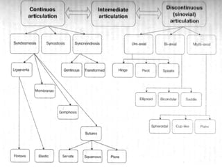
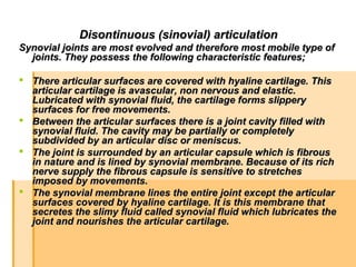
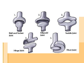
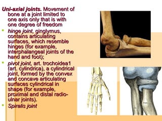
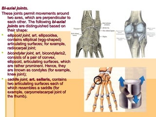
The document provides a comprehensive overview of human joint anatomy, including the classification of articulations such as continuous (synarthroses), intermediate, and discontinuous (synovial) joints. It details the structural characteristics and functions of various joint types, including their mobility and connective tissue components. Additionally, the document covers specific terminology and examples of different joint types, such as hinge, pivot, and ball-and-socket joints.


