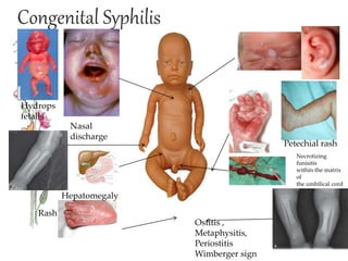
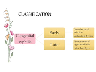
Name : Francis Sharon Jessy
Group : 12
Topic : Congenital Syphilis
Teacher: Pazylova Baktigul
DEFINITION
•syphilis is a sexually transmitted disease caused
by infection with the bacterium treponema
pallidum
• congenital syphilis is defined as disease
transmitted from a pregnant mother to her fetus
Congenital Syphilis
Hepatomegaly
Rash
Ostitis ,
Metaphysitis,
Periostitis
Wimberger sign
Nasal
discharge
Hydrops
fetalis
Petechial rash
Necrotizing
funisitis
within the matrix
of
the umbilical cord
CLASSIFICATION
Congenital
syphilis
Early
Direct bacterial
infection
Within first 2 years
Late
Phenomenon of
hypersensitivity
Later than 2 yrs
PATHOLOGY
• Major fetus- small perivascular inflammatory foci &
lymphocytic infiltrate  reduced growth of parenchymal
cells & fibrosis.
• Placenta- heavy, bulky, pale, greasy.
Placenta HPE-
1. Necrotizing funisitis
2. Villous enlargement
3. Acute villitis
• Stillborn fetus- macerated appearance with collapse of
skull, protuberant abdomen with enlarged liver and
spleen, haemorrhagic bulla.
Early congenital syphilis
• Primary stage – absent (blood borne)
• Baby – Preterm/full term
Early cong.
syphilis
Lack
manifestations -
birth
rhinitis,
pneumonia,
failure to thrive
Classical
presentation -
birth
Wizened, pot
belly, hoarse baby
looking like old
man, withered
brown skin &
runny fissured
nose.
More prone for
intercurrent
infection
Manifestations Of Congenital Syphilis -
Early Congenital Syphilis
• Anemia
• Oedma or wasting
• Fever
• Failure to thrive
• Rhinitis or snaffles is an early symptom
• Profuse mucopurlent discharge and nasal congestion
• Skin eruptions
• Erythematous appearance
• Mucuocutaneous lesions occur around the nose, mouth,
anus and vulva –moist and readily produce fissuring and
bleeding
• Excoriations and ulcers are common in the genital &
perinatal region & on the face
• Around the mouth may lead to radiating scars called
“rhagades”
• Condyloma latum , which are flat, raised plaques with moist
surfaces,occur around the anus and female genitals ;it is
usually found in later infancy, between 6 month 2 yrs.
Late Congenital Syphilis
• Interstitial kerailts
• Pain,photophobia&lacrimation
• Vascular infiltration of deep layers of cornea which
presents a ground - glass appearance& later
become opaque.
• Secondary iridocystitis
Skin Lesions
• Vesicobullous rash, symetrically , palms and soles-
earliest & specific sign.
• Also known as Pemphigus syphiliticus
• Lesions are contagious, also seen around oral cavity,
trunk, buttocks, and genitilia.
• Few weeks later, a papulosquamous rash may appear.
Usually involve the face, mouth, anterior nares, buttocks,
palms & soles.
Diagnosis
• History collection
• Physical examination
-Hepatomegaly
-bone inflammation
For mother
-fluorescent treponemal antibody absorbed test
-Rapid plasma reagin(RPR)
-VDRL
• Bacteriology
• Eye examination
• Lumbar puncture( csf studies- increased protein)
• Radiology – Wimberger's sign( litic lesion in the
upper aspect of tibia)
Complication
• Blindness
• Deafness
• Deformity of the face
• Nervous system problems
Treatment
•Intravenous crystalline penicillin G 100,000-
150,000U//kg/day-im
•Erythromycin 15mg/kg/day for 10 days

Congenital Syphilis - Dermatology

  • 1.
    Name : FrancisSharon Jessy Group : 12 Topic : Congenital Syphilis Teacher: Pazylova Baktigul
  • 2.
    DEFINITION •syphilis is asexually transmitted disease caused by infection with the bacterium treponema pallidum • congenital syphilis is defined as disease transmitted from a pregnant mother to her fetus
  • 3.
    Congenital Syphilis Hepatomegaly Rash Ostitis , Metaphysitis, Periostitis Wimbergersign Nasal discharge Hydrops fetalis Petechial rash Necrotizing funisitis within the matrix of the umbilical cord
  • 4.
    CLASSIFICATION Congenital syphilis Early Direct bacterial infection Within first2 years Late Phenomenon of hypersensitivity Later than 2 yrs
  • 5.
    PATHOLOGY • Major fetus-small perivascular inflammatory foci & lymphocytic infiltrate  reduced growth of parenchymal cells & fibrosis. • Placenta- heavy, bulky, pale, greasy. Placenta HPE- 1. Necrotizing funisitis 2. Villous enlargement 3. Acute villitis • Stillborn fetus- macerated appearance with collapse of skull, protuberant abdomen with enlarged liver and spleen, haemorrhagic bulla.
  • 6.
    Early congenital syphilis •Primary stage – absent (blood borne) • Baby – Preterm/full term Early cong. syphilis Lack manifestations - birth rhinitis, pneumonia, failure to thrive Classical presentation - birth Wizened, pot belly, hoarse baby looking like old man, withered brown skin & runny fissured nose. More prone for intercurrent infection
  • 7.
    Manifestations Of CongenitalSyphilis - Early Congenital Syphilis • Anemia • Oedma or wasting • Fever • Failure to thrive • Rhinitis or snaffles is an early symptom • Profuse mucopurlent discharge and nasal congestion • Skin eruptions • Erythematous appearance
  • 8.
    • Mucuocutaneous lesionsoccur around the nose, mouth, anus and vulva –moist and readily produce fissuring and bleeding • Excoriations and ulcers are common in the genital & perinatal region & on the face • Around the mouth may lead to radiating scars called “rhagades” • Condyloma latum , which are flat, raised plaques with moist surfaces,occur around the anus and female genitals ;it is usually found in later infancy, between 6 month 2 yrs.
  • 9.
    Late Congenital Syphilis •Interstitial kerailts • Pain,photophobia&lacrimation • Vascular infiltration of deep layers of cornea which presents a ground - glass appearance& later become opaque. • Secondary iridocystitis
  • 10.
    Skin Lesions • Vesicobullousrash, symetrically , palms and soles- earliest & specific sign. • Also known as Pemphigus syphiliticus • Lesions are contagious, also seen around oral cavity, trunk, buttocks, and genitilia. • Few weeks later, a papulosquamous rash may appear. Usually involve the face, mouth, anterior nares, buttocks, palms & soles.
  • 12.
    Diagnosis • History collection •Physical examination -Hepatomegaly -bone inflammation For mother -fluorescent treponemal antibody absorbed test -Rapid plasma reagin(RPR) -VDRL
  • 13.
    • Bacteriology • Eyeexamination • Lumbar puncture( csf studies- increased protein) • Radiology – Wimberger's sign( litic lesion in the upper aspect of tibia)
  • 14.
    Complication • Blindness • Deafness •Deformity of the face • Nervous system problems
  • 15.
    Treatment •Intravenous crystalline penicillinG 100,000- 150,000U//kg/day-im •Erythromycin 15mg/kg/day for 10 days