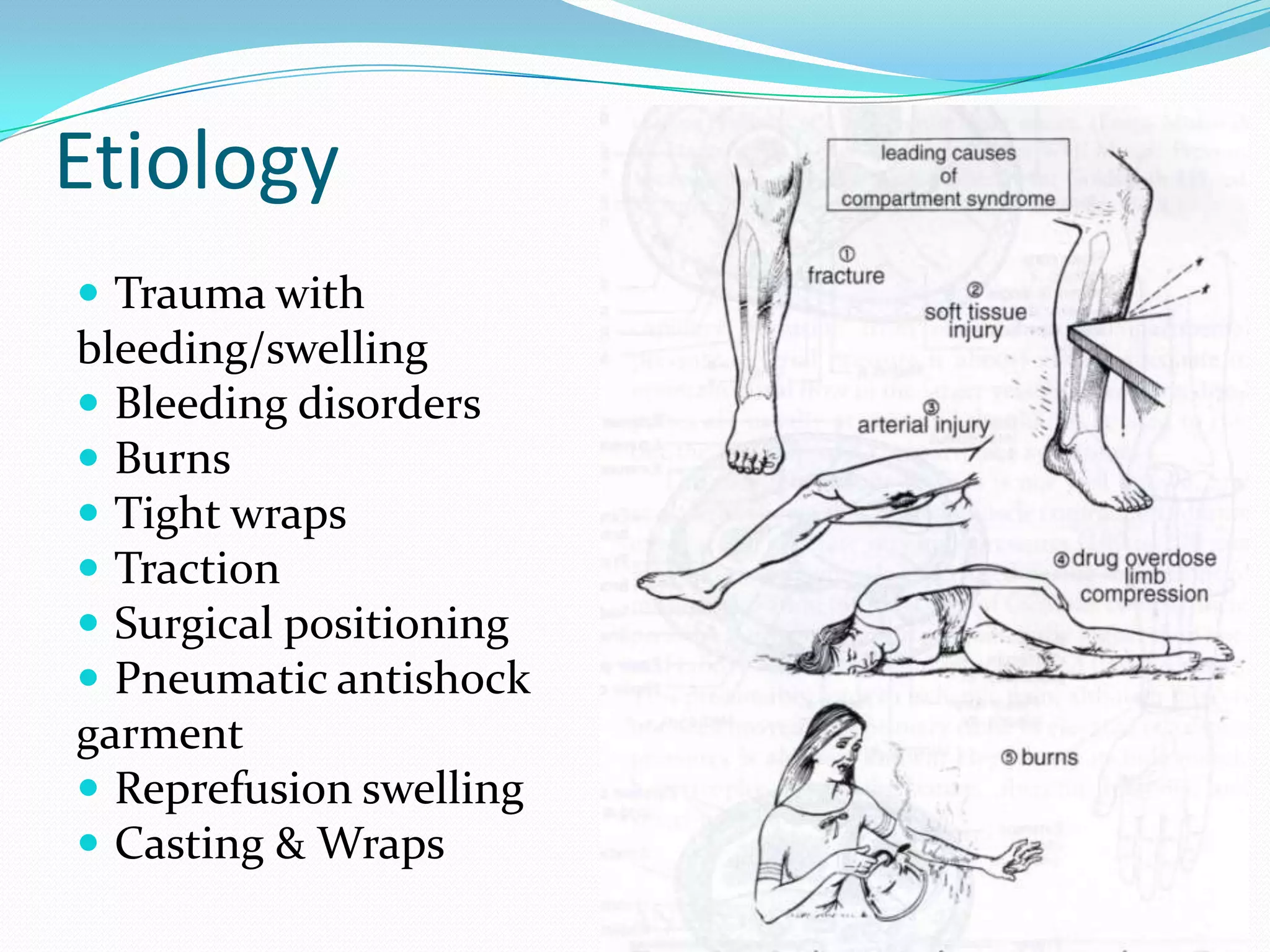
Subhanjan Das
Introduction
These are the associated pathologies other than the loss
of bone continuity which either co exist or originate
due to the fracture.
early diagnosis and aggressive treatment is necessary
to minimize disabilities.
Classification
I.IMMEDIATE
A.Systemic
hypovolaemic Shock
B.Local
injury to
1. major vessels
2. Muscles and tendons
3. Joints
4. viscera
II.EARLY
A.Systemic
1. Hypovolaemic shock
2. ARDS
3. Fat embolism
4. DVT & pulmonary embolism
5. Aseptic traumatic fever
6. Septicaemia
7. Crush syndrome
B. Local
1. Infection
2. Compartment syndrome
III. LATE COMPLICATIONS
A. Related to imperfect union
1. Delayed union
2. Non union
3. Mal union
4. Cross union
B.Others
1. Avascular necrosis
2. Shortening
3. Joint stiffness
4. Sudeck’s dystrophy
5. Osteomyelitis
6. Ischemic contracture
7. Myositis ossificans
8. OA
Hypovolaemic shock
 Commonest cause of death in fractures of major bones
Like pelvis or femur
cause
 External or internal haemorrhage.
 External: compound fractures injuring major vessels of
the LIMB
 Internal: injury to body cavities- chest or pelvis
 Internal is more difficult to diagnose.
 # pelvis (1.5-2 litres) # femur (1-1.5 litres) produces
major haemorrhage.
Prevention
 Early stopping of bleeding
 Avoiding shifting of the patients
 For # pelvis- temporary stabilization with external
fixator
 Emergency angiography and embolisation of bleeding
vessels for deeper vessels.
Management
 Starts even before the cause is established
 Two large bore iv cannulas put
 Infuse 2000 ml of crystalloids (ringer lactate) followed
by colloid (haemaccel) and blood if needed
 Cut down if peripheral vasoconstriction is present
 Localise the site of lesion- if in body cavities, perform
chest aspiration or diagnostic peritonial lewage.
Sometimes a simple x ray is enough.
 Chest bleeding-ICDT
 Abdominal bleeding- laperotomy
ARDS
 Respiratory distress following a trauma
 Cause- not definite. Hypothesized to be by release of
Inflammatory cells and proteinaceous fluid that
accumulate in the alveolar spaces leading to a decrease
in diffusing capacity and hypoxemia. The
microvasculature in dysrupted.
 Onset- 24 hours after injury
 Features:
 Tachypnea
 Laboured breathing
 X- ray- diffused
pulmonary infiltrates
 Arterial Po2 below 50
management
 100% O2 and assisted ventilation
 It takes upto 7 days to get the chest clear
 If not detected early death occurs by multiorgan
failure or cardiorespiratory failure.
Fat Embolism
It is a life threatening complication of fracture where fat
globules occlude the small blood vessels.
Embolism is the process of occlusion of blood vessel by
any material which is brought to the site from
elsewhere by bloodstream.
Pathogenesis
Injury to large bones (e.g. femur) release fat globule
from bone marrow to blood stream. Alternatively fat
can also be released from the adipose tissue.
The fat globules obstruct capillary vasculature of the
lungs.
Also, fat is converted to free fatty acid, which induces
toxic vasculitis followed by thrombosis which obstruct
the microvasculature.
Clinical features
COMMON
Patechial rash of anterior
neck, anterior axillary
fold or conjunctiva
CEREBRAL TYPE
Drowsiness
Restlessness
Disorientation
Coma
PULMONARY TYPE
Tachypnoea
Tachycardia
Respiratory failure
Diagnosis
Retinal artery emboli
Urine: fat globules
CXR: pulmonary infiltration/
Snow storm appearance
Clinical features
management
 Respitarory support
 Heparinisation
 i.v. low mol wt dextran
 Corticosteroid
 Dextrose and alcohol infusion to emulsify fat.
Deep Vein Thrombosis
It is a common complication
originating from altered
hemodynamics in lower
limb and spinal injuries.
Pathology:
pathology
Virchow's triad
1. decreased flow rate of
the blood
2. damage to the blood
vessel wall
3. hypercoagulability
trauma
immobilisation
Venous stasis
thrombosis
Clinical features
Elderly and obese patients are at risk.
Leg swelling
Local redness, warmth
Calf tenderness
Pain in passive dorsiflexion (Homan sign)
Venography shows DVT
 Sequale
1. The venous thrombosis can get dislodged and
produce embolism elsewhere. If it is pulmonary
embolism the condition is life threatening.
Embolism usually occurs within 4-5 days after injury.
2. A late complication of DVT is the post-phlebitic
syndrome, which can manifest itself as edema, pain
or discomfort and skin problems.
Other causes
compression of the veins
physical trauma
cancer
infections
inflammatory diseases
stroke
heart failure
nephrotic syndrome
Risk factor:
Surgery
hospitalization
immobilization
orthopedic casts
economy class syndrome
smoking
Obesity
age
certain drugs (such as estrogen
or erythropoietin)
thrombophilia
pregnancy
postnatal period.
diagnosis
D-dimers
doppler ultrasound
venography
Clinical features
treatment
Prophylaxis
 Active/ passive calf pump
and toe movement
 Elevation
 Deep breathing exercise
 Elastic TED stockings
 Early internal fixation to
provide early mobility.
Management
Complete rest with elevation
thrombolysis
Anticoagulant therapy
graduated compression
stockings (
thromboembolic deterrent
stockings) or
intermittent pneumatic
compression devices.
Respiratory support in case
of pulmonary embolism
Crush syndrome
It is renal failure following
extensive crushing injury
of muscles.
Pathogenesis:
Crushing of muscles causes
entry of myoglobin into
circulation. Myoglobin
precipitates in renal
tubules causing acute
tubular necrosis,
metabolic acidosis &
hperkalemia
Clinical features
(appear within 2-3 days of injury)
Signs of deficient renal function:
Oliguria (Scanty urine)
Apathy
Restlessness
Delirium
Cardiac arrhythmia & failure
Hypothermia
Shock
Treatment
Prophylaxis
Application of tourniquet
and gradual release to
slowly allow the
myoglobin to reach the
kidneys
Treatment
Treated as acute renal
failure.
An increased pressure within enclosed
osteofascial space that reduces capillary per-
fusion below level necessary for tissue
viability; the underlying mechanism is:
- increased volume within space
- decreased space for contents
- combination of both
Compartment syndrome
Etiology
 Trauma with
bleeding/swelling
 Bleeding disorders
 Burns
 Tight wraps
 Traction
 Surgical positioning
 Pneumatic antishock
garment
 Reprefusion swelling
 Casting & Wraps
Pathophysiology:
Increased compartment pressure
leads to increased venous pressure
which decreases A-V gradient resulting
in muscle and nerve ischemia.
Compartments
 Most common
 Forearm
 Leg
 Other compartments
 Hand
 Finger
 Gluteal
 Thigh
 Foot
Diagnosis
 History
 Clinical exam: the Ps
 Compartment pressures
 Laboratory tests
 CPK
 Urine myoglobin
Clinical features
 The six ‘Ps’:
 Pressure: palpation of compartment and its tension or
firmness
 Pain: Exaggerated with passive stretch of the involved
muscles in compartment
 Earliest symptom but inconsistent
 Paresthesia:Peripheral nerve tissue is more sensitive than
muscle to ischemia
 Will progress to anesthesia if pressure not relieved
 Paralysis: late finding
 Pallor
 Pulselessness
Treatment
 Lower leg to level of the heart
 Remove cast
 Split all dressings down to skin
 Fasciotomy if continued clinical findings and/or
elevated compartment pressure
Forearm
Leg Anatomy
Leg Single Incision Technique
Leg Two Incision Technique
Hand Compartments
Foot Compartments
Delayed/ Non union
When a fracture takes more than the usual time to unite
it is said to have gone in delayed union.
When the process of healing stops before completion
the fracture is said to have gone for non union. To
diagnose non union the fracture has to be minimum
six months old.
causes
I. Related to patient
 Old age
 Associated systemic illness: ex. Malignancy
II. Related to fracture
 Distraction at fracture site
Muscle pulling the fragments: ex. # patella
Gravity: ex. # shaft of humerus
 Soft tissue interposition: ex. # shaft of humerus
 Bone loss during fracture: ex. # tibia open type
 Infection from open fracture: ex. # tibia
 Damage to blood supply of # fragment: ex. # scaphoid
 Pathological fracture: ex. # osteomyelitic tibia
III causes related to treatment:
 Inadequate reduction: # shaft of long bones
 Inadequate immobilisation:# shaft of long bones
 Distraction (excessive) during treatment::# shaft of
femur.
types
1. Atrophic: no or minimal callus formation
2. Hypertrophic: callus is present but it does not bridge
the fracture site.
Common sites
Neck of femur
Scaphoid
Lower third of tibia
Lower third of ulna
Lateral condyle of humerus
Clinical features
 Pain
 Deformity
 Abnormal mobility
 Refracture
Radiological findings
Delayed union: inadequate callus, visible fracture line
Non union: ends are rounded, smooth sclerotic.
Medullary cavity may be obliterated. visible fracture
line.
Treatment: Delayed union
1. Most commonly prolonged conservative
management
2. Surgical intervention: bone grafting with or without
internal fixation.
Treatment: non union
Depends upon site and resulting disability. Following are
the options.
1. Bone grafting: commonest.
2. Excision of fragments: when it can be done with
minimal loss of function. A prosthesis may be used
to replace the lost part, eg. In # neck of femur the
head can be replaced with an austin moore
prosthesis.
3. Illizarov menthod
4. No treatment: when there is no disability, eg. #
scaphoid.
Mal union
 When a fracture does not unite in proper position it is
said to have malunited.
Causes:
1. Improper reduction
2. Unchecked muscle pull
3. Excessive communication
Consequences
Deformity
Shortening of limb
Limitation of movements
treatment
1. osteoclasis: refracture, done in children to correct
mild to moderate angular deformities under GA.
2. Redoing the fracture surgically: most common. ORIF
is generally done along with bone grafting.
3. Corrective osteotomy: performed at a site away from
the fracture. Eg. Supracondyle # of humerus.
4. Excision of protruding bone.
 No treatment may be necessary if remodelling occurs.

complicationsoffractures-120127084025-phpapp02.pdf

  • 1.
  • 2.
    Introduction These are theassociated pathologies other than the loss of bone continuity which either co exist or originate due to the fracture. early diagnosis and aggressive treatment is necessary to minimize disabilities.
  • 3.
    Classification I.IMMEDIATE A.Systemic hypovolaemic Shock B.Local injury to 1.major vessels 2. Muscles and tendons 3. Joints 4. viscera
  • 4.
    II.EARLY A.Systemic 1. Hypovolaemic shock 2.ARDS 3. Fat embolism 4. DVT & pulmonary embolism 5. Aseptic traumatic fever 6. Septicaemia 7. Crush syndrome B. Local 1. Infection 2. Compartment syndrome
  • 5.
    III. LATE COMPLICATIONS A.Related to imperfect union 1. Delayed union 2. Non union 3. Mal union 4. Cross union B.Others 1. Avascular necrosis 2. Shortening 3. Joint stiffness 4. Sudeck’s dystrophy 5. Osteomyelitis 6. Ischemic contracture 7. Myositis ossificans 8. OA
  • 6.
    Hypovolaemic shock  Commonestcause of death in fractures of major bones Like pelvis or femur
  • 7.
    cause  External orinternal haemorrhage.  External: compound fractures injuring major vessels of the LIMB  Internal: injury to body cavities- chest or pelvis  Internal is more difficult to diagnose.  # pelvis (1.5-2 litres) # femur (1-1.5 litres) produces major haemorrhage.
  • 8.
    Prevention  Early stoppingof bleeding  Avoiding shifting of the patients  For # pelvis- temporary stabilization with external fixator  Emergency angiography and embolisation of bleeding vessels for deeper vessels.
  • 9.
    Management  Starts evenbefore the cause is established  Two large bore iv cannulas put  Infuse 2000 ml of crystalloids (ringer lactate) followed by colloid (haemaccel) and blood if needed  Cut down if peripheral vasoconstriction is present  Localise the site of lesion- if in body cavities, perform chest aspiration or diagnostic peritonial lewage. Sometimes a simple x ray is enough.  Chest bleeding-ICDT  Abdominal bleeding- laperotomy
  • 10.
    ARDS  Respiratory distressfollowing a trauma  Cause- not definite. Hypothesized to be by release of Inflammatory cells and proteinaceous fluid that accumulate in the alveolar spaces leading to a decrease in diffusing capacity and hypoxemia. The microvasculature in dysrupted.  Onset- 24 hours after injury
  • 11.
     Features:  Tachypnea Laboured breathing  X- ray- diffused pulmonary infiltrates  Arterial Po2 below 50
  • 12.
    management  100% O2and assisted ventilation  It takes upto 7 days to get the chest clear  If not detected early death occurs by multiorgan failure or cardiorespiratory failure.
  • 13.
    Fat Embolism It isa life threatening complication of fracture where fat globules occlude the small blood vessels. Embolism is the process of occlusion of blood vessel by any material which is brought to the site from elsewhere by bloodstream.
  • 14.
    Pathogenesis Injury to largebones (e.g. femur) release fat globule from bone marrow to blood stream. Alternatively fat can also be released from the adipose tissue. The fat globules obstruct capillary vasculature of the lungs. Also, fat is converted to free fatty acid, which induces toxic vasculitis followed by thrombosis which obstruct the microvasculature.
  • 15.
    Clinical features COMMON Patechial rashof anterior neck, anterior axillary fold or conjunctiva CEREBRAL TYPE Drowsiness Restlessness Disorientation Coma PULMONARY TYPE Tachypnoea Tachycardia Respiratory failure
  • 16.
    Diagnosis Retinal artery emboli Urine:fat globules CXR: pulmonary infiltration/ Snow storm appearance Clinical features
  • 17.
    management  Respitarory support Heparinisation  i.v. low mol wt dextran  Corticosteroid  Dextrose and alcohol infusion to emulsify fat.
  • 18.
    Deep Vein Thrombosis Itis a common complication originating from altered hemodynamics in lower limb and spinal injuries. Pathology:
  • 19.
    pathology Virchow's triad 1. decreasedflow rate of the blood 2. damage to the blood vessel wall 3. hypercoagulability trauma immobilisation Venous stasis thrombosis
  • 20.
    Clinical features Elderly andobese patients are at risk. Leg swelling Local redness, warmth Calf tenderness Pain in passive dorsiflexion (Homan sign) Venography shows DVT
  • 21.
     Sequale 1. Thevenous thrombosis can get dislodged and produce embolism elsewhere. If it is pulmonary embolism the condition is life threatening. Embolism usually occurs within 4-5 days after injury. 2. A late complication of DVT is the post-phlebitic syndrome, which can manifest itself as edema, pain or discomfort and skin problems.
  • 22.
    Other causes compression ofthe veins physical trauma cancer infections inflammatory diseases stroke heart failure nephrotic syndrome Risk factor: Surgery hospitalization immobilization orthopedic casts economy class syndrome smoking Obesity age certain drugs (such as estrogen or erythropoietin) thrombophilia pregnancy postnatal period.
  • 23.
  • 24.
    treatment Prophylaxis  Active/ passivecalf pump and toe movement  Elevation  Deep breathing exercise  Elastic TED stockings  Early internal fixation to provide early mobility. Management Complete rest with elevation thrombolysis Anticoagulant therapy graduated compression stockings ( thromboembolic deterrent stockings) or intermittent pneumatic compression devices. Respiratory support in case of pulmonary embolism
  • 25.
    Crush syndrome It isrenal failure following extensive crushing injury of muscles. Pathogenesis: Crushing of muscles causes entry of myoglobin into circulation. Myoglobin precipitates in renal tubules causing acute tubular necrosis, metabolic acidosis & hperkalemia Clinical features (appear within 2-3 days of injury) Signs of deficient renal function: Oliguria (Scanty urine) Apathy Restlessness Delirium Cardiac arrhythmia & failure Hypothermia Shock
  • 26.
    Treatment Prophylaxis Application of tourniquet andgradual release to slowly allow the myoglobin to reach the kidneys Treatment Treated as acute renal failure.
  • 27.
    An increased pressurewithin enclosed osteofascial space that reduces capillary per- fusion below level necessary for tissue viability; the underlying mechanism is: - increased volume within space - decreased space for contents - combination of both Compartment syndrome
  • 28.
    Etiology  Trauma with bleeding/swelling Bleeding disorders  Burns  Tight wraps  Traction  Surgical positioning  Pneumatic antishock garment  Reprefusion swelling  Casting & Wraps
  • 29.
    Pathophysiology: Increased compartment pressure leadsto increased venous pressure which decreases A-V gradient resulting in muscle and nerve ischemia.
  • 30.
    Compartments  Most common Forearm  Leg  Other compartments  Hand  Finger  Gluteal  Thigh  Foot
  • 31.
    Diagnosis  History  Clinicalexam: the Ps  Compartment pressures  Laboratory tests  CPK  Urine myoglobin
  • 32.
    Clinical features  Thesix ‘Ps’:  Pressure: palpation of compartment and its tension or firmness  Pain: Exaggerated with passive stretch of the involved muscles in compartment  Earliest symptom but inconsistent  Paresthesia:Peripheral nerve tissue is more sensitive than muscle to ischemia  Will progress to anesthesia if pressure not relieved  Paralysis: late finding  Pallor  Pulselessness
  • 33.
    Treatment  Lower legto level of the heart  Remove cast  Split all dressings down to skin  Fasciotomy if continued clinical findings and/or elevated compartment pressure
  • 34.
  • 35.
  • 36.
  • 37.
  • 38.
  • 39.
  • 40.
    Delayed/ Non union Whena fracture takes more than the usual time to unite it is said to have gone in delayed union. When the process of healing stops before completion the fracture is said to have gone for non union. To diagnose non union the fracture has to be minimum six months old.
  • 41.
    causes I. Related topatient  Old age  Associated systemic illness: ex. Malignancy II. Related to fracture  Distraction at fracture site Muscle pulling the fragments: ex. # patella Gravity: ex. # shaft of humerus  Soft tissue interposition: ex. # shaft of humerus  Bone loss during fracture: ex. # tibia open type  Infection from open fracture: ex. # tibia  Damage to blood supply of # fragment: ex. # scaphoid  Pathological fracture: ex. # osteomyelitic tibia
  • 42.
    III causes relatedto treatment:  Inadequate reduction: # shaft of long bones  Inadequate immobilisation:# shaft of long bones  Distraction (excessive) during treatment::# shaft of femur.
  • 43.
    types 1. Atrophic: noor minimal callus formation 2. Hypertrophic: callus is present but it does not bridge the fracture site.
  • 44.
    Common sites Neck offemur Scaphoid Lower third of tibia Lower third of ulna Lateral condyle of humerus
  • 45.
    Clinical features  Pain Deformity  Abnormal mobility  Refracture Radiological findings Delayed union: inadequate callus, visible fracture line Non union: ends are rounded, smooth sclerotic. Medullary cavity may be obliterated. visible fracture line.
  • 46.
    Treatment: Delayed union 1.Most commonly prolonged conservative management 2. Surgical intervention: bone grafting with or without internal fixation.
  • 47.
    Treatment: non union Dependsupon site and resulting disability. Following are the options. 1. Bone grafting: commonest. 2. Excision of fragments: when it can be done with minimal loss of function. A prosthesis may be used to replace the lost part, eg. In # neck of femur the head can be replaced with an austin moore prosthesis. 3. Illizarov menthod 4. No treatment: when there is no disability, eg. # scaphoid.
  • 48.
    Mal union  Whena fracture does not unite in proper position it is said to have malunited. Causes: 1. Improper reduction 2. Unchecked muscle pull 3. Excessive communication
  • 49.
  • 50.
    treatment 1. osteoclasis: refracture,done in children to correct mild to moderate angular deformities under GA. 2. Redoing the fracture surgically: most common. ORIF is generally done along with bone grafting. 3. Corrective osteotomy: performed at a site away from the fracture. Eg. Supracondyle # of humerus. 4. Excision of protruding bone.
  • 51.
     No treatmentmay be necessary if remodelling occurs.