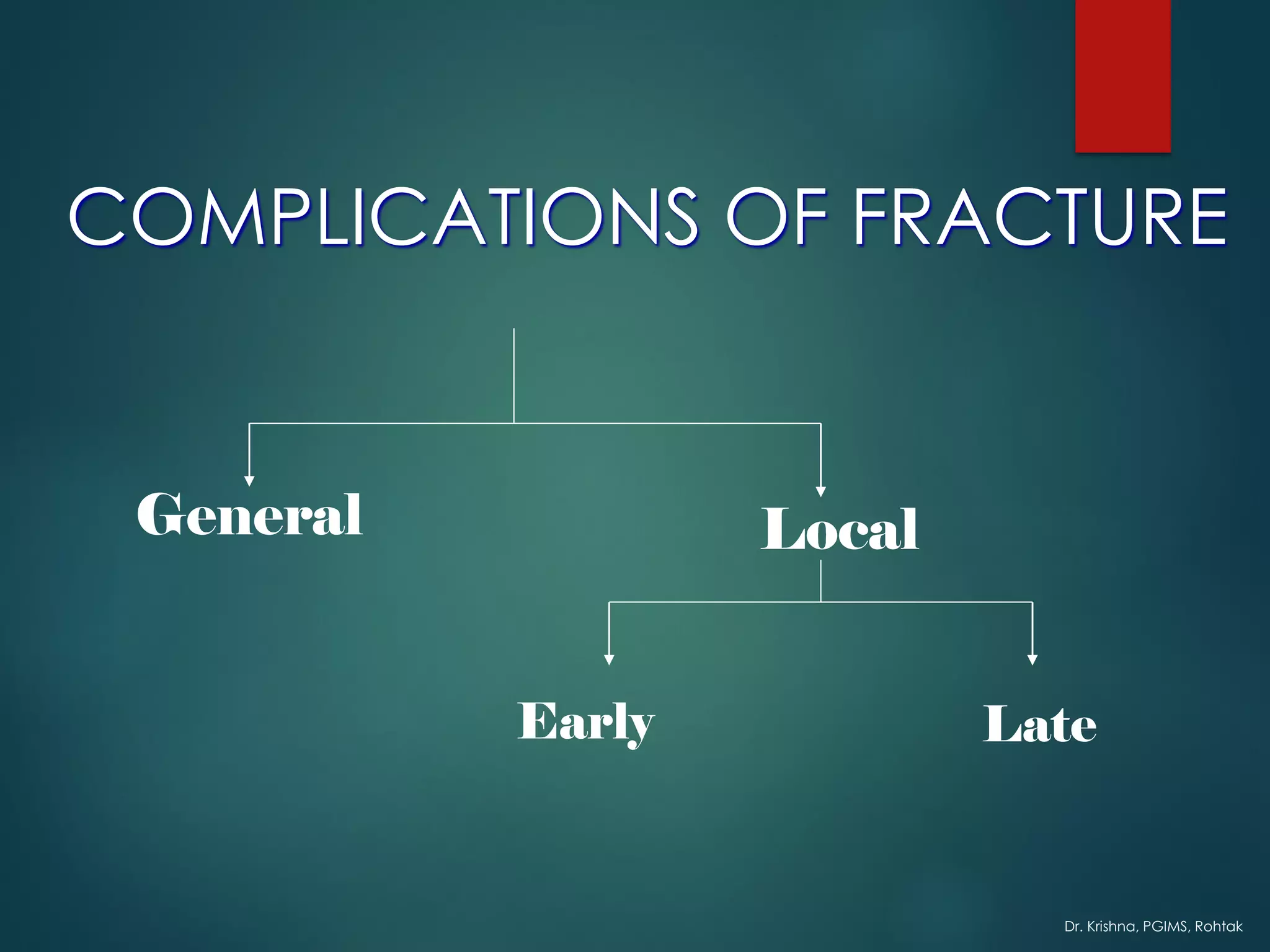
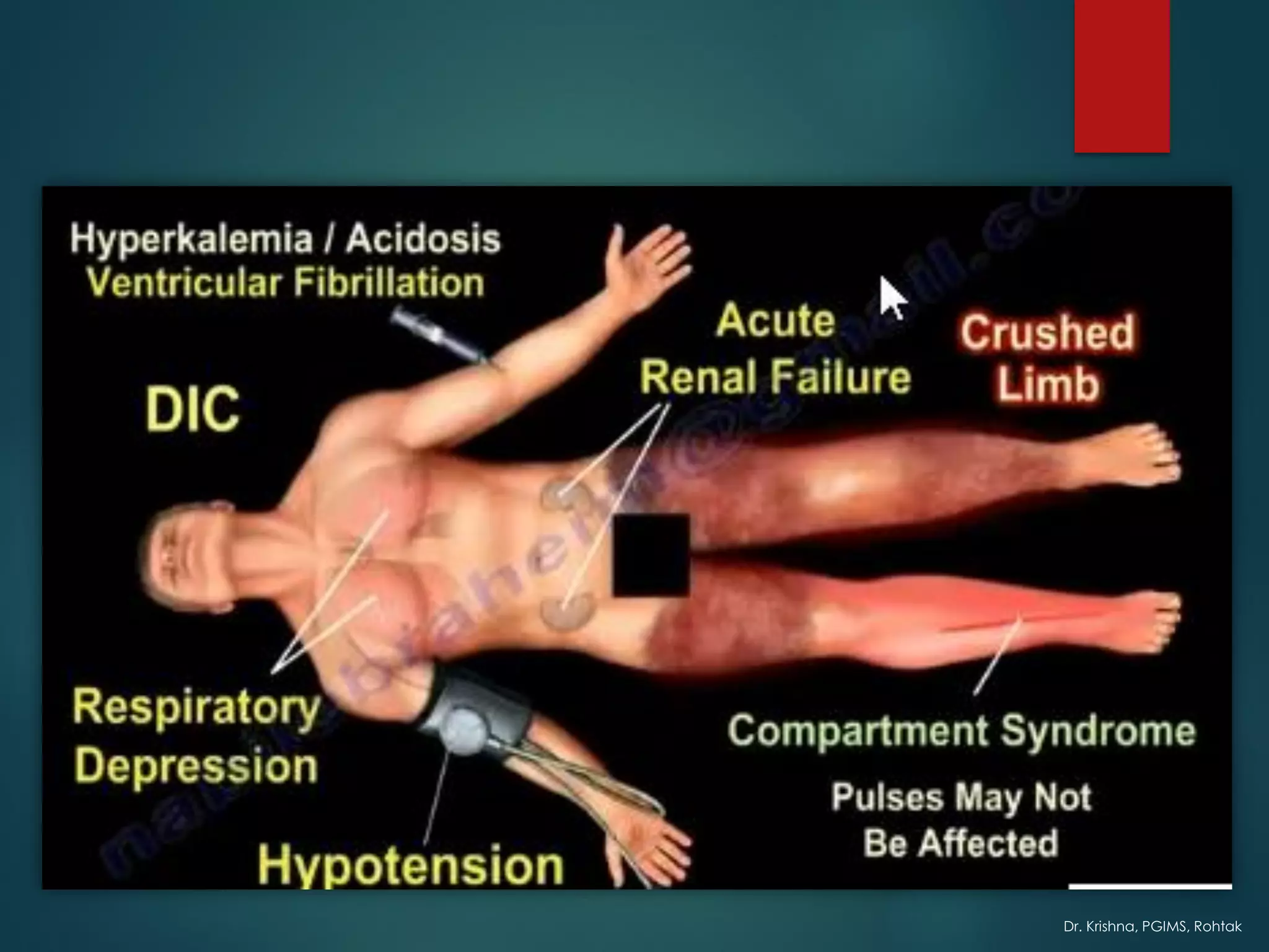
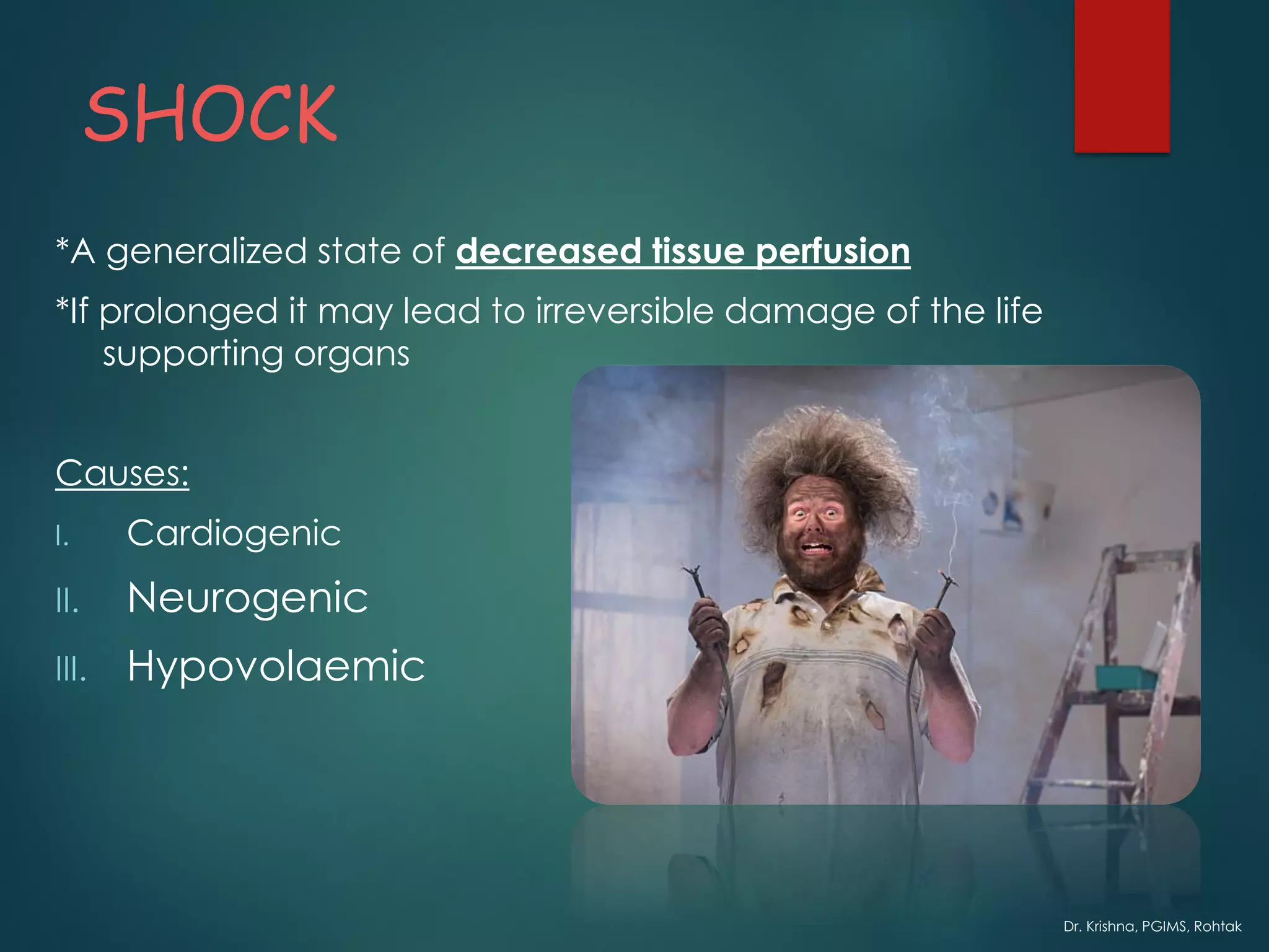
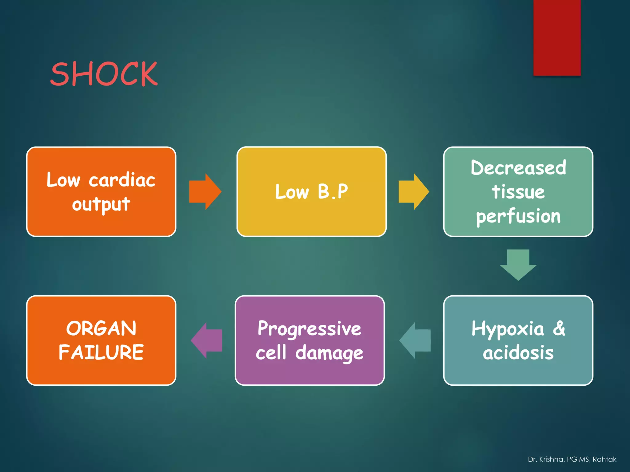
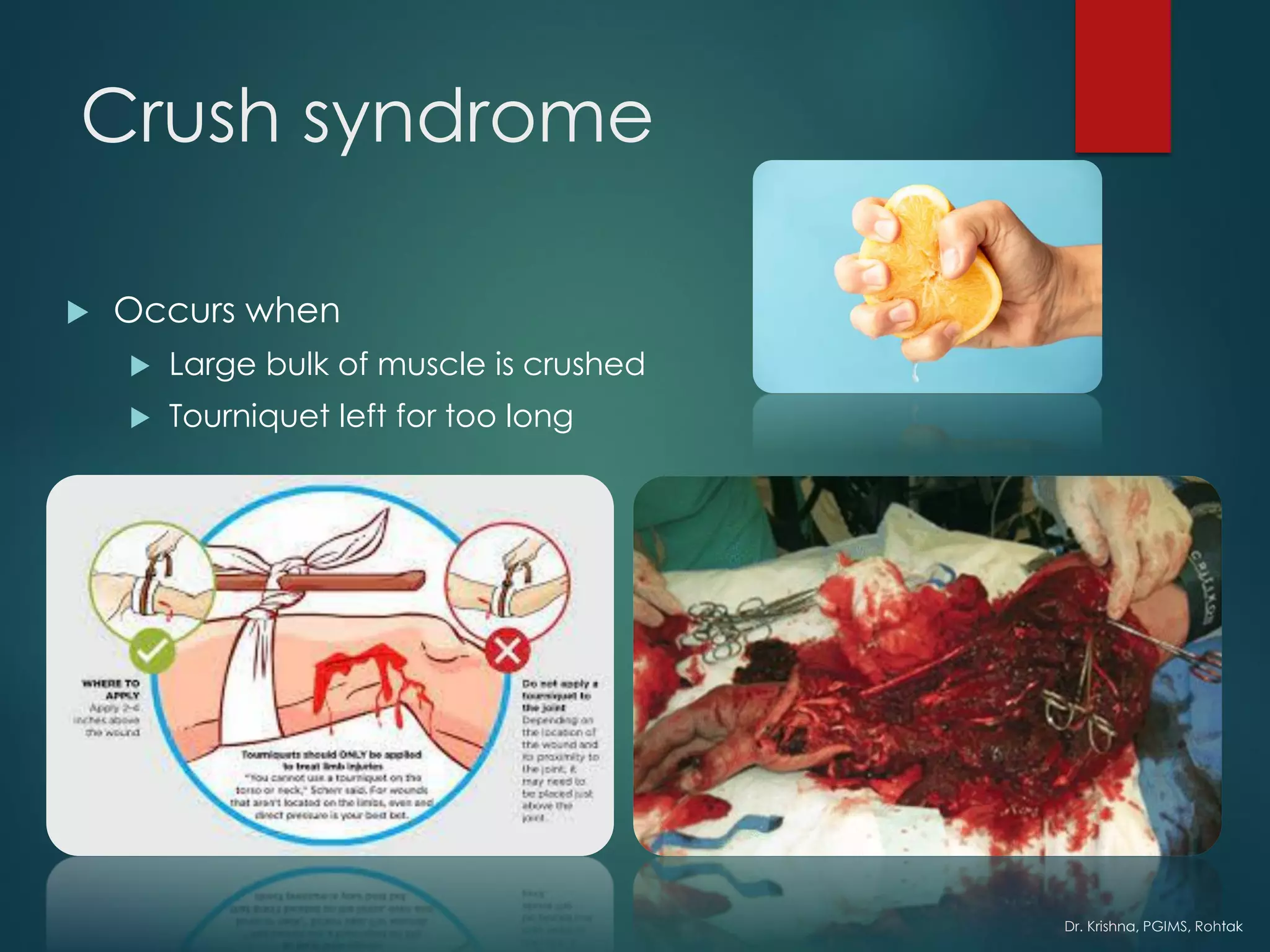
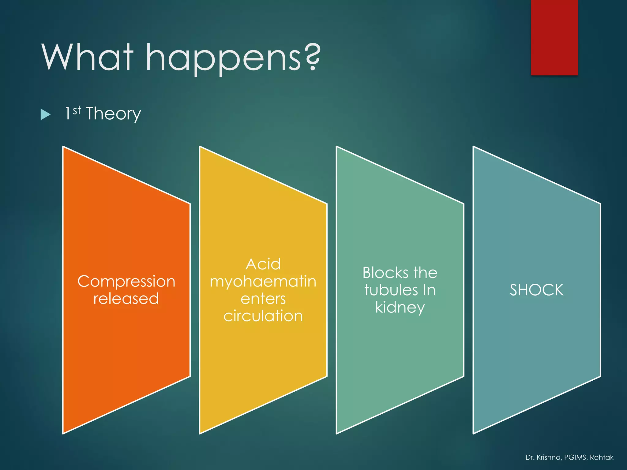
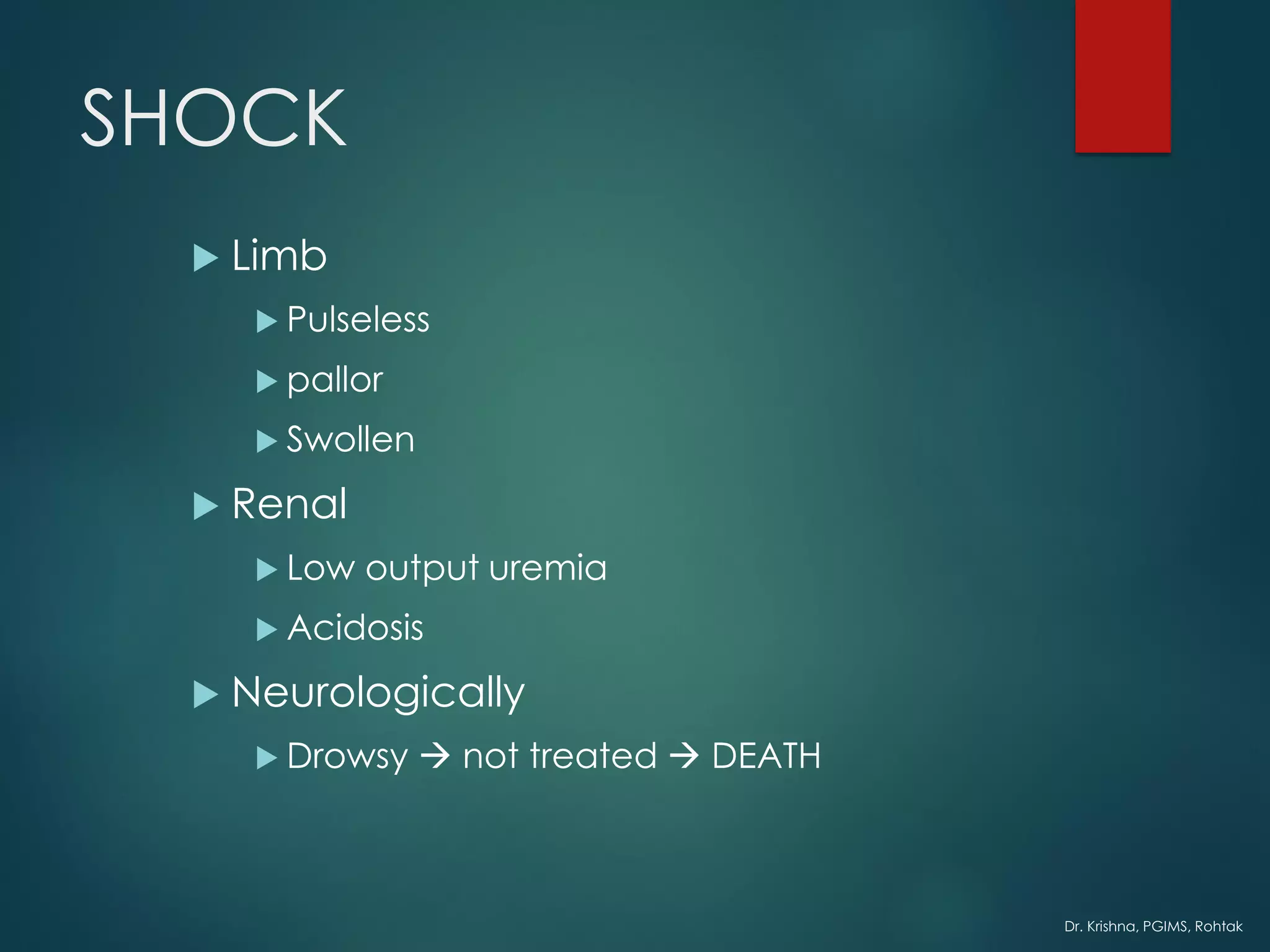
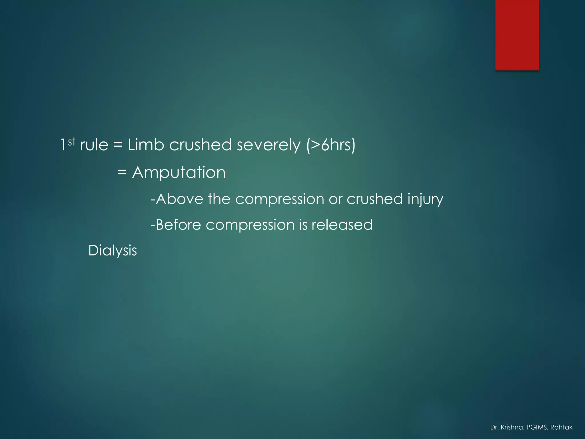
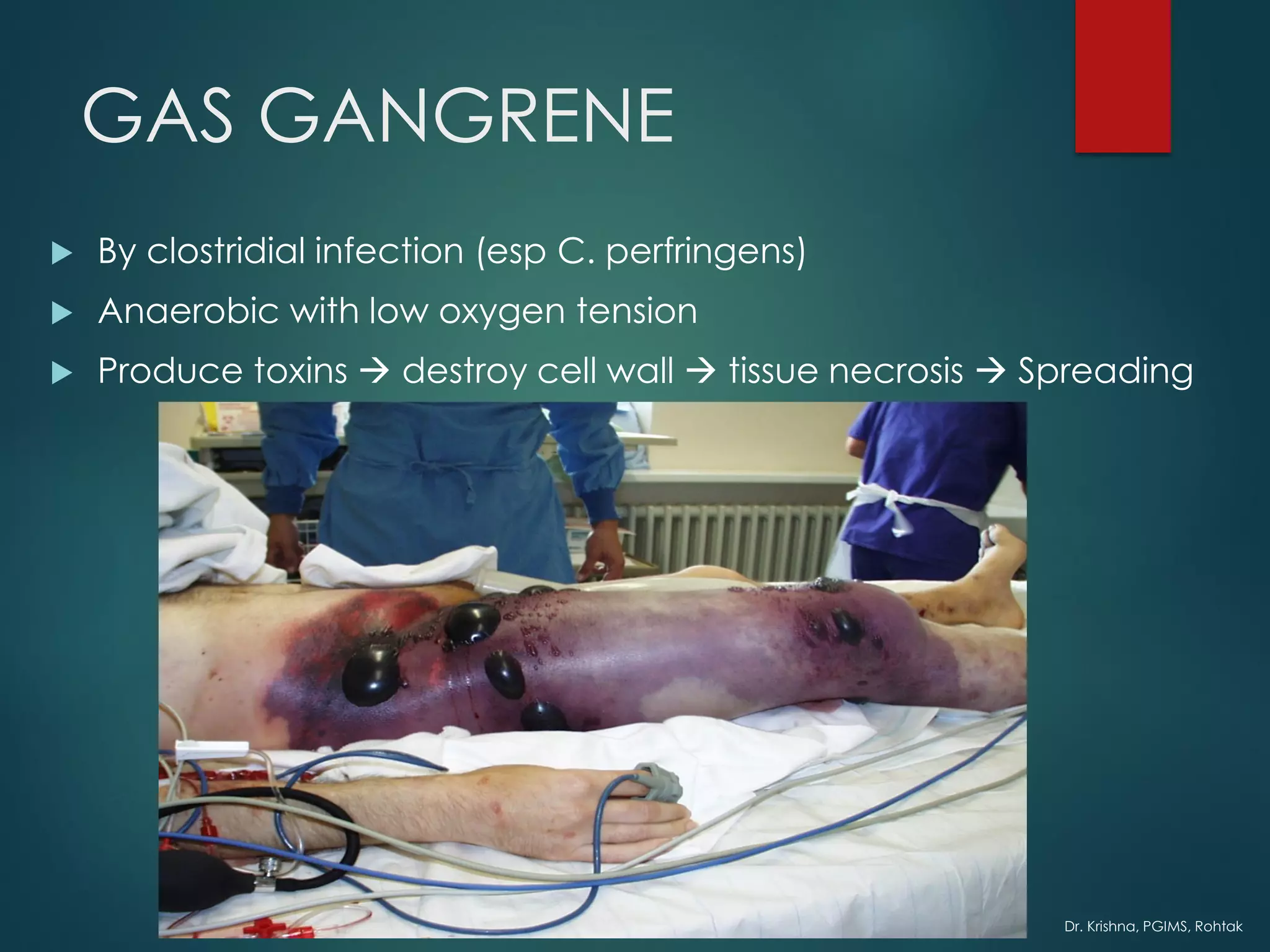
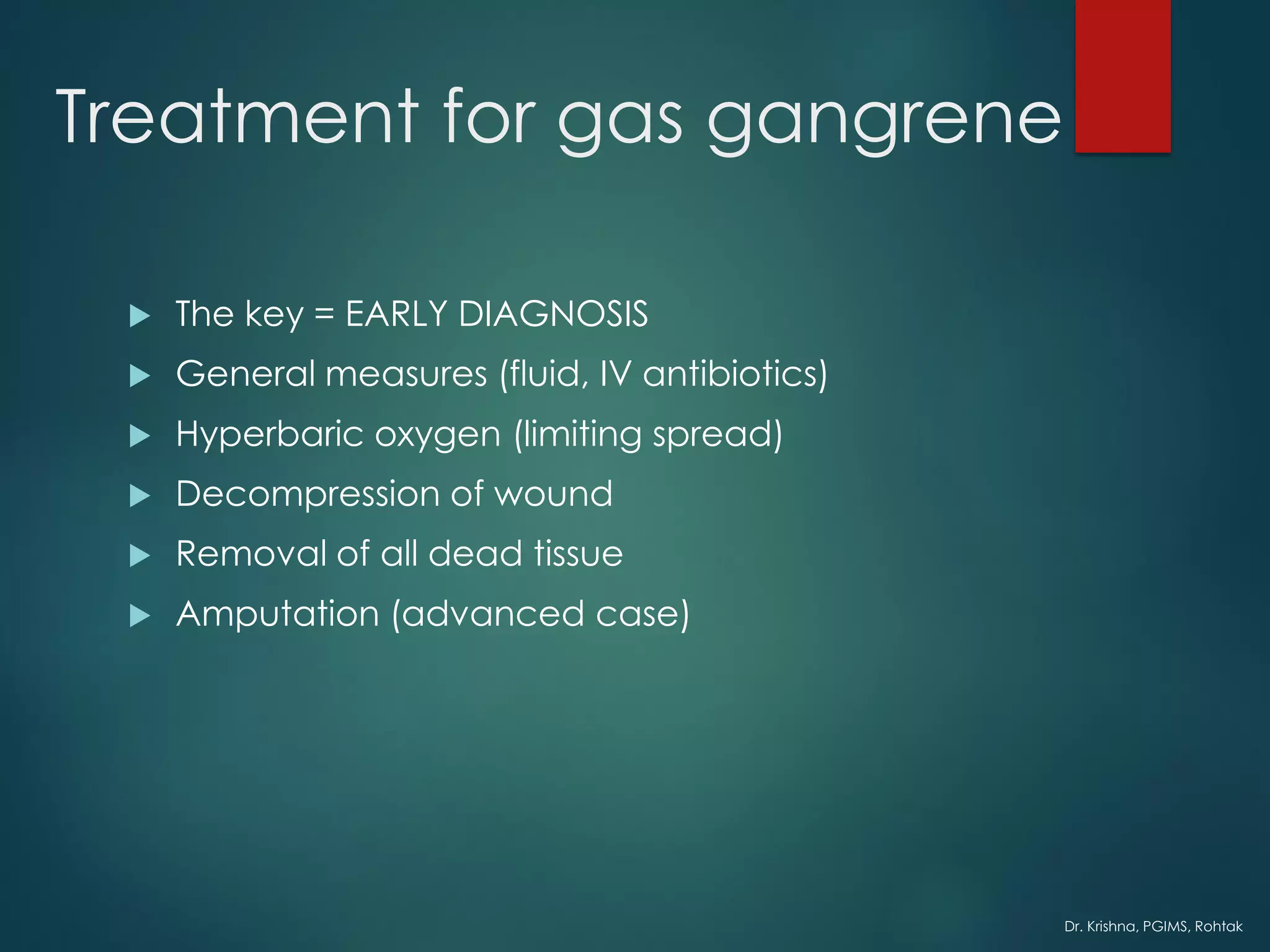
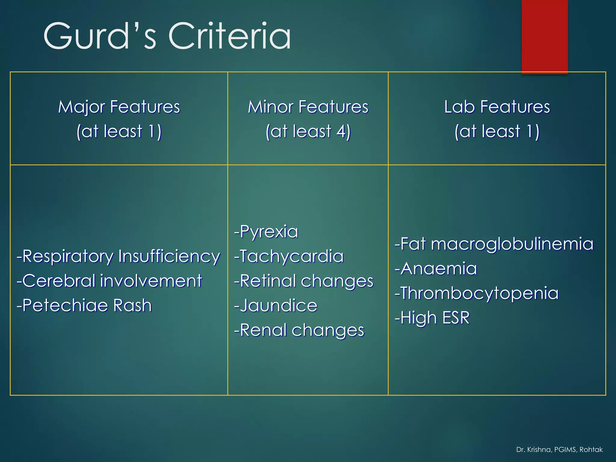
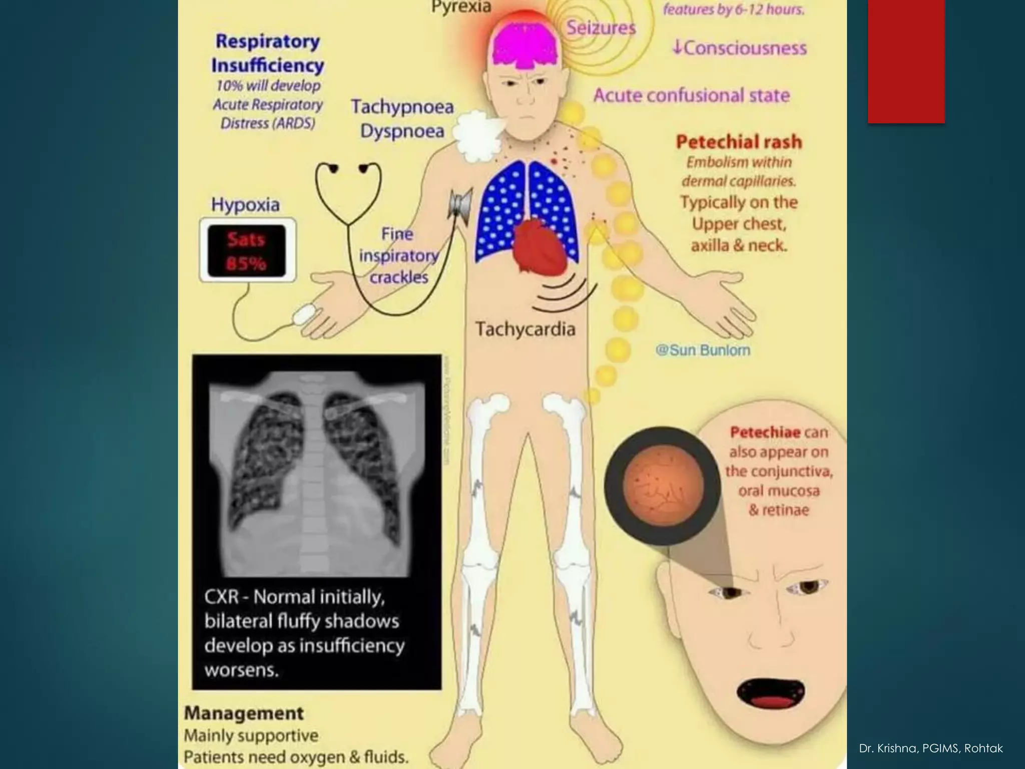
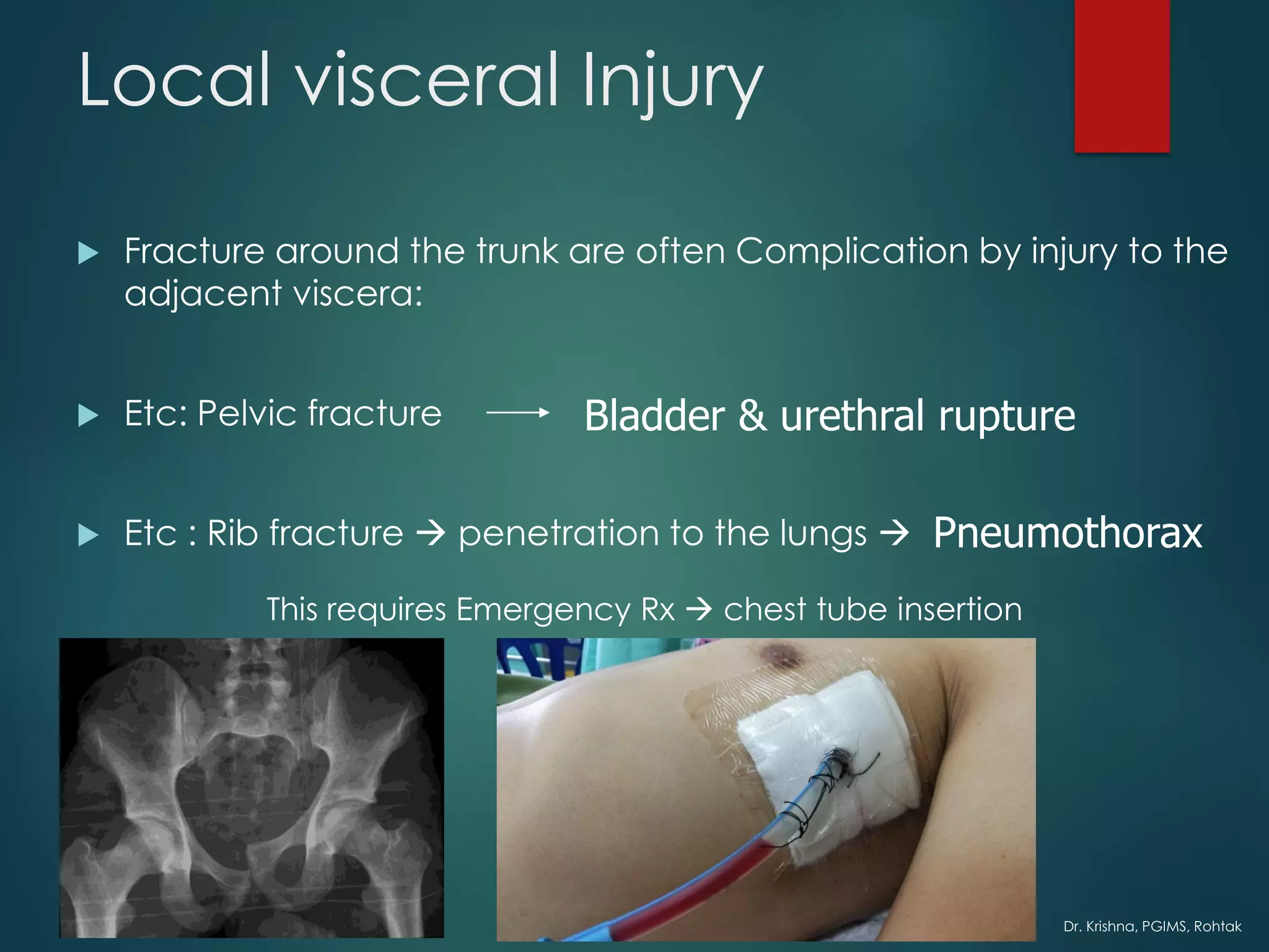
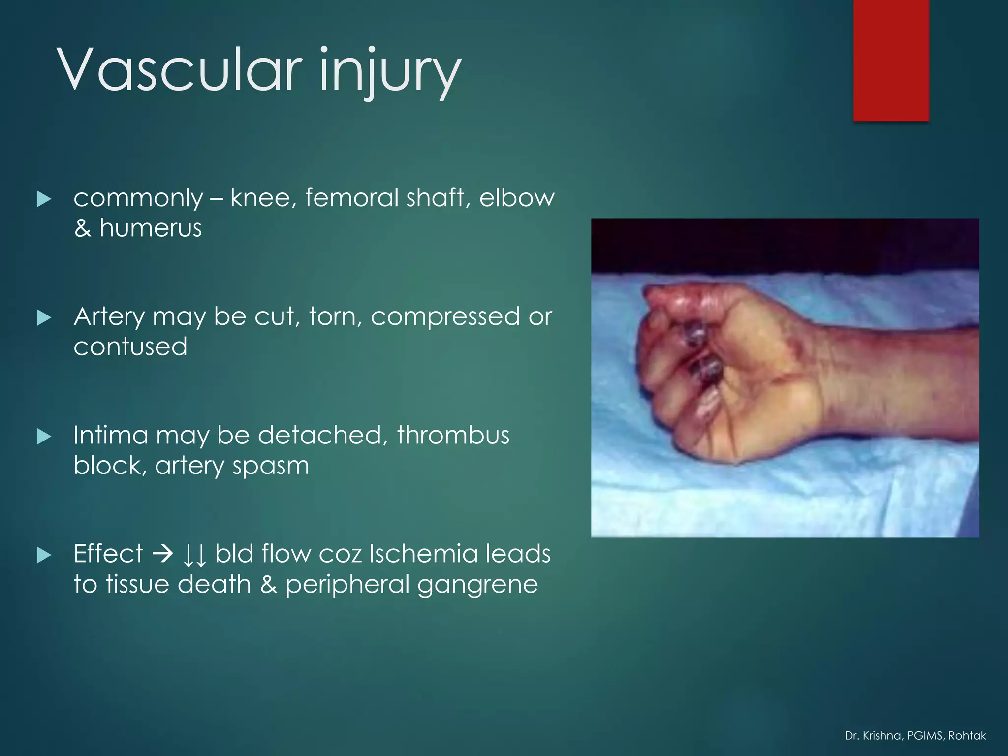
Dr. Krishna, PGIMS, Rohtak
COMPLICATIONS OF FRACTURE
General Local
Early Late
Dr. Krishna, PGIMS, Rohtak
General Complications
Bed sore & other complications of recumbency
Crush Syndrome (rhabdomyolysis)
Diffuse Coagulopathy
Embolism – Pulmonary & Venous Thrombosis & Electrolyte Imbalance
Fat Embolism
Gas gangrene
Hospital related - Complications of anaesthesia & Surgery
Infections incl Tetanus
Jolt (Shock)
Dr. Krishna, PGIMS, Rohtak
SHOCK
*A generalized state of decreased tissue perfusion
*If prolonged it may lead to irreversible damage of the life
supporting organs
Causes:
I. Cardiogenic
II. Neurogenic
III. Hypovolaemic
Dr. Krishna, PGIMS, Rohtak
Low cardiac
output
Low B.P
Decreased
tissue
perfusion
Hypoxia &
acidosis
Progressive
cell damage
ORGAN
FAILURE
SHOCK
Dr. Krishna, PGIMS, Rohtak
Crush syndrome
 Occurs when
 Large bulk of muscle is crushed
 Tourniquet left for too long
Dr. Krishna, PGIMS, Rohtak
What happens?
 1st Theory
Compression
released
Acid
myohaematin
enters
circulation
Blocks the
tubules In
kidney
SHOCK
Dr. Krishna, PGIMS, Rohtak
SHOCK
 Limb
 Pulseless
 pallor
 Swollen
 Renal
 Low output uremia
 Acidosis
 Neurologically
 Drowsy → not treated → DEATH
Dr. Krishna, PGIMS, Rohtak
1st rule = Limb crushed severely (>6hrs)
= Amputation
-Above the compression or crushed injury
-Before compression is released
Dialysis
Dr. Krishna, PGIMS, Rohtak
Venous thrombosis & Pulmonary
Embolism
 Commonest complication of Trauma & Surgery
 Most frequently
 Calf veins
 Less frequent in proximal thigh & pelvic veins
 Pulmonary Embolism
 From Proximal thigh & pelvis
 Incidence = 5% & Fatal = 0.5%
Dr. Krishna, PGIMS, Rohtak
VTE
 The primary cause in surgical
 HYPERCOAGULABILITY of the Blood
 due to activation of Factor X by Thromboplastin from
damaged tissues
 Thrombosis occurs → secondary factors involved
 Stasis
 Pressure
 Prolonged immobility
 Endothelial damage
 Increase in No. & stickiness of platelets
Dr. Krishna, PGIMS, Rohtak
c
Dr. Krishna, PGIMS, Rohtak
VTE - high risk group
 Old age
 Cardiovascular Disease
 Bedridden patient
 Patients undergoing hip arthroplasty
Dr. Krishna, PGIMS, Rohtak
VTE symptoms
 Pain the calf or thigh
 Soft tissue tenderness
 Sudden slight increase in
temperature
 Sudden increase in pulse rate
 Homann’s Sign
positive
Dr. Krishna, PGIMS, Rohtak
Diagnosis of DVT
 USG
 Doppler technique (measure blood
flow)
 Ascending venography (bilaterally)
 Radioactive iodine labelled
fibrinogen(clot)
Dr. Krishna, PGIMS, Rohtak
Pulmonary Embolism
 Difficult to diagnose =only minority have symptoms (chest pain,
dyspnea, hemoptysis)
• So high risk patients should be
examined for pulmonary consolidation
• X-ray
• Lung scintigraphy
• Pulmonary angiography
• Normal D-dimer has almost 100%
negative predictive value (virtually
excludes PE)
• CT
Dr. Krishna, PGIMS, Rohtak
H
A
M
P
T
O
N
’
S
H
U
M
P
Dr. Krishna, PGIMS, Rohtak
Prevention
 Prophylactic treatment
 Foot elevation
 Graduated compression stockings
 Exercise
 Anticoagulant treatment
 Subcut heparin or LMW heparin
Dr. Krishna, PGIMS, Rohtak
Rx of Pulmonary Embolism
 Cardiorespiratory resuscitation
 Vasopressor for shock
 Oxygen
 Large dose heparin (15 000 units)
 Streptokinase (dissolve clot)
 Antibiotics (prevent lung infection)
Dr. Krishna, PGIMS, Rohtak
TETANUS
Tetanus organism live only in dead tissue → exotoxin → blood & lymph
to CNS →anterior horn cell
 Will develop
 Tonic clonic contraction
 Jaw & face (trismus & risus sardonicus)
 Neck & trunk
 Diaphragm & Intercostal muscle → spasm → ASPHYXIA
https://www.youtube.com/watch?v=2baVlK5Uvyc
Dr. Krishna, PGIMS, Rohtak
Prophylaxis
 Active immunization (tetanus toxoid)
 Booster doses (immunized patients)
 Non Immunized patients
 Wound toilet & antibiotics
 If wound contaminated →antitoxin
Dr. Krishna, PGIMS, Rohtak
Treatment for Tetanus
 IV antitoxin
 Heavy Sedation
 Muscle Relaxant drug
 Tracheal Intubation
 Controlled respiration
Dr. Krishna, PGIMS, Rohtak
GAS GANGRENE
 By clostridial infection (esp C. perfringens)
 Anaerobic with low oxygen tension
 Produce toxins → destroy cell wall → tissue necrosis → Spreading
Dr. Krishna, PGIMS, Rohtak
Clinical Features
 Within 24 hours
 Intense pain
 Swelling
 Brownish discharge
 Pulse rate increased
 Characteristic smell sweetish & mousy odor
 Little or no pyrexia
 Gas formation not marked
 Toxaemic → coma → DEATH
Dr. Krishna, PGIMS, Rohtak
Prevention
 Deep penetrating wound should be EXPLORED
 ALL dead tissue → completely EXCISED
 Doubt about tissue viability → leave it OPEN
 No antitoxin
Dr. Krishna, PGIMS, Rohtak
Treatment for gas gangrene
 The key = EARLY DIAGNOSIS
 General measures (fluid, IV antibiotics)
 Hyperbaric oxygen (limiting spread)
 Decompression of wound
 Removal of all dead tissue
 Amputation (advanced case)
Dr. Krishna, PGIMS, Rohtak
FAT EMBOLISM
 Only minority patients with
circulating fat globules will develop
POST TRAUMATIC RESPIRATORY
DYSFUNCTION
 Source of fat emboli = bone
marrow
 Usually in MULTIPLE CLOSED
FRACTURE
 But other condition also reported
(burns, renal infarction,
cardiopulmonary operation)
Dr. Krishna, PGIMS, Rohtak
Closed/open
Fracture
Fat in bone
marrow
escapes
Formation of
fat globules in
vessels
Fat embolus
Stick in
pulmonary
bed
Trigger
clotting
cascade
Dr. Krishna, PGIMS, Rohtak
Features
 After 1-2 days of trauma
 Usually young adults with LL fracture
 Early warning signs
 Rise in temperature & pulse rate
 More pronounced case(classical triad)
 Breathlessness
 Mild mental confusion
 Petechia (chest & conjuntival fold)
Dr. Krishna, PGIMS, Rohtak
 Most severe case
 Marked respiratory distress →coma →ARDS
no definitive test, but hypoxia <60mmHg after major trauma is
suspicious
Dr. Krishna, PGIMS, Rohtak
Gurd’s Criteria
Major Features
(at least 1)
Minor Features
(at least 4)
Lab Features
(at least 1)
-Respiratory Insufficiency
-Cerebral involvement
-Petechiae Rash
-Pyrexia
-Tachycardia
-Retinal changes
-Jaundice
-Renal changes
-Fat macroglobulinemia
-Anaemia
-Thrombocytopenia
-High ESR
Dr. Krishna, PGIMS, Rohtak
Treatment
 Mild case
 Monitoring of blood PO2
 Signs of hypoxia (<8kPa @ 60mmHg)
 Oxygen
 If severe
 ICU with sedation & assisted ventilation
 Swan ganz Catheterization (monitor cardiac Fx)
 Fluid balance
 Supportive
 Heparin-thromboembolism
 Steroids-pulmonary oedema
 Aprotinin - prevent aggregation of chylomicrons
Dr. Krishna, PGIMS, Rohtak
Early (& immediate) Complications
 Local Visceral Injury
 Vascular Injury
 Nerve Injury
 Compartment Syndrome
 Hemarthrosis
 Infection
 Gas gangrene
Dr. Krishna, PGIMS, Rohtak
Local visceral Injury
 Fracture around the trunk are often Complication by injury to the
adjacent viscera:
 Etc: Pelvic fracture
 Etc : Rib fracture → penetration to the lungs → Pneumothorax
Bladder & urethral rupture
This requires Emergency Rx → chest tube insertion
Dr. Krishna, PGIMS, Rohtak
Vascular injury
 commonly – knee, femoral shaft, elbow
& humerus
 Artery may be cut, torn, compressed or
contused
 Intima may be detached, thrombus
block, artery spasm
 Effect → ↓↓ bld flow coz Ischemia leads
to tissue death & peripheral gangrene
Dr. Krishna, PGIMS, Rohtak
Vascular Injury - Clinical features
 Pt with ischemia may have 5 P’s:
- paraesthesia/numbness
- pain
- pallor
- pulselessness
- paralysis
 Investigate if suspect vascular injury : CT Angiogram
Dr. Krishna, PGIMS, Rohtak
Treatment
 Emergency treatment
 All bandages/splints removed
 X-Ray The fracture again
 Circulation reassessed for next half hour
 If no improvement, do vessels exploration
 Suture torn vessels, vein grafting, if thrombosed do
endarterectomy
 Aim: to restore bld flow
Dr. Krishna, PGIMS, Rohtak
Nerve Injury
 Variable degree of motor & sensory loss along the distribution of
the nerve
 May be neurapraxia, axonotmesis or neurotmesis
Dr. Krishna, PGIMS, Rohtak
Nerve Trauma Effect
Axillary Dislocation of shoulder Deltoid paralysis
Radial # of humerus Wrist drop
Median Supracondylar # of humerus Pointing index
Ulnar # medial epicondyl humerus Claw hand
Sciatic Post dislocation of hip Foot drop
Common
peroneal
Knee dislocation # neck of
fibula
Foot drop
Dr. Krishna, PGIMS, Rohtak
 In closed injuries – nerve is seldom severed &
spontaneous recovery should be awaited
 In open fractures – complete lesion (neurotmesis): the
nerve is explored during wound debridement &
repaired
 Acute nerve compression – occur with fracture or
dislocation around the wrist. C/o numbness in median
& ulnar dist. If no improvement >48 hours → after
fracture reduction, do nerve exploration &
decompression

complications of fracture.pdf

  • 1.
    Dr. Krishna, PGIMS,Rohtak COMPLICATIONS OF FRACTURE General Local Early Late
  • 2.
    Dr. Krishna, PGIMS,Rohtak General Complications Bed sore & other complications of recumbency Crush Syndrome (rhabdomyolysis) Diffuse Coagulopathy Embolism – Pulmonary & Venous Thrombosis & Electrolyte Imbalance Fat Embolism Gas gangrene Hospital related - Complications of anaesthesia & Surgery Infections incl Tetanus Jolt (Shock)
  • 3.
  • 4.
    Dr. Krishna, PGIMS,Rohtak SHOCK *A generalized state of decreased tissue perfusion *If prolonged it may lead to irreversible damage of the life supporting organs Causes: I. Cardiogenic II. Neurogenic III. Hypovolaemic
  • 5.
    Dr. Krishna, PGIMS,Rohtak Low cardiac output Low B.P Decreased tissue perfusion Hypoxia & acidosis Progressive cell damage ORGAN FAILURE SHOCK
  • 6.
    Dr. Krishna, PGIMS,Rohtak Crush syndrome  Occurs when  Large bulk of muscle is crushed  Tourniquet left for too long
  • 7.
    Dr. Krishna, PGIMS,Rohtak What happens?  1st Theory Compression released Acid myohaematin enters circulation Blocks the tubules In kidney SHOCK
  • 8.
    Dr. Krishna, PGIMS,Rohtak SHOCK  Limb  Pulseless  pallor  Swollen  Renal  Low output uremia  Acidosis  Neurologically  Drowsy → not treated → DEATH
  • 9.
    Dr. Krishna, PGIMS,Rohtak 1st rule = Limb crushed severely (>6hrs) = Amputation -Above the compression or crushed injury -Before compression is released Dialysis
  • 10.
    Dr. Krishna, PGIMS,Rohtak Venous thrombosis & Pulmonary Embolism  Commonest complication of Trauma & Surgery  Most frequently  Calf veins  Less frequent in proximal thigh & pelvic veins  Pulmonary Embolism  From Proximal thigh & pelvis  Incidence = 5% & Fatal = 0.5%
  • 11.
    Dr. Krishna, PGIMS,Rohtak VTE  The primary cause in surgical  HYPERCOAGULABILITY of the Blood  due to activation of Factor X by Thromboplastin from damaged tissues  Thrombosis occurs → secondary factors involved  Stasis  Pressure  Prolonged immobility  Endothelial damage  Increase in No. & stickiness of platelets
  • 12.
  • 13.
    Dr. Krishna, PGIMS,Rohtak VTE - high risk group  Old age  Cardiovascular Disease  Bedridden patient  Patients undergoing hip arthroplasty
  • 14.
    Dr. Krishna, PGIMS,Rohtak VTE symptoms  Pain the calf or thigh  Soft tissue tenderness  Sudden slight increase in temperature  Sudden increase in pulse rate  Homann’s Sign positive
  • 15.
  • 16.
    Dr. Krishna, PGIMS,Rohtak Diagnosis of DVT  USG  Doppler technique (measure blood flow)  Ascending venography (bilaterally)  Radioactive iodine labelled fibrinogen(clot)
  • 17.
    Dr. Krishna, PGIMS,Rohtak Pulmonary Embolism  Difficult to diagnose =only minority have symptoms (chest pain, dyspnea, hemoptysis) • So high risk patients should be examined for pulmonary consolidation • X-ray • Lung scintigraphy • Pulmonary angiography • Normal D-dimer has almost 100% negative predictive value (virtually excludes PE) • CT
  • 18.
    Dr. Krishna, PGIMS,Rohtak H A M P T O N ’ S H U M P
  • 19.
    Dr. Krishna, PGIMS,Rohtak Prevention  Prophylactic treatment  Foot elevation  Graduated compression stockings  Exercise  Anticoagulant treatment  Subcut heparin or LMW heparin
  • 20.
    Dr. Krishna, PGIMS,Rohtak Rx of Pulmonary Embolism  Cardiorespiratory resuscitation  Vasopressor for shock  Oxygen  Large dose heparin (15 000 units)  Streptokinase (dissolve clot)  Antibiotics (prevent lung infection)
  • 21.
    Dr. Krishna, PGIMS,Rohtak TETANUS Tetanus organism live only in dead tissue → exotoxin → blood & lymph to CNS →anterior horn cell  Will develop  Tonic clonic contraction  Jaw & face (trismus & risus sardonicus)  Neck & trunk  Diaphragm & Intercostal muscle → spasm → ASPHYXIA https://www.youtube.com/watch?v=2baVlK5Uvyc
  • 22.
    Dr. Krishna, PGIMS,Rohtak Prophylaxis  Active immunization (tetanus toxoid)  Booster doses (immunized patients)  Non Immunized patients  Wound toilet & antibiotics  If wound contaminated →antitoxin
  • 23.
    Dr. Krishna, PGIMS,Rohtak Treatment for Tetanus  IV antitoxin  Heavy Sedation  Muscle Relaxant drug  Tracheal Intubation  Controlled respiration
  • 24.
    Dr. Krishna, PGIMS,Rohtak GAS GANGRENE  By clostridial infection (esp C. perfringens)  Anaerobic with low oxygen tension  Produce toxins → destroy cell wall → tissue necrosis → Spreading
  • 25.
    Dr. Krishna, PGIMS,Rohtak Clinical Features  Within 24 hours  Intense pain  Swelling  Brownish discharge  Pulse rate increased  Characteristic smell sweetish & mousy odor  Little or no pyrexia  Gas formation not marked  Toxaemic → coma → DEATH
  • 26.
    Dr. Krishna, PGIMS,Rohtak Prevention  Deep penetrating wound should be EXPLORED  ALL dead tissue → completely EXCISED  Doubt about tissue viability → leave it OPEN  No antitoxin
  • 27.
    Dr. Krishna, PGIMS,Rohtak Treatment for gas gangrene  The key = EARLY DIAGNOSIS  General measures (fluid, IV antibiotics)  Hyperbaric oxygen (limiting spread)  Decompression of wound  Removal of all dead tissue  Amputation (advanced case)
  • 28.
    Dr. Krishna, PGIMS,Rohtak FAT EMBOLISM  Only minority patients with circulating fat globules will develop POST TRAUMATIC RESPIRATORY DYSFUNCTION  Source of fat emboli = bone marrow  Usually in MULTIPLE CLOSED FRACTURE  But other condition also reported (burns, renal infarction, cardiopulmonary operation)
  • 29.
    Dr. Krishna, PGIMS,Rohtak Closed/open Fracture Fat in bone marrow escapes Formation of fat globules in vessels Fat embolus Stick in pulmonary bed Trigger clotting cascade
  • 30.
    Dr. Krishna, PGIMS,Rohtak Features  After 1-2 days of trauma  Usually young adults with LL fracture  Early warning signs  Rise in temperature & pulse rate  More pronounced case(classical triad)  Breathlessness  Mild mental confusion  Petechia (chest & conjuntival fold)
  • 31.
    Dr. Krishna, PGIMS,Rohtak  Most severe case  Marked respiratory distress →coma →ARDS no definitive test, but hypoxia <60mmHg after major trauma is suspicious
  • 32.
    Dr. Krishna, PGIMS,Rohtak Gurd’s Criteria Major Features (at least 1) Minor Features (at least 4) Lab Features (at least 1) -Respiratory Insufficiency -Cerebral involvement -Petechiae Rash -Pyrexia -Tachycardia -Retinal changes -Jaundice -Renal changes -Fat macroglobulinemia -Anaemia -Thrombocytopenia -High ESR
  • 33.
  • 34.
    Dr. Krishna, PGIMS,Rohtak Treatment  Mild case  Monitoring of blood PO2  Signs of hypoxia (<8kPa @ 60mmHg)  Oxygen  If severe  ICU with sedation & assisted ventilation  Swan ganz Catheterization (monitor cardiac Fx)  Fluid balance  Supportive  Heparin-thromboembolism  Steroids-pulmonary oedema  Aprotinin - prevent aggregation of chylomicrons
  • 35.
    Dr. Krishna, PGIMS,Rohtak Early (& immediate) Complications  Local Visceral Injury  Vascular Injury  Nerve Injury  Compartment Syndrome  Hemarthrosis  Infection  Gas gangrene
  • 36.
    Dr. Krishna, PGIMS,Rohtak Local visceral Injury  Fracture around the trunk are often Complication by injury to the adjacent viscera:  Etc: Pelvic fracture  Etc : Rib fracture → penetration to the lungs → Pneumothorax Bladder & urethral rupture This requires Emergency Rx → chest tube insertion
  • 37.
  • 38.
    Dr. Krishna, PGIMS,Rohtak Vascular injury  commonly – knee, femoral shaft, elbow & humerus  Artery may be cut, torn, compressed or contused  Intima may be detached, thrombus block, artery spasm  Effect → ↓↓ bld flow coz Ischemia leads to tissue death & peripheral gangrene
  • 39.
    Dr. Krishna, PGIMS,Rohtak Vascular Injury - Clinical features  Pt with ischemia may have 5 P’s: - paraesthesia/numbness - pain - pallor - pulselessness - paralysis  Investigate if suspect vascular injury : CT Angiogram
  • 40.
    Dr. Krishna, PGIMS,Rohtak Treatment  Emergency treatment  All bandages/splints removed  X-Ray The fracture again  Circulation reassessed for next half hour  If no improvement, do vessels exploration  Suture torn vessels, vein grafting, if thrombosed do endarterectomy  Aim: to restore bld flow
  • 41.
    Dr. Krishna, PGIMS,Rohtak Nerve Injury  Variable degree of motor & sensory loss along the distribution of the nerve  May be neurapraxia, axonotmesis or neurotmesis
  • 42.
  • 43.
    Dr. Krishna, PGIMS,Rohtak Nerve Trauma Effect Axillary Dislocation of shoulder Deltoid paralysis Radial # of humerus Wrist drop Median Supracondylar # of humerus Pointing index Ulnar # medial epicondyl humerus Claw hand Sciatic Post dislocation of hip Foot drop Common peroneal Knee dislocation # neck of fibula Foot drop
  • 44.
    Dr. Krishna, PGIMS,Rohtak  In closed injuries – nerve is seldom severed & spontaneous recovery should be awaited  In open fractures – complete lesion (neurotmesis): the nerve is explored during wound debridement & repaired  Acute nerve compression – occur with fracture or dislocation around the wrist. C/o numbness in median & ulnar dist. If no improvement >48 hours → after fracture reduction, do nerve exploration & decompression