Download to read offline


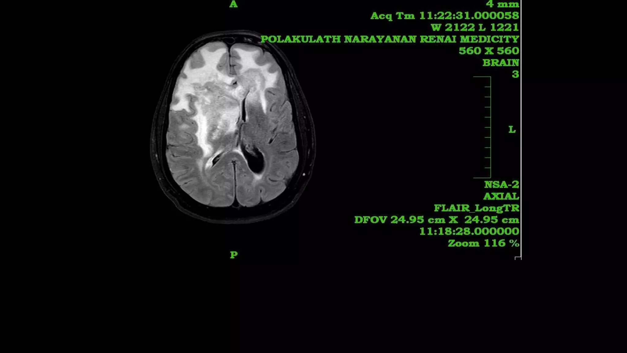


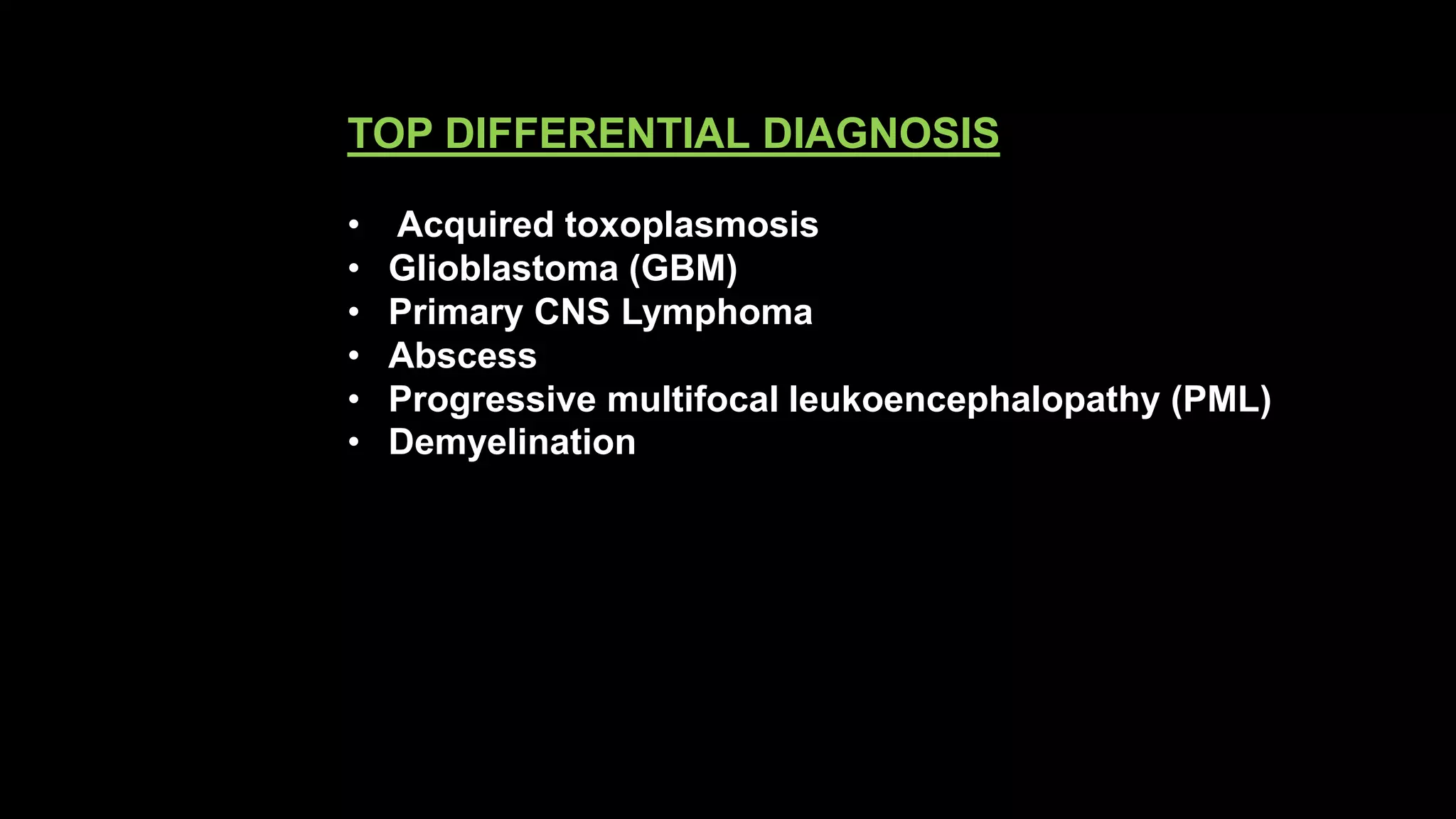


The document discusses the case of a 56-year-old male patient presenting with lesions in the brain. The top differential diagnoses listed are glioblastoma, primary CNS lymphoma, progressive multifocal leukoencephalopathy, and demyelination. Various MRI techniques are discussed for evaluating the lesions. After reviewing the imaging, the final diagnosis is determined to be primary CNS lymphoma, based on the characteristic enhancing lesion seen within the basal ganglia and periventricular white matter. Key features of primary CNS lymphoma on imaging and pathology are described.


