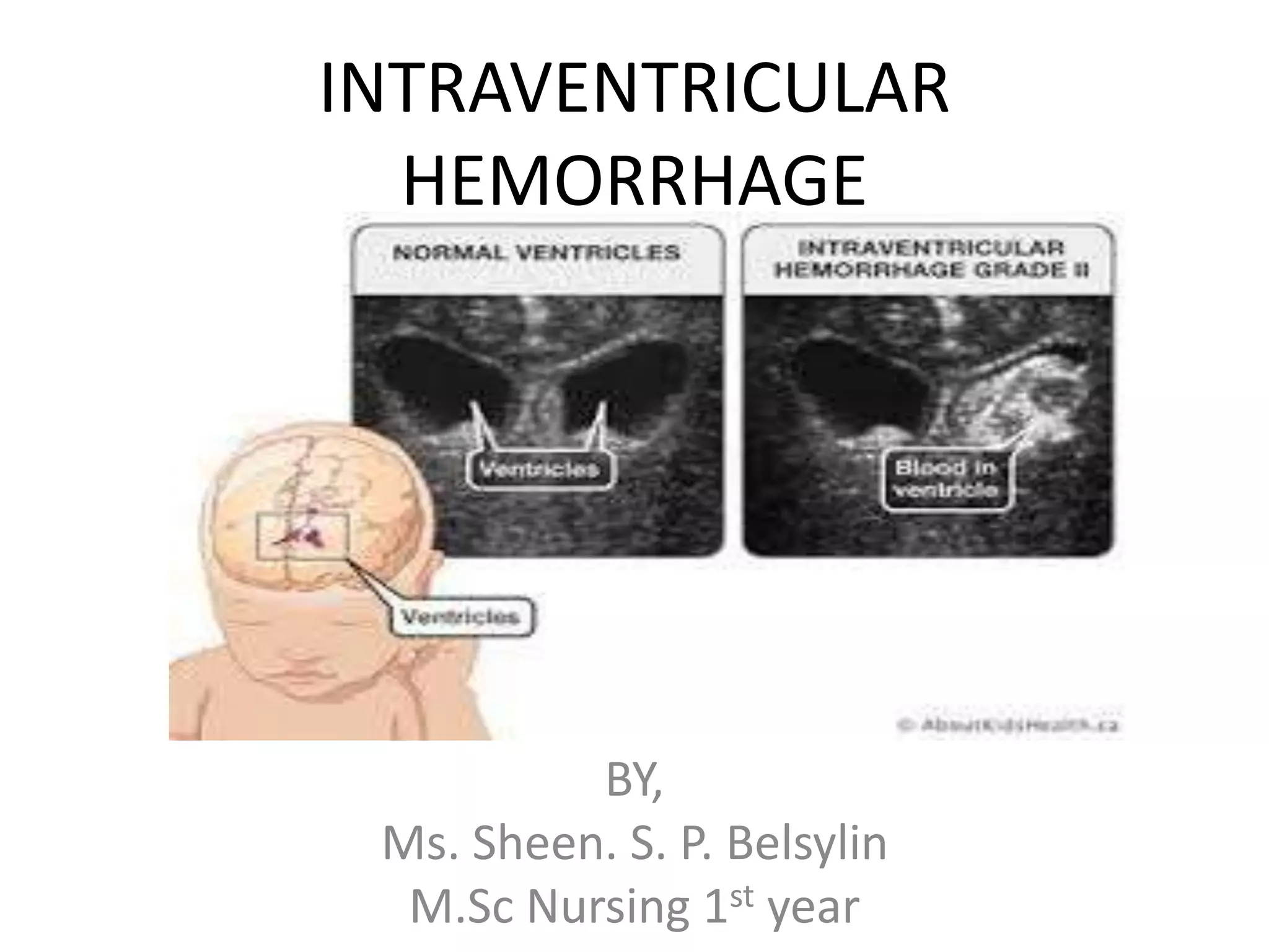
INTRAVENTRICULAR
HEMORRHAGE
BY,
Ms. Sheen. S. P. Belsylin
M.Sc Nursing 1st year
Introduction
• Intraventricular hemorrhage (IVH) of the
newborn is bleeding into the fluid-filled areas
(ventricles) inside the brain.
• The condition occurs most often in babies that
are premature
Causes
IVH is more common in premature babies with:
• Respiratory distress syndrome
• Unstable blood pressure
• Other medical conditions at birth
Infants born more than 10 weeks early are at
highest risk for this type of bleeding.
Grades
1. Grade1:Isolated Germinal Matrix Hemorrhage-
restricted to subependymal region
2. Grade2:IVH without ventricular dilatation-
extension into the normal sized ventricles. Fills
<50% of the volume of ventriclres.
3. Grade3:IVH with ventricular dilatation-
extension into dilated ventricles
4. Grade4:IVH with Parenchymal Extension-
extension into the parenchyma. Blood clots can
form and block the flow of CSF leading to
Hydrocephalus.
Symptoms
There may be no symptoms. The most common
symptoms seen in premature infants include:
• Breathing pauses (apnea)
• Changes in blood pressure and heart rate
• Decreased muscle tone
• Decreased reflexes
• Excessive sleep
• Lethargy
• Weak suck
• Seizures and other abnormal movements
Diagnosis
• All babies born before 30 weeks should have
an ultrasound of the head to screen for IVH.
The test is done in the 1 to 2 weeks of life.
Babies born between 30 to 34 weeks may also
have ultrasound screening if they have
symptoms of the problem.
• A second screening ultrasound may be done
around the time the baby was originally
expected to be born (the due date).
Management
• There is no way to stop bleeding associated with
IVH.
• Effort is made to keep the infant stable and treat
any symptoms.
• For example, a blood transfusion may be given to
improve blood pressure and blood count.
• If fluid builds up to the point that there is concern
about pressure on the brain, a spinal tap may be
done to drain fluid and try to relieve pressure.
• If this helps, surgery may be needed to place a
tube (shunt) in the brain to drain fluid.
Prevention
• Pregnant women who are at high risk of
delivering early should be given corticosteroids.
These drugs can help reduce the baby's risk for
IVH.
• Women who are on medicines that affect
bleeding risks should get vitamin K before
delivery.
• Premature babies whose umbilical cords are not
clamped right away have less risk of IVH.
• Premature babies who are born in a hospital with
a NICU and do not have to be transported after
birth also have less risk of IVH.
Prognosis
• Depends on how premature the baby is and
the grade of the hemorrhage.
• Less than half of babies with lower-grade
bleeding have long term problems.
• Severe bleeding often leads to developmental
delays and problems controlling movement.
Journal information
• A study on Intraventricular Hemorrhage and
Neurodevelopmental Outcomes in Extreme
Preterm Infants revealed that Grade I–II IVH,
even with no documented white matter injury
or other late ultrasound abnormalities, is
associated with adverse neurodevelopmental
outcomes in extremely preterm infants.
Pulmonary Hemorrhage
Definition
• Pulmonary hemorrhage (P-Hem) is an acute,
catastrophic event characterized by discharge
of bloody fluid from the upper respiratory
tract or the endotracheal tube.
• P-Hem is usually massive, is associated with
bleeding in other sites, involves more than
one third of the lungs, and has a high
mortality rate.
Statistics
• The incidence of P-Hem is 1 in 1,000 live
births. P-Hem is present in 7 to 10% of
neonatal autopsies, but up to 80% of
autopsies of very preterm infants.
Etiology
• Prematurity is the factor most commonly associated with P-
Hem
• Factors that favor increased filtration of fluid from
pulmonary capillaries (e.g., low concentration of plasma
proteins, high alveolar surface tension, lung damage,
hypervolemia).
• Other associated factors are those that predispose to
perinatal asphyxia or bleeding disorders, including
o toxemia of pregnancy
o maternal cocaine use
o erythroblastosis fetalis
o breech delivery, hypothermia
o Infection
o Respiratory Distress Syndrome
o administration of exogenous surfactant (in some studies)
o ECMO.
Pathogenesis
Asphyxial insult
Resultant myocardial failure
Increased pulmonary microvascular pressure
Pulmonary edema
Frank bleeding into the pulmonary interstitial and
alveolar spaces.
Clinical features
• Oozing of bloody fluid from the nose and
mouth or endotracheal tube with
• Rapid worsening of the respiratory status
• Cyanosis
• Shock
• Bleeding may be noted from other sites
Diagnosis
• Radiographic findings range from patchy
infiltrates to complete opacification of lung
fields.
• Hematocrit of the P-Hem fluid is usually 15 to
20% less than blood
Management
• Tracheal suction
• Oxygen and positive pressure ventilation.
• To assist in decreasing P-Hem, mean airway
pressure should be increased, either by a
relatively high PEEP (i.e., 6 to 10 cmH2O) or by
high frequency ventilation.
• Correct underlying abnormalities, especially
disorders of coagulation.
• When blood loss is large, prompt blood
transfusion may be needed to maintain an
adequate circulating blood volume.
Journal information
• Pulmonary hemorrhage in premature infants after
treatment with synthetic surfactant: An autopsy evaluation
• This study says that Pulmonary hemorrhage was present in
55% of 159 infants undergoing autopsy
• the incidence was not different in infants treated with
surfactant or air placebo.
• Birth weight was inversely related to the incidence of
pulmonary hemorrhage in both groups.
• Pulmonary pathologic findings significantly associated with
pulmonary hemorrhage included pulmonary interstitial
emphysema and necrotizing laryngotracheitis in both
groups.
• In the surfactant group, patent ductus arteriosus,
intraventricular hemorrhage, and pneumothorax were
significantly more frequent among those who developed
pulmonary hemorrhage.

Intraventricular hemorrhage

  • 1.
    INTRAVENTRICULAR HEMORRHAGE BY, Ms. Sheen. S.P. Belsylin M.Sc Nursing 1st year
  • 2.
    Introduction • Intraventricular hemorrhage(IVH) of the newborn is bleeding into the fluid-filled areas (ventricles) inside the brain. • The condition occurs most often in babies that are premature
  • 3.
    Causes IVH is morecommon in premature babies with: • Respiratory distress syndrome • Unstable blood pressure • Other medical conditions at birth Infants born more than 10 weeks early are at highest risk for this type of bleeding.
  • 4.
    Grades 1. Grade1:Isolated GerminalMatrix Hemorrhage- restricted to subependymal region 2. Grade2:IVH without ventricular dilatation- extension into the normal sized ventricles. Fills <50% of the volume of ventriclres. 3. Grade3:IVH with ventricular dilatation- extension into dilated ventricles 4. Grade4:IVH with Parenchymal Extension- extension into the parenchyma. Blood clots can form and block the flow of CSF leading to Hydrocephalus.
  • 5.
    Symptoms There may beno symptoms. The most common symptoms seen in premature infants include: • Breathing pauses (apnea) • Changes in blood pressure and heart rate • Decreased muscle tone • Decreased reflexes • Excessive sleep • Lethargy • Weak suck • Seizures and other abnormal movements
  • 6.
    Diagnosis • All babiesborn before 30 weeks should have an ultrasound of the head to screen for IVH. The test is done in the 1 to 2 weeks of life. Babies born between 30 to 34 weeks may also have ultrasound screening if they have symptoms of the problem. • A second screening ultrasound may be done around the time the baby was originally expected to be born (the due date).
  • 7.
    Management • There isno way to stop bleeding associated with IVH. • Effort is made to keep the infant stable and treat any symptoms. • For example, a blood transfusion may be given to improve blood pressure and blood count. • If fluid builds up to the point that there is concern about pressure on the brain, a spinal tap may be done to drain fluid and try to relieve pressure. • If this helps, surgery may be needed to place a tube (shunt) in the brain to drain fluid.
  • 8.
    Prevention • Pregnant womenwho are at high risk of delivering early should be given corticosteroids. These drugs can help reduce the baby's risk for IVH. • Women who are on medicines that affect bleeding risks should get vitamin K before delivery. • Premature babies whose umbilical cords are not clamped right away have less risk of IVH. • Premature babies who are born in a hospital with a NICU and do not have to be transported after birth also have less risk of IVH.
  • 9.
    Prognosis • Depends onhow premature the baby is and the grade of the hemorrhage. • Less than half of babies with lower-grade bleeding have long term problems. • Severe bleeding often leads to developmental delays and problems controlling movement.
  • 10.
    Journal information • Astudy on Intraventricular Hemorrhage and Neurodevelopmental Outcomes in Extreme Preterm Infants revealed that Grade I–II IVH, even with no documented white matter injury or other late ultrasound abnormalities, is associated with adverse neurodevelopmental outcomes in extremely preterm infants.
  • 11.
  • 12.
    Definition • Pulmonary hemorrhage(P-Hem) is an acute, catastrophic event characterized by discharge of bloody fluid from the upper respiratory tract or the endotracheal tube. • P-Hem is usually massive, is associated with bleeding in other sites, involves more than one third of the lungs, and has a high mortality rate.
  • 13.
    Statistics • The incidenceof P-Hem is 1 in 1,000 live births. P-Hem is present in 7 to 10% of neonatal autopsies, but up to 80% of autopsies of very preterm infants.
  • 14.
    Etiology • Prematurity isthe factor most commonly associated with P- Hem • Factors that favor increased filtration of fluid from pulmonary capillaries (e.g., low concentration of plasma proteins, high alveolar surface tension, lung damage, hypervolemia). • Other associated factors are those that predispose to perinatal asphyxia or bleeding disorders, including o toxemia of pregnancy o maternal cocaine use o erythroblastosis fetalis o breech delivery, hypothermia o Infection o Respiratory Distress Syndrome o administration of exogenous surfactant (in some studies) o ECMO.
  • 15.
    Pathogenesis Asphyxial insult Resultant myocardialfailure Increased pulmonary microvascular pressure Pulmonary edema Frank bleeding into the pulmonary interstitial and alveolar spaces.
  • 16.
    Clinical features • Oozingof bloody fluid from the nose and mouth or endotracheal tube with • Rapid worsening of the respiratory status • Cyanosis • Shock • Bleeding may be noted from other sites
  • 17.
    Diagnosis • Radiographic findingsrange from patchy infiltrates to complete opacification of lung fields. • Hematocrit of the P-Hem fluid is usually 15 to 20% less than blood
  • 18.
    Management • Tracheal suction •Oxygen and positive pressure ventilation. • To assist in decreasing P-Hem, mean airway pressure should be increased, either by a relatively high PEEP (i.e., 6 to 10 cmH2O) or by high frequency ventilation. • Correct underlying abnormalities, especially disorders of coagulation. • When blood loss is large, prompt blood transfusion may be needed to maintain an adequate circulating blood volume.
  • 19.
    Journal information • Pulmonaryhemorrhage in premature infants after treatment with synthetic surfactant: An autopsy evaluation • This study says that Pulmonary hemorrhage was present in 55% of 159 infants undergoing autopsy • the incidence was not different in infants treated with surfactant or air placebo. • Birth weight was inversely related to the incidence of pulmonary hemorrhage in both groups. • Pulmonary pathologic findings significantly associated with pulmonary hemorrhage included pulmonary interstitial emphysema and necrotizing laryngotracheitis in both groups. • In the surfactant group, patent ductus arteriosus, intraventricular hemorrhage, and pneumothorax were significantly more frequent among those who developed pulmonary hemorrhage.