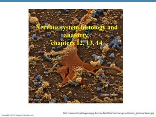
SlideShare a Scribd company logo
Copyright © 2010 Pearson Education, Inc.
Nervous system histology and
anatomy
chapters 12, 13, 14
http://www.eb.tuebingen.mpg.de/core-facilities/microscopy-unit/sem_pictures/axon.jpg
Copyright © 2010 Pearson Education, Inc.
Functions of the Nervous System
1. Sensory input
• Information gathered by sensory receptors about
internal and external changes
3. Integration
• Interpretation of sensory input (CNS)
5. Motor output
• Activation of effector organs (muscles and
glands) produces a response
Copyright © 2010 Pearson Education, Inc.
Anatomical Organization of the Nervous System
• Central nervous system (CNS)
• Brain and spinal cord
• Integration and command center
• Peripheral nervous system (PNS)
• Paired spinal and cranial nerves
• Carries messages to and from the spinal cord and
brain
Copyright © 2010 Pearson Education, Inc. Figure 11 Section 1
The major components and functions of the nervous system
Central Nervous System
The central nervous system
(CNS) consists of the brain and
spinal cord and is responsible
for integrating, processing, and
coordinating sensory data and
motor commands.
Information processing
includes the integration and
distribution of information in
the CNS.
Peripheral Nervous
System
The peripheral
nervous system
(PNS) includes all the
neural tissue outside
the CNS.
The motor division of the
PNS carries motor commands
from the CNS to peripheral
tissues and systems.
includes
The autonomic
nervous system
(ANS) provides
automatic regulation
of smooth muscle,
cardiac muscle,
glands, and adipose
tissue.
The somatic
nervous
system
(SNS)
controls
skeletal
muscle
contractions.
The sensory division of the PNS
brings information to the CNS
from receptors in peripheral
tissues and organs.
Somatic sensory
receptors provide
position, touch,
pressure, pain, and
temperature sensations.
Special sensory
receptors provide
sensations of
smell, taste,
vision, balance,
and hearing.
Visceral sensory receptors
monitor internal organs.
Receptors are sensory structures that detect
changes in the internal or external
environment.
Skeletal
muscle
Effectors are target organs whose
activities change in response to neural
commands.
• Smooth muscle
• Cardiac muscle
• Glands
• Adipose tissue
Copyright © 2010 Pearson Education, Inc.
Histology of Nerve Tissue
• The two cell types of the nervous system are:
• Neurons – excitable cells that transmit electrical
signals
• Supporting cells – cells that surround and wrap
neurons
Copyright © 2010 Pearson Education, Inc.
Neurons (Nerve Cells)
• Structural units of the nervous system
• Composed of a body, axon, and dendrites
• Long-lived (over 100 years)
• Amitotic – once achieve there roles in the system
they loose the ability to divide
• High metabolic rate – require high supply of
oxygen and glucose
• Their plasma membrane function in Electrical signaling
Copyright © 2010 Pearson Education, Inc.
Neuron Cell Body (Perikaryon or Soma)
• Posses all the components needed to produce membrane parts,
synthesize enzymes and neurotransmitters:
• Contains the nucleus and a nucleolus
• rough endoplasmic reticulum/Nissle bodies for protein synthesis
• Golgi apparatus which packages materials into vesicles for
transport
• Numerous mitochondria
• Cytoskeletal elements
• Has no centrioles (hence its amitotic nature)
• Neural stem cells are still found in the adult but are not active
• Cells in the hippocampus (part that is involves memory) can still
dividing
Copyright © 2010 Pearson Education, Inc.
Dendrites
• Short branched processes
• They are the receptive, or input,
regions of the neuron
• Supply big surface for receiving
signals
• Convey incoming messages
towards the cell body.
• Electrical signals are conveyed as
graded potentials (not action
potentials)
Copyright © 2010 Pearson Education, Inc.
Axons: Structure
• The axon contains the same organelles as the cell body with the
exception of Nissle bodies and Golgi apparatus (protein
synthesis and packaging)
• Depends on the cell body to supply proteins
• Each neuron has single axon of uniform diameter
• Initiate in an enlarged area called the axon hillock
• Numerous terminal branches (telodendria)
• Knoblike axon terminals (synaptic knobs or boutons)
• Secretory region of neuron
• Release neurotransmitters to excite or inhibit other cells
• Long axons are called nerve fibers
Copyright © 2010 Pearson Education, Inc.
Axons: Function
• Generate and transmit action potentials – away from the cell
• The signal starts at the junction between the axon hillock and
axon – trigger zone
• Secrete neurotransmitters from the axonal terminals in response
to the impulse arriving along the axon
• Movement along axons (not the signal movement) occurs in
two ways
• Anterograde — toward axonal terminal (mitochondria,
cytoskeletal elements, membrane components for membrane
renewal, enzymes Examples: mitochondria, membrane
components, enzymes
• Retrograde — away from axonal terminal (organelles to be
degraded, signal molecules, viruses, and bacterial toxins like
polio, rabies, herpes simplex)
Copyright © 2010 Pearson Education, Inc.
Supporting Cells: Neuroglia
• Two groups
• In the CNS – astrocytes, microglia, ependymal
cells and oligodendrocytes
• In the PNS – satellite and Schwann cells
Copyright © 2010 Pearson Education, Inc.
Neuroglia in the CNS: Astrocytes
• Most abundant, versatile, and highly branched glial cells
• In some areas of the brain they account for 90% of the
tissue
• They cling to neurons and their synaptic endings, and cover
capillaries
http://www.ucl.ac.uk/biology/images/astrocytes.gifFigure 11.3a
Capillary
Neuron
Astrocyte
Copyright © 2010 Pearson Education, Inc.
Neuroglia in the CNS: Astrocytes functions
• Maintaining blood-brain barrier – preventing free access of
circulating compounds to the CNS.
• The extensions of the astrocytes end in expanded portion
that wraps capillaries
Copyright © 2010 Pearson Education, Inc.
Why are the capillaries in the BBB less permeable?
• Endothelial cells form tight junctions that prevent
solutes movement between cells
• Astrocytes
• Selective transport properties of the endothelial cells
• The BBB
• Helps maintain a stable environment for the brain
• Separates neurons from some bloodborne substances
Copyright © 2010 Pearson Education, Inc.
Blood-Brain Barrier: Functions
• Selective barrier that allows nutrients to pass freely
• Is ineffective against substances that can diffuse through plasma
membranes (ex. Ethanol, caffeine)
• Absent in some areas:
• Ex. - hormones generally do not penetrate the brain from the
blood, so in order to control the rate of hormone secretion
effectively, there are specialized sites where neurons can
"sample" the composition of the circulating blood. At these
sites, the blood-brain barrier is 'leaky‘ (pituitary gland)
• Capillaries of the choroid plexus
• The BBB can break down under certain conditions:
• hypertension, radiation, infection and brain trauma
Copyright © 2010 Pearson Education, Inc.
Example
• In Parkinson’s disease there is a lack in the NT
dopamine (neurons that produce it are either damaged
or dead)
• Dopamine can not be given in a pill or injection
because it can’t cross the BBB
• Instead, a precursor - L-dopa – is given. This is being
transported across the BBB and is being used by
neurons
Copyright © 2010 Pearson Education, Inc.
Neuroglia in the CNS: Astrocytes functions
• Creating a framework for the CNS with microfilaments
that extend from the astrocytes
• Repairing damaged neural tissue- limited structural
repairs that stabilize and prevent further injury.
• In the embryonic brain, the astrocyte appear to be
involve in directing the growth and interconnection of
developing neurons.
• Secrete nerve growth factors that promote neuron
growth and synapse formation
Copyright © 2010 Pearson Education, Inc.
Neuroglia in the CNS: Astrocytes functions
• Control the chemical environment
• Regulating the concentration of sodium, potassium
and carbon dioxide ions
• Providing system for transporting nutrients and
dissolved gasses between capillaries and neurons
• absorb neurotransmitters to prevent excessive
levels in tissue fluid; control ion concentration in
the interstitial fluid
Copyright © 2010 Pearson Education, Inc.
Oligodendrocytes
• Branched cells
• Processes wrap CNS nerve fibers, forming insulating
myelin sheaths
Nerve
fibers
Myelin sheath
Process of
oligodendrocyte
Copyright © 2010 Pearson Education, Inc.
Neuroglia cells in the PNS
• Schwann cells (neurolemmocytes) – surround fibers of
the PNS
• Similar function as the oligodendrocytes
• Satellite cells surround neuron cell bodies in the PNS
• similar function to the astrocytes in the CNS –
controlling the chemical environment
Copyright © 2010 Pearson Education, Inc. Figure 11.3e
(e) Satellite cells and Schwann cells (which
form myelin) surround neurons in the PNS.
Schwann cells
(forming myelin sheath)
Cell body of neuron
Satellite
cells
Nerve fiber
Copyright © 2010 Pearson Education, Inc.
Myelin Sheath: Formation
• Formed by Schwann cells in the PNS
• Schwann cells wraps many times around the axon
• Myelin sheath — concentric layers of Schwann cell
membrane
• Neurilemma—peripheral bulge of Schwann cell
cytoplasm
• Myelin sheath functions :
• Protect the axon
• Electrically insulate fibers from one another
• Increase the speed of nerve impulse transmission
Copyright © 2010 Pearson Education, Inc.
Nodes of Ranvier (Neurofibral Nodes)
• Gaps in the myelin sheath
between adjacent Schwann
cells
• They are the sites where
axon collaterals can emerge
http://www.jdaross.cwc.net/introd13.jpg
Copyright © 2010 Pearson Education, Inc.
Unmyelinated Axons
• Thin nerve fibers are unmyelinated
• One Schwann cell may incompletely enclose 15 or
more unmyelinated axons
Copyright © 2010 Pearson Education, Inc.
Unmyelinated Axons
• A Schwann cell surrounds nerve fibers but coiling does not take place
• Schwann cells partially enclose 15 or more axons
http://www.bu.edu/histology/p/21201loa.htm
Copyright © 2010 Pearson Education, Inc.
Axons of the CNS
• Both myelinated and
unmyelinated fibers are present
• Myelin sheaths are formed by
oligodendrocytes
• Nodes of Ranvier are widely
spaced
Copyright © 2010 Pearson Education, Inc.
Regions of the Brain and Spinal Cord
• White matter – dense collections of myelinated fibers
• Gray matter – mostly soma (cell bodies) and
unmyelinated fibers
Copyright © 2010 Pearson Education, Inc.
CNS: Gray and White Matter
Figure 9.5a
Copyright © 2010 Pearson Education, Inc.
Neurons Cell body locations
• Groups of cell bodies are named according to their
location:
• Most cell bodies are in the CNS in clusters called
nuclei
• Some are in the PNS in clusters called ganglia
Copyright © 2010 Pearson Education, Inc.
Neurons’ Processes location
• The groups of axons are named according to their
locations:
• In the CNS the extensions called tracts
• In the PNS the extensions (axons) are called nerves
Copyright © 2010 Pearson Education, Inc.
Neuron Classification - Structural
• Multipolar — three or more processes
• Many extensions; many dendrites lead toward cell body, one axon leads
away from cell body.
• Most neurons in the CNS and those whose axons carry impulses away
from the CNS are multipolar.
• Bipolar — two processes (axon and dendrite)
• Two extensions; one fused dendrite leads toward cell body, one axon
leads away from cell body
• These are rare and can be found as part of the receptor apparatus in the
eye, ear and olfactory mucosa.
• Unipolar — single, short process
• One very short process from cell body that divides into central and
peripheral processes. Only the distal ends of the peripheral process are
dendrites and the rest act as an axon along with the central process.
• Nearly all neurons that conduct impulses towards the CNS are unipolar.
Copyright © 2010 Pearson Education, Inc.
Neuron Classification - Functional
• Sensory (afferent) - transmit impulses toward the CNS
• Most are unipolar neurons with their bodies in ganglia in
the PNS.
• Location – sensory receptors in the internal organs, skin,
skeletal muscles and joints. They sense changes in the
immediate environment
• Motor (efferent) - carry impulses away from the CNS
• Carry activating impulses from CNS and to the viscera,
body muscles and glands;
• They are multipolar and their cell bodies are in the CNS.
Copyright © 2010 Pearson Education, Inc.
Neuron Classification - Functional
• Interneurons (association neurons) - shuttle signals
through CNS pathways
• Link other neurons together (i.e. sensory neuron to
interneuron to motor neuron).
• All bodies are in CNS and they are multipolar.
Copyright © 2010 Pearson Education, Inc.
Central nervous system
http://www.courses.vcu.edu/DANC291-003/unit%201.htm
Copyright © 2010 Pearson Education, Inc.
3 Major levels of the CNS
• Spinal cord
• involved in walking movement, reflexes of both skeletal
muscles and the autonomic nervous system effectors
• Lower brain/subcortical level
• Subconscious activities of both the autonomic and
somatic nervous system. Involved in several emotional
patterns
• Higher brain/cortical level
• Very large memory storage, place of higher functions
like thoughts, awareness
Copyright © 2010 Pearson Education, Inc.
CNS protection
• Bony protection by the skeleton – brain by skull and
spinal cord by vertebrae
• 3 layers of membrane collectively called meninges
that are found between the bone and the CNS tissue
• Cerebro-spinal fluid – CSF - found in the ventricles,
central canal and subarachnoid space
• Blood brain barrier - BBB
Copyright © 2010 Pearson Education, Inc.
CNS: Physical Support
Figure 9.2a
Copyright © 2010 Pearson Education, Inc.
CNS: Physical Support
Figure 9.2b
Copyright © 2010 Pearson Education, Inc.
Ventricles
• Filled with CSF (cerebrospinal fluid)
• Ventricles continuous w/each other + central canal
of spinal cord
• Lateral Ventricle (#1+2)
• Cerebral Hemisphere
• Third Ventricle
• Diencephalon
• Fourth Ventricle
• Cerebral Aqueduct: connects 3rd and 4th
ventricles
• Connects to central canal of spinal cord &
medulla
3
4
lateral
Copyright © 2010 Pearson Education, Inc.
Choroid Plexuses and CSF
• Clusters of capillaries and the ependymal cells
http://www.sci.uidaho.edu/med532/choroid.htm
Copyright © 2010 Pearson Education, Inc.
CSF production
• The choroid plexus forms tissue fluid filters
• Have ion pumps that selectively pump ions from
the plasma into the ventricles.
• The ions pumps create osmotic pressure that
draws water to the CSF
• the choroid plexus of the lateral ventricles producing the
most.
• The rate of formation is approximately 0.35 ml/min or 500
ml/day; a rate which replaces the total volume of CSF
approximately 2-3 times over in 24 hours.
Copyright © 2010 Pearson Education, Inc.
CSF – cerebrospinal fluid functions
• Liquid cushion for brain and spinal cord
• Nourishes brain
• Removes waste
• Conducts chemical signals between parts of CNS
Copyright © 2010 Pearson Education, Inc. Figure 9.11c
Midbrain
Spinal cord
Pons
Medulla oblongata
Forebrain
Cerebrum
Thalamus
Hypothalamus
Pituitary gland
Brainstem
Diencephalon
Cerebellum
Corpus
callosum
(c) Midsagittal section
Brain: Midsagittal View
Copyright © 2010 Pearson Education, Inc.
http://www.bioon.com/book/biology/whole/image/1/1-6.tif.jpg
Basal nuclei
Cerebrum functional regions
• The cerebrum has three basic regions: cortex
(gray matter), white matter, and basal nuclei
(gray matter)
Copyright © 2010 Pearson Education, Inc.
Layers of the Cerebral Cortex
Figure 9.12a–b
Copyright © 2010 Pearson Education, Inc.
Premotor cortex
(coordinates
voluntary
movements)
Primary somatosensory
cortex (somesthetic sensations
and proprioception)
Sensory association
areas (integration of
sensory information)
Primary motor cortex
(voluntary movement)
Central sulcus
Prefrontal
association
areas (idea and
plan for voluntary
movement, thoughts,
personality)
Broca’s area
(speech formation)
Limbic association
cortex (emotions,
learning, and memory)
Olfactory cortex
(smell)
Visual association
areas (higher vision
processing)
Wernicke’s area
(language
comprehension)
Auditory
association
areas
Primary auditory
cortex (hearing)
Primary visual cortex
(vision)
Figure 9.14
Functional Areas of Cerebrum
Copyright © 2010 Pearson Education, Inc.
Beneath the cerebrum – basal nuclei
• Are masses of gray matter that are embedded in white
matter of cerebrum
• Function in the subconscious control of skeletal
muscle tone
• The coordination of learned movement patterns
(walking, lifting)
Copyright © 2010 Pearson Education, Inc.
White Matter of the Cerebrum
• Association fibers
• Connections within one hemisphere
• Commissural fibers connecting two hemispheres:
●
corpus callosum
●
anterior commissure
• Projection fibers link cerebral cortex with:
●
diencephalon, brain stem, cerebellum, and
spinal cord
Copyright © 2010 Pearson Education, Inc.
Diencephalon
• Consists of three structures
• thalamus
• Hypothalamus
• epithalamus
• Encloses the third ventricle
Copyright © 2010 Pearson Education, Inc.
Epithalamus
• Most dorsal portion of the diencephalon; forms roof of
the third ventricle
• Pineal gland – extends from the posterior border and
secretes melatonin
• Melatonin – a hormone involved with sleep
regulation, sleep-wake cycles, and mood
• Choroid plexus – a structure that secretes cerebral
spinal fluid (CSF)
Copyright © 2010 Pearson Education, Inc.
Brain Stem
• Consists of three regions –
• midbrain,
• Pons
• medulla oblongata
• Controls automatic behaviors necessary for survival
(breathing, digestion, heart rate, blood pressure)
• Provides the pathway for tracts between higher and lower
brain centers
• Associated with 10 of the 12 pairs of cranial nerves
Copyright © 2010 Pearson Education, Inc.
Cerebellar Processing
• Cerebellum receives impulses of the intent to initiate
voluntary muscle contraction
• Proprioceptors and visual signals “inform” the
cerebellum of the body’s condition
• Cerebellar cortex calculates the best way to perform a
movement
• A “blueprint” of coordinated movement is sent to the
cerebral motor cortex
Copyright © 2010 Pearson Education, Inc.
Spinal cord
Copyright © 2010 Pearson Education, Inc. Figure 12.32
Somatic
sensory
neuron
Dorsal root (sensory)
Dorsal root ganglion
Visceral
sensory
neuron
Somatic
motor neuron
Spinal nerve
Ventral root
(motor)
Ventral horn
(motor neurons)
Dorsal horn (interneurons)
Visceral
motor
neuron
Interneurons receiving input from somatic sensory neurons
Interneurons receiving input from visceral sensory neurons
Visceral motor (autonomic) neurons
Somatic motor neurons
Gray Matter: Organization
Copyright © 2010 Pearson Education, Inc.
White Matter in the Spinal Cord
• Composed of both myelinated and unmyelinated fibers that run
in three directions
• Ascending – to higher centers; sensory
• Descending – to lower levels from brain or higher places in
the cord; motor
• Transversely – from one side of the cord to the other
• Divided into three funiculi (columns) – posterior, lateral, and
anterior
• Each funiculus contains several fiber tracks with similar
destination and function
• Fiber tract names reveal their origin and destination
• Fiber tracts are composed of axons with similar functions
Copyright © 2010 Pearson Education, Inc.
Peripheral nervous system
Copyright © 2010 Pearson Education, Inc.
Peripheral nervous system components
• Includes nerves and ganglia
• Most nerves are mixtures of afferent and efferent fibers
and somatic and autonomic (visceral) fibers
• Pure sensory (afferent) or motor (efferent) nerves are rare
• Peripheral nerves classified as cranial or spinal nerves
• Ganglia contain neuron cell bodies associated with
nerves
• Dorsal root ganglia (sensory, somatic) (Chapter 12)
• Autonomic ganglia (motor, visceral) (Chapter 14)
Copyright © 2010 Pearson Education, Inc.
Structure of a Nerve
• Nerve – cordlike organ of the
PNS consisting of peripheral
axons enclosed by connective
tissue
• Connective tissue coverings
include:
• Endoneurium – loose
connective tissue that
surrounds axons
• Perineurium – coarse
connective tissue that
bundles fibers into fascicles
• Epineurium – tough fibrous
sheath around a nerve
Copyright © 2010 Pearson Education, Inc.
Spinal Nerves: Rami
• The short spinal nerves branch into three or four mixed,
distal rami
• Dorsal ramus – innervate skin of the back and deep
back muscle
• Ventral ramus – form plexuses. The ventral rami of
T2-T12 do not form plexuses and innervate
intercostal spaces and muscles of the trunk
• Tiny meningeal branch – innervate the meninges and
blood vessels within the vertebral canal
• Rami communicantes at the base of the ventral rami
in the thoracic region that contain autonomic nerve
fibers
Copyright © 2010 Pearson Education, Inc.
Nerve Plexuses
• All ventral rami except T2-T12 form nerve networks
called plexuses
• Plexuses are found in the cervical, brachial, lumbar, and
sacral regions
• Each resulting branch of a plexus contains fibers from
several spinal nerves
• Fibers travel to the periphery via several different routes
• Each muscle receives a nerve supply from more than one
spinal nerve
• Damage to one spinal segment cannot completely
paralyze a muscle
Copyright © 2010 Pearson Education, Inc.
Plexus Main spinal nerves Regions innervated Major nerves
Cervical C1-C5 Skin and muscles of head &
neck. Superior chest and
shoulder
Phrenic (diaphragm)
Brachial C5-C8, T1 Shoulder and upper limbs Axillary
Musculocutaneous
Radial
Median
Ulnar
Lumbar L1-L4 Antero-lateral abdominal
wall, external genitalia, part
of lower limbs
Femoral
Sacral L4-L5, S1-S4 Buttocks, perineum, lower
limbs
Sciatic
Copyright © 2010 Pearson Education, Inc.
• Neurons in the nervous system link together to form
circuits with specific functions.
http://www.thefullwiki.org/Nervous_System
Copyright © 2010 Pearson Education, Inc.
Nervous system functions
• Motor functions
• Control contraction of skeletal muscles – somatic nervous
system
• Control contractions of smooth and cardiac muscle –
autonomic nervous system
• Control of glands function – autonomic nervous system
• Sensory and integrative functions
• Process incoming information to ensure appropriate motor
response
●
More than 99% of the incoming information is classified
as irrelevant and unimportant
Copyright © 2010 Pearson Education, Inc.
The nervous system - the study approach in this class
• Our approach will be to cover functional systems:
• Sensory pathways - Somatic and autonomic
• Special senses
• Motor pathways – somatic and autonomic
• All system will cover both the central and
peripheral nervous systems

More Related Content

What's hot

Nervous tissue histology
Nervous tissue  histology Nervous tissue  histology
Nervous tissue histology
Dr. Devi Shankar
 
Central Nervous System 1
Central Nervous System 1Central Nervous System 1
Central Nervous System 1MBBS IMS MSU
 
B.Sc.(Micro+Biotech) II Animal & Plant Physiology Unit 4.1 Neurones & The Act...
B.Sc.(Micro+Biotech) II Animal & Plant Physiology Unit 4.1 Neurones & The Act...B.Sc.(Micro+Biotech) II Animal & Plant Physiology Unit 4.1 Neurones & The Act...
B.Sc.(Micro+Biotech) II Animal & Plant Physiology Unit 4.1 Neurones & The Act...
Rai University
 
Urinary system
Urinary systemUrinary system
Urinary system
sidath sanjeewa
 
General Physiology - The nervous system, basic functions of synapses
General Physiology - The nervous system, basic functions of synapsesGeneral Physiology - The nervous system, basic functions of synapses
General Physiology - The nervous system, basic functions of synapses
Hamzeh AlBattikhi
 
Nervous system anatomy and physiology
Nervous system anatomy and physiologyNervous system anatomy and physiology
Nervous system anatomy and physiology
Manthan Kapadiya
 
Brain
BrainBrain
Nervous System
Nervous SystemNervous System
Nervous Systemlevouge777
 
Physiology of Endocrine System
Physiology of Endocrine System Physiology of Endocrine System
Physiology of Endocrine System
PRANJAL SHARMA
 
Spinal Cord Dev
Spinal Cord DevSpinal Cord Dev
Spinal Cord DevAnan
 
Nervous system
Nervous systemNervous system
Nervous system
BobbyPabores1
 
histologic structure of nervous system
histologic structure of nervous systemhistologic structure of nervous system
histologic structure of nervous system
wayan sugiritama
 
Histology of Cerebellum
Histology of CerebellumHistology of Cerebellum
Histology of Cerebellum
Rashmi Priyem Saravanan
 
Histology of special senses
Histology of special sensesHistology of special senses
Histology of special senses
mgmcri1234
 
Cell Histology, Structure and Function of Cell,
Cell Histology, Structure and Function of Cell,Cell Histology, Structure and Function of Cell,
Cell Histology, Structure and Function of Cell,
Dr. Shahbaz Ahmad PT Chaudhary
 
Histology of ear
Histology of earHistology of ear
Histology of ear
emebet chimdi
 
Nervous system (neuron & ion channels)
Nervous system (neuron & ion channels)Nervous system (neuron & ion channels)
Nervous system (neuron & ion channels)
Rohit Paswan
 
Histology of Female Reproductive System.pptx
Histology of Female Reproductive System.pptxHistology of Female Reproductive System.pptx
Histology of Female Reproductive System.pptx
Dr Abdul Qayyum Khan
 

What's hot (20)

Nervous tissue histology
Nervous tissue  histology Nervous tissue  histology
Nervous tissue histology
 
Central Nervous System 1
Central Nervous System 1Central Nervous System 1
Central Nervous System 1
 
B.Sc.(Micro+Biotech) II Animal & Plant Physiology Unit 4.1 Neurones & The Act...
B.Sc.(Micro+Biotech) II Animal & Plant Physiology Unit 4.1 Neurones & The Act...B.Sc.(Micro+Biotech) II Animal & Plant Physiology Unit 4.1 Neurones & The Act...
B.Sc.(Micro+Biotech) II Animal & Plant Physiology Unit 4.1 Neurones & The Act...
 
Sensory system
Sensory systemSensory system
Sensory system
 
Urinary system
Urinary systemUrinary system
Urinary system
 
General Physiology - The nervous system, basic functions of synapses
General Physiology - The nervous system, basic functions of synapsesGeneral Physiology - The nervous system, basic functions of synapses
General Physiology - The nervous system, basic functions of synapses
 
Nervous system anatomy and physiology
Nervous system anatomy and physiologyNervous system anatomy and physiology
Nervous system anatomy and physiology
 
Brain
BrainBrain
Brain
 
Nervous System
Nervous SystemNervous System
Nervous System
 
Physiology of Endocrine System
Physiology of Endocrine System Physiology of Endocrine System
Physiology of Endocrine System
 
Spinal Cord Dev
Spinal Cord DevSpinal Cord Dev
Spinal Cord Dev
 
Liver pancreas
Liver pancreasLiver pancreas
Liver pancreas
 
Nervous system
Nervous systemNervous system
Nervous system
 
histologic structure of nervous system
histologic structure of nervous systemhistologic structure of nervous system
histologic structure of nervous system
 
Histology of Cerebellum
Histology of CerebellumHistology of Cerebellum
Histology of Cerebellum
 
Histology of special senses
Histology of special sensesHistology of special senses
Histology of special senses
 
Cell Histology, Structure and Function of Cell,
Cell Histology, Structure and Function of Cell,Cell Histology, Structure and Function of Cell,
Cell Histology, Structure and Function of Cell,
 
Histology of ear
Histology of earHistology of ear
Histology of ear
 
Nervous system (neuron & ion channels)
Nervous system (neuron & ion channels)Nervous system (neuron & ion channels)
Nervous system (neuron & ion channels)
 
Histology of Female Reproductive System.pptx
Histology of Female Reproductive System.pptxHistology of Female Reproductive System.pptx
Histology of Female Reproductive System.pptx
 

Similar to Cns histology and anatomy

Ch 13_lecture_presentation
 Ch 13_lecture_presentation Ch 13_lecture_presentation
Ch 13_lecture_presentationTheSlaps
 
cells of the ns
cells of the nscells of the ns
cells of the ns
PriyakanthS
 
INTRODUCTION TO NEUROSCIENCE-PART 1.pptx
INTRODUCTION TO NEUROSCIENCE-PART 1.pptxINTRODUCTION TO NEUROSCIENCE-PART 1.pptx
INTRODUCTION TO NEUROSCIENCE-PART 1.pptx
iqranaz71
 
Ch13lecturepresentation 140918213440-phpapp02
Ch13lecturepresentation 140918213440-phpapp02Ch13lecturepresentation 140918213440-phpapp02
Ch13lecturepresentation 140918213440-phpapp02
Cleophas Rwemera
 
Biological bases of human behaviour (complete) 2
Biological  bases  of human  behaviour (complete) 2Biological  bases  of human  behaviour (complete) 2
Biological bases of human behaviour (complete) 2
PoornimaSingh35
 
Brain anatomy
Brain anatomyBrain anatomy
Brain anatomy
Ayshah Hashimi
 
Brain anatomy
Brain anatomyBrain anatomy
Brain anatomy
Ayshah Hashimi
 
NEURON physiology carrer medical college and hospital
NEURON  physiology carrer medical college and hospitalNEURON  physiology carrer medical college and hospital
NEURON physiology carrer medical college and hospital
rashmimishra129
 
Neurons
NeuronsNeurons
Neurons
Numan Ijaz
 
163 ch 08_lecture_presentation
163 ch 08_lecture_presentation163 ch 08_lecture_presentation
163 ch 08_lecture_presentationgwrandall
 
Nervous Tissues.pptx
Nervous Tissues.pptxNervous Tissues.pptx
Nervous Tissues.pptx
AsifBahadurAli
 
_ch_11_Neural Tissue_lecture_presentation.ppt
_ch_11_Neural Tissue_lecture_presentation.ppt_ch_11_Neural Tissue_lecture_presentation.ppt
_ch_11_Neural Tissue_lecture_presentation.ppt
ismaeljemal1
 
Nueron ppt
Nueron pptNueron ppt
Nueron ppt
naseemmeeran
 
Cellular organization of the nervous system
Cellular organization of the nervous systemCellular organization of the nervous system
Cellular organization of the nervous systemDavis Mburu
 
PsychologyChapter02
PsychologyChapter02PsychologyChapter02
PsychologyChapter02
drellen
 
Anatomy nervous tissue chap 13
Anatomy nervous tissue   chap 13Anatomy nervous tissue   chap 13
Anatomy nervous tissue chap 13Diore Solidum
 
20200829190340_PPT1 Chemical and Biology
20200829190340_PPT1 Chemical and Biology20200829190340_PPT1 Chemical and Biology
20200829190340_PPT1 Chemical and Biology
dona663283
 
Anatomy and physiology of brain ppt, its organization
Anatomy and physiology of brain ppt,  its organizationAnatomy and physiology of brain ppt,  its organization
Anatomy and physiology of brain ppt, its organization
Jyotirmayee Mandal
 
Nerve and muscle
Nerve and muscleNerve and muscle
Nerve and muscle
laraib jameel
 

Similar to Cns histology and anatomy (20)

Ch 13_lecture_presentation
 Ch 13_lecture_presentation Ch 13_lecture_presentation
Ch 13_lecture_presentation
 
cells of the ns
cells of the nscells of the ns
cells of the ns
 
INTRODUCTION TO NEUROSCIENCE-PART 1.pptx
INTRODUCTION TO NEUROSCIENCE-PART 1.pptxINTRODUCTION TO NEUROSCIENCE-PART 1.pptx
INTRODUCTION TO NEUROSCIENCE-PART 1.pptx
 
CNS.pdf
CNS.pdfCNS.pdf
CNS.pdf
 
Ch13lecturepresentation 140918213440-phpapp02
Ch13lecturepresentation 140918213440-phpapp02Ch13lecturepresentation 140918213440-phpapp02
Ch13lecturepresentation 140918213440-phpapp02
 
Biological bases of human behaviour (complete) 2
Biological  bases  of human  behaviour (complete) 2Biological  bases  of human  behaviour (complete) 2
Biological bases of human behaviour (complete) 2
 
Brain anatomy
Brain anatomyBrain anatomy
Brain anatomy
 
Brain anatomy
Brain anatomyBrain anatomy
Brain anatomy
 
NEURON physiology carrer medical college and hospital
NEURON  physiology carrer medical college and hospitalNEURON  physiology carrer medical college and hospital
NEURON physiology carrer medical college and hospital
 
Neurons
NeuronsNeurons
Neurons
 
163 ch 08_lecture_presentation
163 ch 08_lecture_presentation163 ch 08_lecture_presentation
163 ch 08_lecture_presentation
 
Nervous Tissues.pptx
Nervous Tissues.pptxNervous Tissues.pptx
Nervous Tissues.pptx
 
_ch_11_Neural Tissue_lecture_presentation.ppt
_ch_11_Neural Tissue_lecture_presentation.ppt_ch_11_Neural Tissue_lecture_presentation.ppt
_ch_11_Neural Tissue_lecture_presentation.ppt
 
Nueron ppt
Nueron pptNueron ppt
Nueron ppt
 
Cellular organization of the nervous system
Cellular organization of the nervous systemCellular organization of the nervous system
Cellular organization of the nervous system
 
PsychologyChapter02
PsychologyChapter02PsychologyChapter02
PsychologyChapter02
 
Anatomy nervous tissue chap 13
Anatomy nervous tissue   chap 13Anatomy nervous tissue   chap 13
Anatomy nervous tissue chap 13
 
20200829190340_PPT1 Chemical and Biology
20200829190340_PPT1 Chemical and Biology20200829190340_PPT1 Chemical and Biology
20200829190340_PPT1 Chemical and Biology
 
Anatomy and physiology of brain ppt, its organization
Anatomy and physiology of brain ppt,  its organizationAnatomy and physiology of brain ppt,  its organization
Anatomy and physiology of brain ppt, its organization
 
Nerve and muscle
Nerve and muscleNerve and muscle
Nerve and muscle
 

Recently uploaded

Additional Benefits for Employee Website.pdf
Additional Benefits for Employee Website.pdfAdditional Benefits for Employee Website.pdf
Additional Benefits for Employee Website.pdf
joachimlavalley1
 
Chapter 3 - Islamic Banking Products and Services.pptx
Chapter 3 - Islamic Banking Products and Services.pptxChapter 3 - Islamic Banking Products and Services.pptx
Chapter 3 - Islamic Banking Products and Services.pptx
Mohd Adib Abd Muin, Senior Lecturer at Universiti Utara Malaysia
 
The French Revolution Class 9 Study Material pdf free download
The French Revolution Class 9 Study Material pdf free downloadThe French Revolution Class 9 Study Material pdf free download
The French Revolution Class 9 Study Material pdf free download
Vivekanand Anglo Vedic Academy
 
Unit 2- Research Aptitude (UGC NET Paper I).pdf
Unit 2- Research Aptitude (UGC NET Paper I).pdfUnit 2- Research Aptitude (UGC NET Paper I).pdf
Unit 2- Research Aptitude (UGC NET Paper I).pdf
Thiyagu K
 
Basic phrases for greeting and assisting costumers
Basic phrases for greeting and assisting costumersBasic phrases for greeting and assisting costumers
Basic phrases for greeting and assisting costumers
PedroFerreira53928
 
The Challenger.pdf DNHS Official Publication
The Challenger.pdf DNHS Official PublicationThe Challenger.pdf DNHS Official Publication
The Challenger.pdf DNHS Official Publication
Delapenabediema
 
Cambridge International AS A Level Biology Coursebook - EBook (MaryFosbery J...
Cambridge International AS  A Level Biology Coursebook - EBook (MaryFosbery J...Cambridge International AS  A Level Biology Coursebook - EBook (MaryFosbery J...
Cambridge International AS A Level Biology Coursebook - EBook (MaryFosbery J...
AzmatAli747758
 
Supporting (UKRI) OA monographs at Salford.pptx
Supporting (UKRI) OA monographs at Salford.pptxSupporting (UKRI) OA monographs at Salford.pptx
Supporting (UKRI) OA monographs at Salford.pptx
Jisc
 
The Roman Empire A Historical Colossus.pdf
The Roman Empire A Historical Colossus.pdfThe Roman Empire A Historical Colossus.pdf
The Roman Empire A Historical Colossus.pdf
kaushalkr1407
 
Model Attribute Check Company Auto Property
Model Attribute  Check Company Auto PropertyModel Attribute  Check Company Auto Property
Model Attribute Check Company Auto Property
Celine George
 
1.4 modern child centered education - mahatma gandhi-2.pptx
1.4 modern child centered education - mahatma gandhi-2.pptx1.4 modern child centered education - mahatma gandhi-2.pptx
1.4 modern child centered education - mahatma gandhi-2.pptx
JosvitaDsouza2
 
Unit 8 - Information and Communication Technology (Paper I).pdf
Unit 8 - Information and Communication Technology (Paper I).pdfUnit 8 - Information and Communication Technology (Paper I).pdf
Unit 8 - Information and Communication Technology (Paper I).pdf
Thiyagu K
 
Overview on Edible Vaccine: Pros & Cons with Mechanism
Overview on Edible Vaccine: Pros & Cons with MechanismOverview on Edible Vaccine: Pros & Cons with Mechanism
Overview on Edible Vaccine: Pros & Cons with Mechanism
DeeptiGupta154
 
The geography of Taylor Swift - some ideas
The geography of Taylor Swift - some ideasThe geography of Taylor Swift - some ideas
The geography of Taylor Swift - some ideas
GeoBlogs
 
Sha'Carri Richardson Presentation 202345
Sha'Carri Richardson Presentation 202345Sha'Carri Richardson Presentation 202345
Sha'Carri Richardson Presentation 202345
beazzy04
 
The Art Pastor's Guide to Sabbath | Steve Thomason
The Art Pastor's Guide to Sabbath | Steve ThomasonThe Art Pastor's Guide to Sabbath | Steve Thomason
The Art Pastor's Guide to Sabbath | Steve Thomason
Steve Thomason
 
aaaaaaaaaaaaaaaaaaaaaaaaaaaaaaaaaaaaaaaaa
aaaaaaaaaaaaaaaaaaaaaaaaaaaaaaaaaaaaaaaaaaaaaaaaaaaaaaaaaaaaaaaaaaaaaaaaaaaaaaaaaa
aaaaaaaaaaaaaaaaaaaaaaaaaaaaaaaaaaaaaaaaa
siemaillard
 
GIÁO ÁN DẠY THÊM (KẾ HOẠCH BÀI BUỔI 2) - TIẾNG ANH 8 GLOBAL SUCCESS (2 CỘT) N...
GIÁO ÁN DẠY THÊM (KẾ HOẠCH BÀI BUỔI 2) - TIẾNG ANH 8 GLOBAL SUCCESS (2 CỘT) N...GIÁO ÁN DẠY THÊM (KẾ HOẠCH BÀI BUỔI 2) - TIẾNG ANH 8 GLOBAL SUCCESS (2 CỘT) N...
GIÁO ÁN DẠY THÊM (KẾ HOẠCH BÀI BUỔI 2) - TIẾNG ANH 8 GLOBAL SUCCESS (2 CỘT) N...
Nguyen Thanh Tu Collection
 
Digital Tools and AI for Teaching Learning and Research
Digital Tools and AI for Teaching Learning and ResearchDigital Tools and AI for Teaching Learning and Research
Digital Tools and AI for Teaching Learning and Research
Vikramjit Singh
 
Thesis Statement for students diagnonsed withADHD.ppt
Thesis Statement for students diagnonsed withADHD.pptThesis Statement for students diagnonsed withADHD.ppt
Thesis Statement for students diagnonsed withADHD.ppt
EverAndrsGuerraGuerr
 

Recently uploaded (20)

Additional Benefits for Employee Website.pdf
Additional Benefits for Employee Website.pdfAdditional Benefits for Employee Website.pdf
Additional Benefits for Employee Website.pdf
 
Chapter 3 - Islamic Banking Products and Services.pptx
Chapter 3 - Islamic Banking Products and Services.pptxChapter 3 - Islamic Banking Products and Services.pptx
Chapter 3 - Islamic Banking Products and Services.pptx
 
The French Revolution Class 9 Study Material pdf free download
The French Revolution Class 9 Study Material pdf free downloadThe French Revolution Class 9 Study Material pdf free download
The French Revolution Class 9 Study Material pdf free download
 
Unit 2- Research Aptitude (UGC NET Paper I).pdf
Unit 2- Research Aptitude (UGC NET Paper I).pdfUnit 2- Research Aptitude (UGC NET Paper I).pdf
Unit 2- Research Aptitude (UGC NET Paper I).pdf
 
Basic phrases for greeting and assisting costumers
Basic phrases for greeting and assisting costumersBasic phrases for greeting and assisting costumers
Basic phrases for greeting and assisting costumers
 
The Challenger.pdf DNHS Official Publication
The Challenger.pdf DNHS Official PublicationThe Challenger.pdf DNHS Official Publication
The Challenger.pdf DNHS Official Publication
 
Cambridge International AS A Level Biology Coursebook - EBook (MaryFosbery J...
Cambridge International AS  A Level Biology Coursebook - EBook (MaryFosbery J...Cambridge International AS  A Level Biology Coursebook - EBook (MaryFosbery J...
Cambridge International AS A Level Biology Coursebook - EBook (MaryFosbery J...
 
Supporting (UKRI) OA monographs at Salford.pptx
Supporting (UKRI) OA monographs at Salford.pptxSupporting (UKRI) OA monographs at Salford.pptx
Supporting (UKRI) OA monographs at Salford.pptx
 
The Roman Empire A Historical Colossus.pdf
The Roman Empire A Historical Colossus.pdfThe Roman Empire A Historical Colossus.pdf
The Roman Empire A Historical Colossus.pdf
 
Model Attribute Check Company Auto Property
Model Attribute  Check Company Auto PropertyModel Attribute  Check Company Auto Property
Model Attribute Check Company Auto Property
 
1.4 modern child centered education - mahatma gandhi-2.pptx
1.4 modern child centered education - mahatma gandhi-2.pptx1.4 modern child centered education - mahatma gandhi-2.pptx
1.4 modern child centered education - mahatma gandhi-2.pptx
 
Unit 8 - Information and Communication Technology (Paper I).pdf
Unit 8 - Information and Communication Technology (Paper I).pdfUnit 8 - Information and Communication Technology (Paper I).pdf
Unit 8 - Information and Communication Technology (Paper I).pdf
 
Overview on Edible Vaccine: Pros & Cons with Mechanism
Overview on Edible Vaccine: Pros & Cons with MechanismOverview on Edible Vaccine: Pros & Cons with Mechanism
Overview on Edible Vaccine: Pros & Cons with Mechanism
 
The geography of Taylor Swift - some ideas
The geography of Taylor Swift - some ideasThe geography of Taylor Swift - some ideas
The geography of Taylor Swift - some ideas
 
Sha'Carri Richardson Presentation 202345
Sha'Carri Richardson Presentation 202345Sha'Carri Richardson Presentation 202345
Sha'Carri Richardson Presentation 202345
 
The Art Pastor's Guide to Sabbath | Steve Thomason
The Art Pastor's Guide to Sabbath | Steve ThomasonThe Art Pastor's Guide to Sabbath | Steve Thomason
The Art Pastor's Guide to Sabbath | Steve Thomason
 
aaaaaaaaaaaaaaaaaaaaaaaaaaaaaaaaaaaaaaaaa
aaaaaaaaaaaaaaaaaaaaaaaaaaaaaaaaaaaaaaaaaaaaaaaaaaaaaaaaaaaaaaaaaaaaaaaaaaaaaaaaaa
aaaaaaaaaaaaaaaaaaaaaaaaaaaaaaaaaaaaaaaaa
 
GIÁO ÁN DẠY THÊM (KẾ HOẠCH BÀI BUỔI 2) - TIẾNG ANH 8 GLOBAL SUCCESS (2 CỘT) N...
GIÁO ÁN DẠY THÊM (KẾ HOẠCH BÀI BUỔI 2) - TIẾNG ANH 8 GLOBAL SUCCESS (2 CỘT) N...GIÁO ÁN DẠY THÊM (KẾ HOẠCH BÀI BUỔI 2) - TIẾNG ANH 8 GLOBAL SUCCESS (2 CỘT) N...
GIÁO ÁN DẠY THÊM (KẾ HOẠCH BÀI BUỔI 2) - TIẾNG ANH 8 GLOBAL SUCCESS (2 CỘT) N...
 
Digital Tools and AI for Teaching Learning and Research
Digital Tools and AI for Teaching Learning and ResearchDigital Tools and AI for Teaching Learning and Research
Digital Tools and AI for Teaching Learning and Research
 
Thesis Statement for students diagnonsed withADHD.ppt
Thesis Statement for students diagnonsed withADHD.pptThesis Statement for students diagnonsed withADHD.ppt
Thesis Statement for students diagnonsed withADHD.ppt
 

Cns histology and anatomy

  • 1. Copyright © 2010 Pearson Education, Inc. Nervous system histology and anatomy chapters 12, 13, 14 http://www.eb.tuebingen.mpg.de/core-facilities/microscopy-unit/sem_pictures/axon.jpg
  • 2. Copyright © 2010 Pearson Education, Inc. Functions of the Nervous System 1. Sensory input • Information gathered by sensory receptors about internal and external changes 3. Integration • Interpretation of sensory input (CNS) 5. Motor output • Activation of effector organs (muscles and glands) produces a response
  • 3. Copyright © 2010 Pearson Education, Inc. Anatomical Organization of the Nervous System • Central nervous system (CNS) • Brain and spinal cord • Integration and command center • Peripheral nervous system (PNS) • Paired spinal and cranial nerves • Carries messages to and from the spinal cord and brain
  • 4. Copyright © 2010 Pearson Education, Inc. Figure 11 Section 1 The major components and functions of the nervous system Central Nervous System The central nervous system (CNS) consists of the brain and spinal cord and is responsible for integrating, processing, and coordinating sensory data and motor commands. Information processing includes the integration and distribution of information in the CNS. Peripheral Nervous System The peripheral nervous system (PNS) includes all the neural tissue outside the CNS. The motor division of the PNS carries motor commands from the CNS to peripheral tissues and systems. includes The autonomic nervous system (ANS) provides automatic regulation of smooth muscle, cardiac muscle, glands, and adipose tissue. The somatic nervous system (SNS) controls skeletal muscle contractions. The sensory division of the PNS brings information to the CNS from receptors in peripheral tissues and organs. Somatic sensory receptors provide position, touch, pressure, pain, and temperature sensations. Special sensory receptors provide sensations of smell, taste, vision, balance, and hearing. Visceral sensory receptors monitor internal organs. Receptors are sensory structures that detect changes in the internal or external environment. Skeletal muscle Effectors are target organs whose activities change in response to neural commands. • Smooth muscle • Cardiac muscle • Glands • Adipose tissue
  • 5. Copyright © 2010 Pearson Education, Inc. Histology of Nerve Tissue • The two cell types of the nervous system are: • Neurons – excitable cells that transmit electrical signals • Supporting cells – cells that surround and wrap neurons
  • 6. Copyright © 2010 Pearson Education, Inc. Neurons (Nerve Cells) • Structural units of the nervous system • Composed of a body, axon, and dendrites • Long-lived (over 100 years) • Amitotic – once achieve there roles in the system they loose the ability to divide • High metabolic rate – require high supply of oxygen and glucose • Their plasma membrane function in Electrical signaling
  • 7. Copyright © 2010 Pearson Education, Inc. Neuron Cell Body (Perikaryon or Soma) • Posses all the components needed to produce membrane parts, synthesize enzymes and neurotransmitters: • Contains the nucleus and a nucleolus • rough endoplasmic reticulum/Nissle bodies for protein synthesis • Golgi apparatus which packages materials into vesicles for transport • Numerous mitochondria • Cytoskeletal elements • Has no centrioles (hence its amitotic nature) • Neural stem cells are still found in the adult but are not active • Cells in the hippocampus (part that is involves memory) can still dividing
  • 8. Copyright © 2010 Pearson Education, Inc. Dendrites • Short branched processes • They are the receptive, or input, regions of the neuron • Supply big surface for receiving signals • Convey incoming messages towards the cell body. • Electrical signals are conveyed as graded potentials (not action potentials)
  • 9. Copyright © 2010 Pearson Education, Inc. Axons: Structure • The axon contains the same organelles as the cell body with the exception of Nissle bodies and Golgi apparatus (protein synthesis and packaging) • Depends on the cell body to supply proteins • Each neuron has single axon of uniform diameter • Initiate in an enlarged area called the axon hillock • Numerous terminal branches (telodendria) • Knoblike axon terminals (synaptic knobs or boutons) • Secretory region of neuron • Release neurotransmitters to excite or inhibit other cells • Long axons are called nerve fibers
  • 10. Copyright © 2010 Pearson Education, Inc. Axons: Function • Generate and transmit action potentials – away from the cell • The signal starts at the junction between the axon hillock and axon – trigger zone • Secrete neurotransmitters from the axonal terminals in response to the impulse arriving along the axon • Movement along axons (not the signal movement) occurs in two ways • Anterograde — toward axonal terminal (mitochondria, cytoskeletal elements, membrane components for membrane renewal, enzymes Examples: mitochondria, membrane components, enzymes • Retrograde — away from axonal terminal (organelles to be degraded, signal molecules, viruses, and bacterial toxins like polio, rabies, herpes simplex)
  • 11. Copyright © 2010 Pearson Education, Inc. Supporting Cells: Neuroglia • Two groups • In the CNS – astrocytes, microglia, ependymal cells and oligodendrocytes • In the PNS – satellite and Schwann cells
  • 12. Copyright © 2010 Pearson Education, Inc.
  • 13. Copyright © 2010 Pearson Education, Inc. Neuroglia in the CNS: Astrocytes • Most abundant, versatile, and highly branched glial cells • In some areas of the brain they account for 90% of the tissue • They cling to neurons and their synaptic endings, and cover capillaries http://www.ucl.ac.uk/biology/images/astrocytes.gifFigure 11.3a Capillary Neuron Astrocyte
  • 14. Copyright © 2010 Pearson Education, Inc. Neuroglia in the CNS: Astrocytes functions • Maintaining blood-brain barrier – preventing free access of circulating compounds to the CNS. • The extensions of the astrocytes end in expanded portion that wraps capillaries
  • 15. Copyright © 2010 Pearson Education, Inc. Why are the capillaries in the BBB less permeable? • Endothelial cells form tight junctions that prevent solutes movement between cells • Astrocytes • Selective transport properties of the endothelial cells • The BBB • Helps maintain a stable environment for the brain • Separates neurons from some bloodborne substances
  • 16. Copyright © 2010 Pearson Education, Inc. Blood-Brain Barrier: Functions • Selective barrier that allows nutrients to pass freely • Is ineffective against substances that can diffuse through plasma membranes (ex. Ethanol, caffeine) • Absent in some areas: • Ex. - hormones generally do not penetrate the brain from the blood, so in order to control the rate of hormone secretion effectively, there are specialized sites where neurons can "sample" the composition of the circulating blood. At these sites, the blood-brain barrier is 'leaky‘ (pituitary gland) • Capillaries of the choroid plexus • The BBB can break down under certain conditions: • hypertension, radiation, infection and brain trauma
  • 17. Copyright © 2010 Pearson Education, Inc. Example • In Parkinson’s disease there is a lack in the NT dopamine (neurons that produce it are either damaged or dead) • Dopamine can not be given in a pill or injection because it can’t cross the BBB • Instead, a precursor - L-dopa – is given. This is being transported across the BBB and is being used by neurons
  • 18. Copyright © 2010 Pearson Education, Inc. Neuroglia in the CNS: Astrocytes functions • Creating a framework for the CNS with microfilaments that extend from the astrocytes • Repairing damaged neural tissue- limited structural repairs that stabilize and prevent further injury. • In the embryonic brain, the astrocyte appear to be involve in directing the growth and interconnection of developing neurons. • Secrete nerve growth factors that promote neuron growth and synapse formation
  • 19. Copyright © 2010 Pearson Education, Inc. Neuroglia in the CNS: Astrocytes functions • Control the chemical environment • Regulating the concentration of sodium, potassium and carbon dioxide ions • Providing system for transporting nutrients and dissolved gasses between capillaries and neurons • absorb neurotransmitters to prevent excessive levels in tissue fluid; control ion concentration in the interstitial fluid
  • 20. Copyright © 2010 Pearson Education, Inc. Oligodendrocytes • Branched cells • Processes wrap CNS nerve fibers, forming insulating myelin sheaths Nerve fibers Myelin sheath Process of oligodendrocyte
  • 21. Copyright © 2010 Pearson Education, Inc. Neuroglia cells in the PNS • Schwann cells (neurolemmocytes) – surround fibers of the PNS • Similar function as the oligodendrocytes • Satellite cells surround neuron cell bodies in the PNS • similar function to the astrocytes in the CNS – controlling the chemical environment
  • 22. Copyright © 2010 Pearson Education, Inc. Figure 11.3e (e) Satellite cells and Schwann cells (which form myelin) surround neurons in the PNS. Schwann cells (forming myelin sheath) Cell body of neuron Satellite cells Nerve fiber
  • 23. Copyright © 2010 Pearson Education, Inc. Myelin Sheath: Formation • Formed by Schwann cells in the PNS • Schwann cells wraps many times around the axon • Myelin sheath — concentric layers of Schwann cell membrane • Neurilemma—peripheral bulge of Schwann cell cytoplasm • Myelin sheath functions : • Protect the axon • Electrically insulate fibers from one another • Increase the speed of nerve impulse transmission
  • 24. Copyright © 2010 Pearson Education, Inc. Nodes of Ranvier (Neurofibral Nodes) • Gaps in the myelin sheath between adjacent Schwann cells • They are the sites where axon collaterals can emerge http://www.jdaross.cwc.net/introd13.jpg
  • 25. Copyright © 2010 Pearson Education, Inc. Unmyelinated Axons • Thin nerve fibers are unmyelinated • One Schwann cell may incompletely enclose 15 or more unmyelinated axons
  • 26. Copyright © 2010 Pearson Education, Inc. Unmyelinated Axons • A Schwann cell surrounds nerve fibers but coiling does not take place • Schwann cells partially enclose 15 or more axons http://www.bu.edu/histology/p/21201loa.htm
  • 27. Copyright © 2010 Pearson Education, Inc. Axons of the CNS • Both myelinated and unmyelinated fibers are present • Myelin sheaths are formed by oligodendrocytes • Nodes of Ranvier are widely spaced
  • 28. Copyright © 2010 Pearson Education, Inc. Regions of the Brain and Spinal Cord • White matter – dense collections of myelinated fibers • Gray matter – mostly soma (cell bodies) and unmyelinated fibers
  • 29. Copyright © 2010 Pearson Education, Inc. CNS: Gray and White Matter Figure 9.5a
  • 30. Copyright © 2010 Pearson Education, Inc. Neurons Cell body locations • Groups of cell bodies are named according to their location: • Most cell bodies are in the CNS in clusters called nuclei • Some are in the PNS in clusters called ganglia
  • 31. Copyright © 2010 Pearson Education, Inc. Neurons’ Processes location • The groups of axons are named according to their locations: • In the CNS the extensions called tracts • In the PNS the extensions (axons) are called nerves
  • 32. Copyright © 2010 Pearson Education, Inc. Neuron Classification - Structural • Multipolar — three or more processes • Many extensions; many dendrites lead toward cell body, one axon leads away from cell body. • Most neurons in the CNS and those whose axons carry impulses away from the CNS are multipolar. • Bipolar — two processes (axon and dendrite) • Two extensions; one fused dendrite leads toward cell body, one axon leads away from cell body • These are rare and can be found as part of the receptor apparatus in the eye, ear and olfactory mucosa. • Unipolar — single, short process • One very short process from cell body that divides into central and peripheral processes. Only the distal ends of the peripheral process are dendrites and the rest act as an axon along with the central process. • Nearly all neurons that conduct impulses towards the CNS are unipolar.
  • 33. Copyright © 2010 Pearson Education, Inc. Neuron Classification - Functional • Sensory (afferent) - transmit impulses toward the CNS • Most are unipolar neurons with their bodies in ganglia in the PNS. • Location – sensory receptors in the internal organs, skin, skeletal muscles and joints. They sense changes in the immediate environment • Motor (efferent) - carry impulses away from the CNS • Carry activating impulses from CNS and to the viscera, body muscles and glands; • They are multipolar and their cell bodies are in the CNS.
  • 34. Copyright © 2010 Pearson Education, Inc. Neuron Classification - Functional • Interneurons (association neurons) - shuttle signals through CNS pathways • Link other neurons together (i.e. sensory neuron to interneuron to motor neuron). • All bodies are in CNS and they are multipolar.
  • 35. Copyright © 2010 Pearson Education, Inc. Central nervous system http://www.courses.vcu.edu/DANC291-003/unit%201.htm
  • 36. Copyright © 2010 Pearson Education, Inc. 3 Major levels of the CNS • Spinal cord • involved in walking movement, reflexes of both skeletal muscles and the autonomic nervous system effectors • Lower brain/subcortical level • Subconscious activities of both the autonomic and somatic nervous system. Involved in several emotional patterns • Higher brain/cortical level • Very large memory storage, place of higher functions like thoughts, awareness
  • 37. Copyright © 2010 Pearson Education, Inc. CNS protection • Bony protection by the skeleton – brain by skull and spinal cord by vertebrae • 3 layers of membrane collectively called meninges that are found between the bone and the CNS tissue • Cerebro-spinal fluid – CSF - found in the ventricles, central canal and subarachnoid space • Blood brain barrier - BBB
  • 38. Copyright © 2010 Pearson Education, Inc. CNS: Physical Support Figure 9.2a
  • 39. Copyright © 2010 Pearson Education, Inc. CNS: Physical Support Figure 9.2b
  • 40. Copyright © 2010 Pearson Education, Inc. Ventricles • Filled with CSF (cerebrospinal fluid) • Ventricles continuous w/each other + central canal of spinal cord • Lateral Ventricle (#1+2) • Cerebral Hemisphere • Third Ventricle • Diencephalon • Fourth Ventricle • Cerebral Aqueduct: connects 3rd and 4th ventricles • Connects to central canal of spinal cord & medulla 3 4 lateral
  • 41. Copyright © 2010 Pearson Education, Inc. Choroid Plexuses and CSF • Clusters of capillaries and the ependymal cells http://www.sci.uidaho.edu/med532/choroid.htm
  • 42. Copyright © 2010 Pearson Education, Inc. CSF production • The choroid plexus forms tissue fluid filters • Have ion pumps that selectively pump ions from the plasma into the ventricles. • The ions pumps create osmotic pressure that draws water to the CSF • the choroid plexus of the lateral ventricles producing the most. • The rate of formation is approximately 0.35 ml/min or 500 ml/day; a rate which replaces the total volume of CSF approximately 2-3 times over in 24 hours.
  • 43. Copyright © 2010 Pearson Education, Inc. CSF – cerebrospinal fluid functions • Liquid cushion for brain and spinal cord • Nourishes brain • Removes waste • Conducts chemical signals between parts of CNS
  • 44. Copyright © 2010 Pearson Education, Inc. Figure 9.11c Midbrain Spinal cord Pons Medulla oblongata Forebrain Cerebrum Thalamus Hypothalamus Pituitary gland Brainstem Diencephalon Cerebellum Corpus callosum (c) Midsagittal section Brain: Midsagittal View
  • 45. Copyright © 2010 Pearson Education, Inc. http://www.bioon.com/book/biology/whole/image/1/1-6.tif.jpg Basal nuclei Cerebrum functional regions • The cerebrum has three basic regions: cortex (gray matter), white matter, and basal nuclei (gray matter)
  • 46. Copyright © 2010 Pearson Education, Inc. Layers of the Cerebral Cortex Figure 9.12a–b
  • 47. Copyright © 2010 Pearson Education, Inc. Premotor cortex (coordinates voluntary movements) Primary somatosensory cortex (somesthetic sensations and proprioception) Sensory association areas (integration of sensory information) Primary motor cortex (voluntary movement) Central sulcus Prefrontal association areas (idea and plan for voluntary movement, thoughts, personality) Broca’s area (speech formation) Limbic association cortex (emotions, learning, and memory) Olfactory cortex (smell) Visual association areas (higher vision processing) Wernicke’s area (language comprehension) Auditory association areas Primary auditory cortex (hearing) Primary visual cortex (vision) Figure 9.14 Functional Areas of Cerebrum
  • 48. Copyright © 2010 Pearson Education, Inc. Beneath the cerebrum – basal nuclei • Are masses of gray matter that are embedded in white matter of cerebrum • Function in the subconscious control of skeletal muscle tone • The coordination of learned movement patterns (walking, lifting)
  • 49. Copyright © 2010 Pearson Education, Inc. White Matter of the Cerebrum • Association fibers • Connections within one hemisphere • Commissural fibers connecting two hemispheres: ● corpus callosum ● anterior commissure • Projection fibers link cerebral cortex with: ● diencephalon, brain stem, cerebellum, and spinal cord
  • 50. Copyright © 2010 Pearson Education, Inc. Diencephalon • Consists of three structures • thalamus • Hypothalamus • epithalamus • Encloses the third ventricle
  • 51. Copyright © 2010 Pearson Education, Inc. Epithalamus • Most dorsal portion of the diencephalon; forms roof of the third ventricle • Pineal gland – extends from the posterior border and secretes melatonin • Melatonin – a hormone involved with sleep regulation, sleep-wake cycles, and mood • Choroid plexus – a structure that secretes cerebral spinal fluid (CSF)
  • 52. Copyright © 2010 Pearson Education, Inc. Brain Stem • Consists of three regions – • midbrain, • Pons • medulla oblongata • Controls automatic behaviors necessary for survival (breathing, digestion, heart rate, blood pressure) • Provides the pathway for tracts between higher and lower brain centers • Associated with 10 of the 12 pairs of cranial nerves
  • 53. Copyright © 2010 Pearson Education, Inc. Cerebellar Processing • Cerebellum receives impulses of the intent to initiate voluntary muscle contraction • Proprioceptors and visual signals “inform” the cerebellum of the body’s condition • Cerebellar cortex calculates the best way to perform a movement • A “blueprint” of coordinated movement is sent to the cerebral motor cortex
  • 54. Copyright © 2010 Pearson Education, Inc. Spinal cord
  • 55. Copyright © 2010 Pearson Education, Inc. Figure 12.32 Somatic sensory neuron Dorsal root (sensory) Dorsal root ganglion Visceral sensory neuron Somatic motor neuron Spinal nerve Ventral root (motor) Ventral horn (motor neurons) Dorsal horn (interneurons) Visceral motor neuron Interneurons receiving input from somatic sensory neurons Interneurons receiving input from visceral sensory neurons Visceral motor (autonomic) neurons Somatic motor neurons Gray Matter: Organization
  • 56. Copyright © 2010 Pearson Education, Inc. White Matter in the Spinal Cord • Composed of both myelinated and unmyelinated fibers that run in three directions • Ascending – to higher centers; sensory • Descending – to lower levels from brain or higher places in the cord; motor • Transversely – from one side of the cord to the other • Divided into three funiculi (columns) – posterior, lateral, and anterior • Each funiculus contains several fiber tracks with similar destination and function • Fiber tract names reveal their origin and destination • Fiber tracts are composed of axons with similar functions
  • 57. Copyright © 2010 Pearson Education, Inc. Peripheral nervous system
  • 58. Copyright © 2010 Pearson Education, Inc. Peripheral nervous system components • Includes nerves and ganglia • Most nerves are mixtures of afferent and efferent fibers and somatic and autonomic (visceral) fibers • Pure sensory (afferent) or motor (efferent) nerves are rare • Peripheral nerves classified as cranial or spinal nerves • Ganglia contain neuron cell bodies associated with nerves • Dorsal root ganglia (sensory, somatic) (Chapter 12) • Autonomic ganglia (motor, visceral) (Chapter 14)
  • 59. Copyright © 2010 Pearson Education, Inc. Structure of a Nerve • Nerve – cordlike organ of the PNS consisting of peripheral axons enclosed by connective tissue • Connective tissue coverings include: • Endoneurium – loose connective tissue that surrounds axons • Perineurium – coarse connective tissue that bundles fibers into fascicles • Epineurium – tough fibrous sheath around a nerve
  • 60. Copyright © 2010 Pearson Education, Inc. Spinal Nerves: Rami • The short spinal nerves branch into three or four mixed, distal rami • Dorsal ramus – innervate skin of the back and deep back muscle • Ventral ramus – form plexuses. The ventral rami of T2-T12 do not form plexuses and innervate intercostal spaces and muscles of the trunk • Tiny meningeal branch – innervate the meninges and blood vessels within the vertebral canal • Rami communicantes at the base of the ventral rami in the thoracic region that contain autonomic nerve fibers
  • 61. Copyright © 2010 Pearson Education, Inc. Nerve Plexuses • All ventral rami except T2-T12 form nerve networks called plexuses • Plexuses are found in the cervical, brachial, lumbar, and sacral regions • Each resulting branch of a plexus contains fibers from several spinal nerves • Fibers travel to the periphery via several different routes • Each muscle receives a nerve supply from more than one spinal nerve • Damage to one spinal segment cannot completely paralyze a muscle
  • 62. Copyright © 2010 Pearson Education, Inc. Plexus Main spinal nerves Regions innervated Major nerves Cervical C1-C5 Skin and muscles of head & neck. Superior chest and shoulder Phrenic (diaphragm) Brachial C5-C8, T1 Shoulder and upper limbs Axillary Musculocutaneous Radial Median Ulnar Lumbar L1-L4 Antero-lateral abdominal wall, external genitalia, part of lower limbs Femoral Sacral L4-L5, S1-S4 Buttocks, perineum, lower limbs Sciatic
  • 63. Copyright © 2010 Pearson Education, Inc. • Neurons in the nervous system link together to form circuits with specific functions. http://www.thefullwiki.org/Nervous_System
  • 64. Copyright © 2010 Pearson Education, Inc. Nervous system functions • Motor functions • Control contraction of skeletal muscles – somatic nervous system • Control contractions of smooth and cardiac muscle – autonomic nervous system • Control of glands function – autonomic nervous system • Sensory and integrative functions • Process incoming information to ensure appropriate motor response ● More than 99% of the incoming information is classified as irrelevant and unimportant
  • 65. Copyright © 2010 Pearson Education, Inc.
  • 66. Copyright © 2010 Pearson Education, Inc. The nervous system - the study approach in this class • Our approach will be to cover functional systems: • Sensory pathways - Somatic and autonomic • Special senses • Motor pathways – somatic and autonomic • All system will cover both the central and peripheral nervous systems

Editor's Notes

  1. <number>
  2. <number>