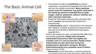
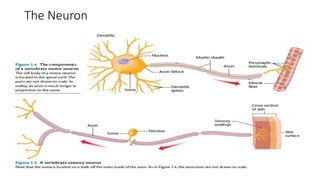
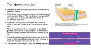
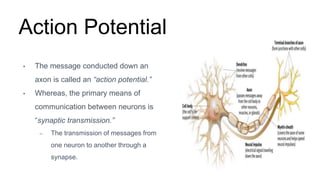
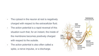
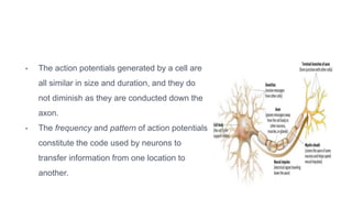
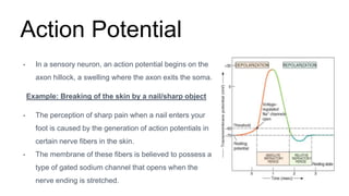
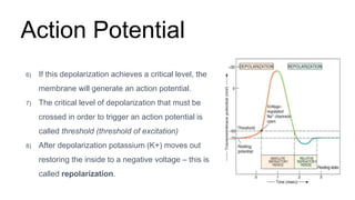
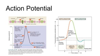
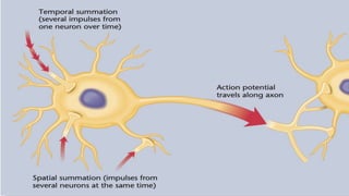
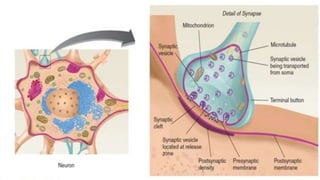
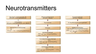
SlideShare a Scribd company logo
1 of 49
INTRODUCTION TO NEUROSCIENCE
The human brain has 100 billion neurons, each neuron
connected to 10,000 other neurons. Sitting on your shoulders is
the most complicated object in the known universe.
-Michio Kaku, American Physicist.
The Basic Animal Cell
• The surface of a cell is its membrane (or plasma
membrane), a structure that separates the inside of the
cell from the outside environment. Most chemicals
cannot cross the membrane, but protein channels in
the membrane permit a controlled flow of water,
oxygen, sodium, potassium, calcium, chloride, and
other important chemicals.
• Except for mammalian red blood cells, all animal cells
have a nucleus, the structure that contains the
chromosomes.
• A mitochondrion (plural: mitochondria) is the structure
that metabolic activities, providing the energy that the
cell uses for all activities. Mitochondria have genes
separate from
those in the nucleus of a cell, and mitochondria differ from
one another genetically. People with overactive
mitochondria tend to burn their fuel rapidly and
overheat, even in a cool environment. People whose
mitochondria are less active than normal are
predisposed to depression and pains. Mutated
mitochondrial genes are a possible cause of autism
(Aoki & Cortese, 2016).
• Ribosomes are the sites within a cell that synthesize
The Neuron
• All neurons include a soma (cell body), and most also have dendrites, an axon, and
presynaptic terminals. The tiniest neurons lack axons, and some lack well-defined
dendrites.
• A motor neuron, with its soma in the spinal cord, receives excitation through its dendrites
and conducts impulses along its axon to a muscle.
• A sensory neuron is specialized at one end to be highly sensitive to a particular type of
stimulation, such as light, sound, or touch.
• Dendrites are branching fibers that get narrower near their ends. The dendrite’s surface
is lined with specialized synaptic receptors, at which the dendrite receives information
from other neurons. The greater the surface area of a dendrite, the more information it
can receive.
• Many dendrites contain dendritic spines, short outgrowths that increase the surface area
available for synapses.
• The axon is a thin fiber of constant diameter. The axon conveys an impulse toward other
neurons, an organ, or a muscle. Axons can be more than a meter in length, as in the case
of axons from your spinal cord to your feet. The length of an axon is enormous in
comparison to its width, and in comparison.
• Many vertebrate axons are covered with an insulating material
called a myelin sheath with interruptions known as nodes of
Ranvier (RAHN-vee-ay).
• Although a neuron can have many dendrites, it can have only one
axon, but the axon may have branches. The end of each branch
has a swelling, called a presynaptic terminal, also known as an
end bulb or bouton (French for “button”). At that point the axon
releases chemicals that cross through the junction between that
neuron and another cell.
• Other terms associated with neurons are afferent, efferent, and
intrinsic.
• An afferent axon brings information into a structure; an efferent
axon carries information away from a structure.
• Every sensory neuron is an afferent to the rest of the nervous
system, and every motor neuron is an efferent from the
nervous system.
• Within the nervous system, a given neuron is an efferent from one
structure and an afferent to another. You can remember that
efferent starts with e as in exit; afferent starts with a as in admit.
• If a cell’s dendrites and axon are entirely contained within a single
structure, the cell is an interneuron or intrinsic neuron of that
structure. For example, an intrinsic neuron of the thalamus has its
Varying Types of Neurons
• Neurons vary enormously
in size, shape, and
function.
• The shape of a neuron
determines its connections
with other cells and
thereby determines its
function. For example, the
widely branching dendrites
of the Purkinje cell in the
cerebellum enable it to
receive input from up to
200,000 other neurons.
• By contrast, bipolar
neurons in the retina have
only short branches, and
some receive input from as
few as two other cells.
Glia
Neuroglial cells—usually referred to simply as glial cells or glia—are quite different
from nerve cells.
The major distinction is that glia do not participate directly in synaptic interactions and
electrical signaling, although their supportive functions help define synaptic contacts and
maintain the signaling abilities of neurons.
Although glial cells also have complex processes extending from their cell bodies, they are
generally smaller than neurons, and they lack axons and dendrites.
Glial roles that are well-established include:
1. Maintaining the ionic environment of nerve cells,
2. Modulating the rate of nerve signal propagation,
3. Modulating synaptic action by controlling the uptake of neurotransmitters,
4. Providing a scaffold for some aspects of neural development,
5. Aiding in (or preventing, in some instances) recovery from neural injury.
There are three types of glial cells in the mature central
nervous system: astrocytes, oligodendrocytes,
and microglial cells.
1. Astrocytes, which are restricted to the brain
and spinal cord, have elaborate local processes that
give these cells a starlike appearance. The major
function of astrocytes is to maintain, in a variety of
ways, an appropriate chemical environment for
neuronal signaling.
2. Oligodendrocytes, which are also restricted to the
central nervous system, lay down a laminated, lipid-rich
wrapping called myelin around some, but not all,
axons. Myelin has important effects on the speed
of action potential conduction.
3. In the peripheral nervous system, the cells that
elaborate myelin are called Schwann cells.
4. Microglial cells are primarily scavenger cells that
remove cellular debris from sites of injury or normal cell
turnover.
The Blood-Brain Barrier
• The brain, like any other organ, needs to receive nutrients from the blood, many
chemicals cannot cross from the blood to the brain (Hagenbuch, Gao, & Meier,
2002). The mechanism that excludes most chemicals from the vertebrate brain
is known as the blood–brain barrier.
• WHY DO WE NEED IT?
• When a virus invades a cell, mechanisms within the cell extrude virus particles
through the membrane so that the immune system can find them. When the
immune system cells discover a virus, they kill it and the cell that contains it.
• This plan works fine if the virus-infected cell is, for example, a skin cell or a
blood cell, which the body replaces easily. However, with few exceptions, the
vertebrate brain does not replace damaged neurons. To minimize the risk of
irreparable brain damage, the body lines the brain’s blood vessels with tightly
packed cells that keep out most viruses, bacteria, and harmful chemicals.
How the Blood-Brain
Barrier works
The blood–brain barrier depends on the endothelial cells that
form the walls of the capillaries (Bundgaard, 1986; Rapoport &
Robinson, 1986).
Outside the brain, such cells are separated by small gaps, but
in the brain, they are joined so tightly that they block viruses,
bacteria, and other harmful chemicals from passage.
If the blood–brain barrier is such a good defense, then
why don’t we have similar walls around all our other
organs?
The barrier keeps out useful chemicals as well as harmful ones.
Those useful chemicals include all fuels and amino acids, the
building blocks for proteins. For these chemicals to cross the
blood–brain barrier, the brain needs special mechanisms not
found in the rest of the body.
• No special mechanism is required for small, uncharged molecules such as
oxygen and carbon dioxide that cross through cell walls freely. Also,
molecules that dissolve in the fats of the membrane cross easily.
Examples include vitamins A and D and all the drugs that affect the brain—from
antidepressants and other psychiatric drugs to illegal drugs such as heroin.
How fast a drug takes effect depends largely on how readily it dissolves in fats
and therefore crosses the blood–brain barrier.
• Water crosses through special protein channels in the wall of the endothelial
cells (Amiry-Moghaddam & Ottersen, 2003). For certain other chemicals, the
brain uses active transport, a protein-mediated process that expends energy
to pump chemicals from the blood into the brain.
• Chemicals that are actively transported into the brain include glucose (the
brain’s main fuel), amino acids (the building blocks of proteins), purines,
choline, a few vitamins, and iron (Abbott, Rönnback, & Hansson, 2006;
Jones & Shusta, 2007).
COMMUNICATION WITHIN NEURONS
The Nerve Impulse
• Messages in a neuron develop from disturbances of the
resting potential.
• All parts of a neuron are covered by a membrane about 8
nanometers (nm) thick. The membrane is composed of
two layers (free to float relative to each other) of
phospholipid molecules.
• Embedded among the phospholipids are cylindrical
protein molecules through which certain chemicals can
pass.
• When at rest, the membrane maintains an electrical
gradient, also known as polarization—a difference in
electrical charge between the inside and outside of the
cell.
• The electrical potential inside the membrane is slightly
negative with respect to the outside, mainly because of
negatively charged proteins inside the cell.
• This difference in voltage is called the resting potential.
Forces Acting on Sodium and Potassium Ions
• If charged ions could flow freely across the membrane, the membrane would depolarize, eliminating the
negative potential inside.
• However, the membrane has selective permeability. That is, some chemicals pass through it more freely
than others do.
• When the membrane is at rest, the sodium and potassium channels are closed, permitting almost no flow of
sodium and only a small flow of potassium.
• Certain types of stimulation can open these channels, permitting freer flow of either or both ions.
• The sodium–potassium pump, a protein complex, repeatedly transports three sodium ions out of the cell
while drawing two potassium ions into it. The sodium–potassium pump is an active transport that requires
energy. membrane than inside, and potassium ions are more concentrated inside than outside.
• The sodium–potassium pump is effective only because of the selective permeability of the membrane,
which prevents the sodium ions that were pumped out of the neuron from leaking right back in again.
• When sodium ions are pumped out, they stay out. However, some of the potassium ions in the neuron
slowly leak out, carrying a positive charge with them.
The Neuron at Rest: Resting Potential
• If charged ions could flow freely across the membrane, the membrane would
depolarize at once.
– SInce the membrane is selectively permeable; that is, some chemicals can
pass through it more freely than others can.
• Due to its selective permeability, the membrane of a neuron maintains an
electrical gradient, a difference in electrical charge between the inside and
outside of the cell.
• This positive outside/negative inside electric polarization is called resting
potential (approximately -70mV).
Why is a Resting Potential Necessary
• The body invests much energy to operate the sodium–potassium pump,
which maintains the resting potential. Why is it worth so much energy? The
resting potential prepares the neuron to respond rapidly.
• Because the membrane did its work in advance by maintaining the
concentration gradient for sodium, the cell is prepared to respond
vigorously to a stimulus.
• Compare the resting potential of a neuron to a poised bow and arrow: An
archer who pulls the bow in advance is ready to fire at the appropriate
moment. The neuron uses the same strategy. The resting potential
remains stable until the neuron is stimulated.
Action Potential
• The message conducted down an
axon is called an “action potential.”
• Whereas, the primary means of
communication between neurons is
“synaptic transmission.”
– The transmission of messages from
one neuron to another through a
synapse.
• The cytosol in the neuron at rest is negatively
charged with respect to the extracellular fluid.
• The action potential is a rapid reversal of this
situation such that, for an instant, the inside of
the membrane becomes positively charged
with respect to the outside.
• The action potential is also often called a
spike, a nerve impulse, or a discharge.
• The action potentials generated by a cell are
all similar in size and duration, and they do
not diminish as they are conducted down the
axon.
• The frequency and pattern of action potentials
constitute the code used by neurons to
transfer information from one location to
another.
Action Potential
• In a sensory neuron, an action potential begins on the
axon hillock, a swelling where the axon exits the soma.
Example: Breaking of the skin by a nail/sharp object
• The perception of sharp pain when a nail enters your
foot is caused by the generation of action potentials in
certain nerve fibers in the skin.
• The membrane of these fibers is believed to possess a
type of gated sodium channel that opens when the
nerve ending is stretched.
Action Potential
1) The nail enters the skin.
2) The membrane of the nerve fibers in the skin is stretched.
3) Na+ permeable channels open.
4) Because of the large concentration gradient and the negative
charge of the cytosol, Na+ ions enter the fiber through these
channels.
5) The entry of Na+ depolarizes the membrane.
– the cytoplasmic (inside) surface of the membrane
becomes less negative.
Action Potential
6) If this depolarization achieves a critical level, the
membrane will generate an action potential.
7) The critical level of depolarization that must be
crossed in order to trigger an action potential is
called threshold (threshold of excitation)
8) After depolarization potassium (K+) moves out
restoring the inside to a negative voltage – this is
called repolarization.
Action Potential
Action potentials are caused by depolarization of the
membrane beyond threshold.
The rapid depolarization and repolarization produce a
pattern called a spike.
Action Potential
Absolute Refractory Period
• The period immediately after an action potential
when another action potential cannot occur.
• The refractory period is the time during which a
neuron resists further action potentials while it
recharges.
Relative Refractory Period
• The period following absolute refractory period
when a neuron will only respond to a stronger
than normal impulse.
All-or-No Law
• A neuron either fires or it does not.
• When it does fire, it will always produce an impulse of the same strength.
• Intensity of a stimulus is coded by the frequency of action potentials.
– The size, amplitude and velocity of an action potential are independent of
the intensity of the stimulus that initiated it.
– Neurons don’t fire faster instead more neurons are fired and fired more
often.
The Neural Impulse
• Each point along the membrane regenerates the action potential in much the
same way that it was generated initially.
• During the action potential, sodium ions enters a point on the axon.
• Temporarily, that location is positively charged in comparison with neighboring
areas along the axon.
• The positive ions flow down the axon and across the membrane.
• The positive charges now inside the membrane slightly depolarize the adjacent
areas of the membrane, causing the next area to reach its threshold and open
the voltage-gated sodium channels.
The Neural Impulse
The Neural Impulse
• Therefore, the membrane regenerates the action potential at that point.
• In this manner, the action potential travels like a wave along the axon.
• The term propagation of the action potential describes the transmission of
an action potential down an axon.
Communication
Between Neurons:
Synapses
Overview
• Synaptic transmission is a comprehensive and fascinating topic.
• The actions of psychoactive drugs, the causes of mental disorders, the
neural bases of learning and memory – indeed, all the operations of the
nervous system – cannot be understood without knowledge of synaptic
transmission.
Concept of Synapse
• In the late 1800s, Cajal anatomically demonstrated a narrow gap
separating one neuron from another.
• In 1906, Sherrington physiologically demonstrated that communication
between one neuron and the next differs from communication along a
single axon.
– He inferred a specialized gap between neurons and introduced the term
synapse to describe it.
Synapse
• The synapse is where the nerve impulse passes from one cell to the next.
• The electrical signal (the action potential) stops and a chemical signal takes
over to cross the gap between the cells – the chemical messenger is called a
neurotransmitter.
• The neurotransmitter crosses the gap by diffusion, which creates a small delay.
• The advantage of using neurotransmitter is that the nerve impulse can be given
some more specificity.
• Neurotransmitters can also control the operation of the nervous system by
inhibition or excitation.
Properties of a Synapse
• Sherrington conducted his research on reflexes (automatic muscular
responses to stimuli) and observed several properties of reflexes
suggesting special processes at the junctions between neurons:
a) Reflexes are slower than conduction along an axon.
b) Several weak stimuli presented at slightly different times (temporal summation)
or slightly different locations (spatial summation) produce a stronger reflex than
a single stimulus does .
c) When one set of muscles becomes excited, a different set becomes relaxed .
Structure of Synapses
• Synapses are junctions between the terminal buttons at the ends of the
axonal branches of one neuron and the membrane of another.
• Synapses can occur in three places: on dendrites, on the soma, and on
other axons.
– These synapses are referred to as a axodendritic, axosomatic, and axoaxonic.
– Axodendritic synapses can occur on the smooth surface of a dendrite or on
dendritic spines (small protrusions that stud the dendrites of several types of
large neurons in the brain).
Structure of Synapses
• Synaptic cleft – The space between the presynaptic membrane and the
postsynaptic membrane. This is approximately 20 to 40 nm wide. (A
nanometer (nm) is one billionth of a meter.)
• Synaptic vesicle – A small, hollow, bead-like structure found in terminal
buttons: contains molecules of a neurotransmitter
• Release zone – A region of the interior of the presynaptic membrane of a
synapse to which synaptic vesicles attach and release their
neurotransmitter into the synaptic cleft.
Sequence of Events at a
Synapse
1) The neuron synthesizes chemicals that serve as neurotransmitters. It
synthesizes the smaller neurotransmitters in the axon terminals and
neuropeptides in the cell body.
2) The neuron transports the neuropeptides that were formed in the cell body
to the axon terminals or to the dendrites. (Neuropeptides are released from
multiple sites in the cell.)
3) Action potentials travel down the axon. At the presynaptic terminal, an
action potential enables calcium to enter the cell. Calcium releases
neurotransmitters from the terminals (exocytosis) and into the synaptic
cleft.
Sequence of Events at a
Synapse
4) The released molecules diffuse across the cleft, attach to receptors, and
alter the activity of the postsynaptic neuron.
5) The neurotransmitter molecules separate from their receptors. Depending
on the neurotransmitter, it may be converted into inactive chemicals.
6) The neurotransmitter molecules may be taken back into the presynaptic
neuron for recycling or may diffuse away. In some cases, empty vesicles
are returned to the cell body.
7) Some postsynaptic cells send reverse messages to control the further
release of neurotransmitter by presynaptic cells.
Sequence of Events at a
Synapse
Neurotransmitters
• At a synapse, a neuron releases chemicals that affect another neuron. Those chemicals are known
as neurotransmitters. A hundred or so chemicals are known or suspected to be neurotransmitters,
Neurotransmitters
Neurotransmitters
• Each pathway above begins with substances found in the diet.
• Acetylcholine is synthesized from choline, which is abundant in milk, eggs, and
peanuts.
• The amino acids phenylalanine and tyrosine, present in virtually any protein, are
precursors of dopamine, norepinephrine, and epinephrine.
• The amino acid tryptophan, the precursor to serotonin, crosses the blood-brain
barrier by a special transport system that it shares with other large amino acids.
Neurotransmitters
• The amount of tryptophan in the diet controls the amount of serotonin in the brain, so your
serotonin levels rise after you eat foods rich in tryptophan, such as soy, and fall after
something low in tryptophan, such as maize (corn).
• Another way to increase tryptophan entry to the brain is to decrease consumption of
phenylalanine or increase consumption of carbohydrates.
• Carbohydrates increase the release of insulin, a hormone, which takes several of the
competing amino acids out of the bloodstream and into body cells, thus decreasing the
competition against tryptophan.

More Related Content

Similar to INTRODUCTION TO NEUROSCIENCE-PART 1.pptx

The nervous system
The nervous systemThe nervous system
The nervous systemMariam Ohanyan
 
Nervous Tissues.pptx
Nervous Tissues.pptxNervous Tissues.pptx
Nervous Tissues.pptxAsifBahadurAli
 
Nervous system.pptx
Nervous system.pptxNervous system.pptx
Nervous system.pptxRohit Sharma
 
Neurons and the glia.pptx
Neurons and the glia.pptxNeurons and the glia.pptx
Neurons and the glia.pptxSabaJahan2
 
NEURON SUPPORTIVE CELLS OR ANS
NEURON SUPPORTIVE CELLS OR ANSNEURON SUPPORTIVE CELLS OR ANS
NEURON SUPPORTIVE CELLS OR ANSoptometry student
 
NERVE CELLS FINAL( NEURON AND GLIAL CELLS.pptx FOR NURSING STUDENTS
NERVE CELLS FINAL( NEURON AND GLIAL CELLS.pptx FOR NURSING STUDENTSNERVE CELLS FINAL( NEURON AND GLIAL CELLS.pptx FOR NURSING STUDENTS
NERVE CELLS FINAL( NEURON AND GLIAL CELLS.pptx FOR NURSING STUDENTSWINCY THIRUMURUGAN
 
vertebrate and invertebrate nervous system physiology.pptx
vertebrate and invertebrate nervous system physiology.pptxvertebrate and invertebrate nervous system physiology.pptx
vertebrate and invertebrate nervous system physiology.pptxAdomatiOresto
 
Report in biology (nervous system)
Report in biology (nervous system)Report in biology (nervous system)
Report in biology (nervous system)Mary Tuazon
 
The nervous system
The nervous systemThe nervous system
The nervous systemSmiley Rathy
 
Nervous System,Neuron, Glial cells
Nervous System,Neuron, Glial cellsNervous System,Neuron, Glial cells
Nervous System,Neuron, Glial cellssrinupappu
 
Anatomy and physiology of brain ppt, its organization
Anatomy and physiology of brain ppt,  its organizationAnatomy and physiology of brain ppt,  its organization
Anatomy and physiology of brain ppt, its organizationJyotirmayee Mandal
 
Nervous tissue
Nervous tissueNervous tissue
Nervous tissueBithikaBaidya
 
Neuroglial cells _neurons
Neuroglial cells _neuronsNeuroglial cells _neurons
Neuroglial cells _neuronsPersonal
 
Nerve and muscle
Nerve and muscleNerve and muscle
Nerve and musclelaraib jameel
 
Simple information about nervous cell
Simple information about nervous cellSimple information about nervous cell
Simple information about nervous cellMohamed Ali Radwan
 
About Neuron, Glia cells and Neurotransmitters.pdf
About Neuron, Glia cells and Neurotransmitters.pdfAbout Neuron, Glia cells and Neurotransmitters.pdf
About Neuron, Glia cells and Neurotransmitters.pdfSELF-EXPLANATORY
 
Bilogical basis of psychology
Bilogical basis of psychologyBilogical basis of psychology
Bilogical basis of psychologymaheshpingali
 
Cns histology and anatomy
Cns histology and anatomyCns histology and anatomy
Cns histology and anatomyporpgp
 

Similar to INTRODUCTION TO NEUROSCIENCE-PART 1.pptx (20)

The nervous system
The nervous systemThe nervous system
The nervous system
 
Nervous Tissues.pptx
Nervous Tissues.pptxNervous Tissues.pptx
Nervous Tissues.pptx
 
Nervous system.pptx
Nervous system.pptxNervous system.pptx
Nervous system.pptx
 
Neurons and the glia.pptx
Neurons and the glia.pptxNeurons and the glia.pptx
Neurons and the glia.pptx
 
NEURON SUPPORTIVE CELLS OR ANS
NEURON SUPPORTIVE CELLS OR ANSNEURON SUPPORTIVE CELLS OR ANS
NEURON SUPPORTIVE CELLS OR ANS
 
NERVE CELLS FINAL( NEURON AND GLIAL CELLS.pptx FOR NURSING STUDENTS
NERVE CELLS FINAL( NEURON AND GLIAL CELLS.pptx FOR NURSING STUDENTSNERVE CELLS FINAL( NEURON AND GLIAL CELLS.pptx FOR NURSING STUDENTS
NERVE CELLS FINAL( NEURON AND GLIAL CELLS.pptx FOR NURSING STUDENTS
 
vertebrate and invertebrate nervous system physiology.pptx
vertebrate and invertebrate nervous system physiology.pptxvertebrate and invertebrate nervous system physiology.pptx
vertebrate and invertebrate nervous system physiology.pptx
 
Report in biology (nervous system)
Report in biology (nervous system)Report in biology (nervous system)
Report in biology (nervous system)
 
The nervous system
The nervous systemThe nervous system
The nervous system
 
Nervous System,Neuron, Glial cells
Nervous System,Neuron, Glial cellsNervous System,Neuron, Glial cells
Nervous System,Neuron, Glial cells
 
Anatomy and physiology of brain ppt, its organization
Anatomy and physiology of brain ppt,  its organizationAnatomy and physiology of brain ppt,  its organization
Anatomy and physiology of brain ppt, its organization
 
Nervous tissue
Nervous tissueNervous tissue
Nervous tissue
 
Neuroglial cells _neurons
Neuroglial cells _neuronsNeuroglial cells _neurons
Neuroglial cells _neurons
 
Nueron ppt
Nueron pptNueron ppt
Nueron ppt
 
Nerve and muscle
Nerve and muscleNerve and muscle
Nerve and muscle
 
Simple information about nervous cell
Simple information about nervous cellSimple information about nervous cell
Simple information about nervous cell
 
About Neuron, Glia cells and Neurotransmitters.pdf
About Neuron, Glia cells and Neurotransmitters.pdfAbout Neuron, Glia cells and Neurotransmitters.pdf
About Neuron, Glia cells and Neurotransmitters.pdf
 
Neurons and its structure
Neurons and its structureNeurons and its structure
Neurons and its structure
 
Bilogical basis of psychology
Bilogical basis of psychologyBilogical basis of psychology
Bilogical basis of psychology
 
Cns histology and anatomy
Cns histology and anatomyCns histology and anatomy
Cns histology and anatomy
 

Recently uploaded

VIP Call Girls Mumbai Arpita 9910780858 Independent Escort Service Mumbai
VIP Call Girls Mumbai Arpita 9910780858 Independent Escort Service MumbaiVIP Call Girls Mumbai Arpita 9910780858 Independent Escort Service Mumbai
VIP Call Girls Mumbai Arpita 9910780858 Independent Escort Service Mumbaisonalikaur4
 
Call Girls Whitefield Just Call 7001305949 Top Class Call Girl Service Available
Call Girls Whitefield Just Call 7001305949 Top Class Call Girl Service AvailableCall Girls Whitefield Just Call 7001305949 Top Class Call Girl Service Available
Call Girls Whitefield Just Call 7001305949 Top Class Call Girl Service Availablenarwatsonia7
 
Call Girl Bangalore Nandini 7001305949 Independent Escort Service Bangalore
Call Girl Bangalore Nandini 7001305949 Independent Escort Service BangaloreCall Girl Bangalore Nandini 7001305949 Independent Escort Service Bangalore
Call Girl Bangalore Nandini 7001305949 Independent Escort Service Bangalorenarwatsonia7
 
Russian Call Girls in Pune Riya 9907093804 Short 1500 Night 6000 Best call gi...
Russian Call Girls in Pune Riya 9907093804 Short 1500 Night 6000 Best call gi...Russian Call Girls in Pune Riya 9907093804 Short 1500 Night 6000 Best call gi...
Russian Call Girls in Pune Riya 9907093804 Short 1500 Night 6000 Best call gi...Miss joya
 
Call Girls Jayanagar Just Call 7001305949 Top Class Call Girl Service Available
Call Girls Jayanagar Just Call 7001305949 Top Class Call Girl Service AvailableCall Girls Jayanagar Just Call 7001305949 Top Class Call Girl Service Available
Call Girls Jayanagar Just Call 7001305949 Top Class Call Girl Service Availablenarwatsonia7
 
High Profile Call Girls Jaipur Vani 8445551418 Independent Escort Service Jaipur
High Profile Call Girls Jaipur Vani 8445551418 Independent Escort Service JaipurHigh Profile Call Girls Jaipur Vani 8445551418 Independent Escort Service Jaipur
High Profile Call Girls Jaipur Vani 8445551418 Independent Escort Service Jaipurparulsinha
 
Call Girls Kanakapura Road Just Call 7001305949 Top Class Call Girl Service A...
Call Girls Kanakapura Road Just Call 7001305949 Top Class Call Girl Service A...Call Girls Kanakapura Road Just Call 7001305949 Top Class Call Girl Service A...
Call Girls Kanakapura Road Just Call 7001305949 Top Class Call Girl Service A...narwatsonia7
 
Low Rate Call Girls Mumbai Suman 9910780858 Independent Escort Service Mumbai
Low Rate Call Girls Mumbai Suman 9910780858 Independent Escort Service MumbaiLow Rate Call Girls Mumbai Suman 9910780858 Independent Escort Service Mumbai
Low Rate Call Girls Mumbai Suman 9910780858 Independent Escort Service Mumbaisonalikaur4
 
Russian Call Girl Brookfield - 7001305949 Escorts Service 50% Off with Cash O...
Russian Call Girl Brookfield - 7001305949 Escorts Service 50% Off with Cash O...Russian Call Girl Brookfield - 7001305949 Escorts Service 50% Off with Cash O...
Russian Call Girl Brookfield - 7001305949 Escorts Service 50% Off with Cash O...narwatsonia7
 
Call Girls Service in Bommanahalli - 7001305949 with real photos and phone nu...
Call Girls Service in Bommanahalli - 7001305949 with real photos and phone nu...Call Girls Service in Bommanahalli - 7001305949 with real photos and phone nu...
Call Girls Service in Bommanahalli - 7001305949 with real photos and phone nu...narwatsonia7
 
College Call Girls Vyasarpadi Whatsapp 7001305949 Independent Escort Service
College Call Girls Vyasarpadi Whatsapp 7001305949 Independent Escort ServiceCollege Call Girls Vyasarpadi Whatsapp 7001305949 Independent Escort Service
College Call Girls Vyasarpadi Whatsapp 7001305949 Independent Escort ServiceNehru place Escorts
 
Low Rate Call Girls Pune Esha 9907093804 Short 1500 Night 6000 Best call girl...
Low Rate Call Girls Pune Esha 9907093804 Short 1500 Night 6000 Best call girl...Low Rate Call Girls Pune Esha 9907093804 Short 1500 Night 6000 Best call girl...
Low Rate Call Girls Pune Esha 9907093804 Short 1500 Night 6000 Best call girl...Miss joya
 
Russian Call Girls Chickpet - 7001305949 Booking and charges genuine rate for...
Russian Call Girls Chickpet - 7001305949 Booking and charges genuine rate for...Russian Call Girls Chickpet - 7001305949 Booking and charges genuine rate for...
Russian Call Girls Chickpet - 7001305949 Booking and charges genuine rate for...narwatsonia7
 
Bangalore Call Girls Majestic đź“ž 9907093804 High Profile Service 100% Safe
Bangalore Call Girls Majestic đź“ž 9907093804 High Profile Service 100% SafeBangalore Call Girls Majestic đź“ž 9907093804 High Profile Service 100% Safe
Bangalore Call Girls Majestic đź“ž 9907093804 High Profile Service 100% Safenarwatsonia7
 
Call Girls Service In Shyam Nagar Whatsapp 8445551418 Independent Escort Service
Call Girls Service In Shyam Nagar Whatsapp 8445551418 Independent Escort ServiceCall Girls Service In Shyam Nagar Whatsapp 8445551418 Independent Escort Service
Call Girls Service In Shyam Nagar Whatsapp 8445551418 Independent Escort Serviceparulsinha
 
Asthma Review - GINA guidelines summary 2024
Asthma Review - GINA guidelines summary 2024Asthma Review - GINA guidelines summary 2024
Asthma Review - GINA guidelines summary 2024Gabriel Guevara MD
 
Call Girls Hsr Layout Just Call 7001305949 Top Class Call Girl Service Available
Call Girls Hsr Layout Just Call 7001305949 Top Class Call Girl Service AvailableCall Girls Hsr Layout Just Call 7001305949 Top Class Call Girl Service Available
Call Girls Hsr Layout Just Call 7001305949 Top Class Call Girl Service Availablenarwatsonia7
 
Bangalore Call Girls Marathahalli đź“ž 9907093804 High Profile Service 100% Safe
Bangalore Call Girls Marathahalli đź“ž 9907093804 High Profile Service 100% SafeBangalore Call Girls Marathahalli đź“ž 9907093804 High Profile Service 100% Safe
Bangalore Call Girls Marathahalli đź“ž 9907093804 High Profile Service 100% Safenarwatsonia7
 

Recently uploaded (20)

VIP Call Girls Mumbai Arpita 9910780858 Independent Escort Service Mumbai
VIP Call Girls Mumbai Arpita 9910780858 Independent Escort Service MumbaiVIP Call Girls Mumbai Arpita 9910780858 Independent Escort Service Mumbai
VIP Call Girls Mumbai Arpita 9910780858 Independent Escort Service Mumbai
 
Call Girls Whitefield Just Call 7001305949 Top Class Call Girl Service Available
Call Girls Whitefield Just Call 7001305949 Top Class Call Girl Service AvailableCall Girls Whitefield Just Call 7001305949 Top Class Call Girl Service Available
Call Girls Whitefield Just Call 7001305949 Top Class Call Girl Service Available
 
Call Girl Bangalore Nandini 7001305949 Independent Escort Service Bangalore
Call Girl Bangalore Nandini 7001305949 Independent Escort Service BangaloreCall Girl Bangalore Nandini 7001305949 Independent Escort Service Bangalore
Call Girl Bangalore Nandini 7001305949 Independent Escort Service Bangalore
 
Russian Call Girls in Pune Riya 9907093804 Short 1500 Night 6000 Best call gi...
Russian Call Girls in Pune Riya 9907093804 Short 1500 Night 6000 Best call gi...Russian Call Girls in Pune Riya 9907093804 Short 1500 Night 6000 Best call gi...
Russian Call Girls in Pune Riya 9907093804 Short 1500 Night 6000 Best call gi...
 
Call Girls Jayanagar Just Call 7001305949 Top Class Call Girl Service Available
Call Girls Jayanagar Just Call 7001305949 Top Class Call Girl Service AvailableCall Girls Jayanagar Just Call 7001305949 Top Class Call Girl Service Available
Call Girls Jayanagar Just Call 7001305949 Top Class Call Girl Service Available
 
High Profile Call Girls Jaipur Vani 8445551418 Independent Escort Service Jaipur
High Profile Call Girls Jaipur Vani 8445551418 Independent Escort Service JaipurHigh Profile Call Girls Jaipur Vani 8445551418 Independent Escort Service Jaipur
High Profile Call Girls Jaipur Vani 8445551418 Independent Escort Service Jaipur
 
Call Girls Kanakapura Road Just Call 7001305949 Top Class Call Girl Service A...
Call Girls Kanakapura Road Just Call 7001305949 Top Class Call Girl Service A...Call Girls Kanakapura Road Just Call 7001305949 Top Class Call Girl Service A...
Call Girls Kanakapura Road Just Call 7001305949 Top Class Call Girl Service A...
 
Low Rate Call Girls Mumbai Suman 9910780858 Independent Escort Service Mumbai
Low Rate Call Girls Mumbai Suman 9910780858 Independent Escort Service MumbaiLow Rate Call Girls Mumbai Suman 9910780858 Independent Escort Service Mumbai
Low Rate Call Girls Mumbai Suman 9910780858 Independent Escort Service Mumbai
 
Russian Call Girl Brookfield - 7001305949 Escorts Service 50% Off with Cash O...
Russian Call Girl Brookfield - 7001305949 Escorts Service 50% Off with Cash O...Russian Call Girl Brookfield - 7001305949 Escorts Service 50% Off with Cash O...
Russian Call Girl Brookfield - 7001305949 Escorts Service 50% Off with Cash O...
 
Call Girls Service in Bommanahalli - 7001305949 with real photos and phone nu...
Call Girls Service in Bommanahalli - 7001305949 with real photos and phone nu...Call Girls Service in Bommanahalli - 7001305949 with real photos and phone nu...
Call Girls Service in Bommanahalli - 7001305949 with real photos and phone nu...
 
College Call Girls Vyasarpadi Whatsapp 7001305949 Independent Escort Service
College Call Girls Vyasarpadi Whatsapp 7001305949 Independent Escort ServiceCollege Call Girls Vyasarpadi Whatsapp 7001305949 Independent Escort Service
College Call Girls Vyasarpadi Whatsapp 7001305949 Independent Escort Service
 
Low Rate Call Girls Pune Esha 9907093804 Short 1500 Night 6000 Best call girl...
Low Rate Call Girls Pune Esha 9907093804 Short 1500 Night 6000 Best call girl...Low Rate Call Girls Pune Esha 9907093804 Short 1500 Night 6000 Best call girl...
Low Rate Call Girls Pune Esha 9907093804 Short 1500 Night 6000 Best call girl...
 
Russian Call Girls Chickpet - 7001305949 Booking and charges genuine rate for...
Russian Call Girls Chickpet - 7001305949 Booking and charges genuine rate for...Russian Call Girls Chickpet - 7001305949 Booking and charges genuine rate for...
Russian Call Girls Chickpet - 7001305949 Booking and charges genuine rate for...
 
Bangalore Call Girls Majestic đź“ž 9907093804 High Profile Service 100% Safe
Bangalore Call Girls Majestic đź“ž 9907093804 High Profile Service 100% SafeBangalore Call Girls Majestic đź“ž 9907093804 High Profile Service 100% Safe
Bangalore Call Girls Majestic đź“ž 9907093804 High Profile Service 100% Safe
 
Call Girls Service In Shyam Nagar Whatsapp 8445551418 Independent Escort Service
Call Girls Service In Shyam Nagar Whatsapp 8445551418 Independent Escort ServiceCall Girls Service In Shyam Nagar Whatsapp 8445551418 Independent Escort Service
Call Girls Service In Shyam Nagar Whatsapp 8445551418 Independent Escort Service
 
Russian Call Girls in Delhi Tanvi ➡️ 9711199012 💋📞 Independent Escort Service...
Russian Call Girls in Delhi Tanvi ➡️ 9711199012 💋📞 Independent Escort Service...Russian Call Girls in Delhi Tanvi ➡️ 9711199012 💋📞 Independent Escort Service...
Russian Call Girls in Delhi Tanvi ➡️ 9711199012 💋📞 Independent Escort Service...
 
sauth delhi call girls in Bhajanpura 🔝 9953056974 🔝 escort Service
sauth delhi call girls in Bhajanpura 🔝 9953056974 🔝 escort Servicesauth delhi call girls in Bhajanpura 🔝 9953056974 🔝 escort Service
sauth delhi call girls in Bhajanpura 🔝 9953056974 🔝 escort Service
 
Asthma Review - GINA guidelines summary 2024
Asthma Review - GINA guidelines summary 2024Asthma Review - GINA guidelines summary 2024
Asthma Review - GINA guidelines summary 2024
 
Call Girls Hsr Layout Just Call 7001305949 Top Class Call Girl Service Available
Call Girls Hsr Layout Just Call 7001305949 Top Class Call Girl Service AvailableCall Girls Hsr Layout Just Call 7001305949 Top Class Call Girl Service Available
Call Girls Hsr Layout Just Call 7001305949 Top Class Call Girl Service Available
 
Bangalore Call Girls Marathahalli đź“ž 9907093804 High Profile Service 100% Safe
Bangalore Call Girls Marathahalli đź“ž 9907093804 High Profile Service 100% SafeBangalore Call Girls Marathahalli đź“ž 9907093804 High Profile Service 100% Safe
Bangalore Call Girls Marathahalli đź“ž 9907093804 High Profile Service 100% Safe
 

INTRODUCTION TO NEUROSCIENCE-PART 1.pptx

  • 2. The human brain has 100 billion neurons, each neuron connected to 10,000 other neurons. Sitting on your shoulders is the most complicated object in the known universe. -Michio Kaku, American Physicist.
  • 3. The Basic Animal Cell • The surface of a cell is its membrane (or plasma membrane), a structure that separates the inside of the cell from the outside environment. Most chemicals cannot cross the membrane, but protein channels in the membrane permit a controlled flow of water, oxygen, sodium, potassium, calcium, chloride, and other important chemicals. • Except for mammalian red blood cells, all animal cells have a nucleus, the structure that contains the chromosomes. • A mitochondrion (plural: mitochondria) is the structure that metabolic activities, providing the energy that the cell uses for all activities. Mitochondria have genes separate from those in the nucleus of a cell, and mitochondria differ from one another genetically. People with overactive mitochondria tend to burn their fuel rapidly and overheat, even in a cool environment. People whose mitochondria are less active than normal are predisposed to depression and pains. Mutated mitochondrial genes are a possible cause of autism (Aoki & Cortese, 2016). • Ribosomes are the sites within a cell that synthesize
  • 5. • All neurons include a soma (cell body), and most also have dendrites, an axon, and presynaptic terminals. The tiniest neurons lack axons, and some lack well-defined dendrites. • A motor neuron, with its soma in the spinal cord, receives excitation through its dendrites and conducts impulses along its axon to a muscle. • A sensory neuron is specialized at one end to be highly sensitive to a particular type of stimulation, such as light, sound, or touch. • Dendrites are branching fibers that get narrower near their ends. The dendrite’s surface is lined with specialized synaptic receptors, at which the dendrite receives information from other neurons. The greater the surface area of a dendrite, the more information it can receive. • Many dendrites contain dendritic spines, short outgrowths that increase the surface area available for synapses. • The axon is a thin fiber of constant diameter. The axon conveys an impulse toward other neurons, an organ, or a muscle. Axons can be more than a meter in length, as in the case of axons from your spinal cord to your feet. The length of an axon is enormous in comparison to its width, and in comparison.
  • 6. • Many vertebrate axons are covered with an insulating material called a myelin sheath with interruptions known as nodes of Ranvier (RAHN-vee-ay). • Although a neuron can have many dendrites, it can have only one axon, but the axon may have branches. The end of each branch has a swelling, called a presynaptic terminal, also known as an end bulb or bouton (French for “button”). At that point the axon releases chemicals that cross through the junction between that neuron and another cell. • Other terms associated with neurons are afferent, efferent, and intrinsic. • An afferent axon brings information into a structure; an efferent axon carries information away from a structure. • Every sensory neuron is an afferent to the rest of the nervous system, and every motor neuron is an efferent from the nervous system. • Within the nervous system, a given neuron is an efferent from one structure and an afferent to another. You can remember that efferent starts with e as in exit; afferent starts with a as in admit. • If a cell’s dendrites and axon are entirely contained within a single structure, the cell is an interneuron or intrinsic neuron of that structure. For example, an intrinsic neuron of the thalamus has its
  • 7. Varying Types of Neurons • Neurons vary enormously in size, shape, and function. • The shape of a neuron determines its connections with other cells and thereby determines its function. For example, the widely branching dendrites of the Purkinje cell in the cerebellum enable it to receive input from up to 200,000 other neurons. • By contrast, bipolar neurons in the retina have only short branches, and some receive input from as few as two other cells.
  • 9. Neuroglial cells—usually referred to simply as glial cells or glia—are quite different from nerve cells. The major distinction is that glia do not participate directly in synaptic interactions and electrical signaling, although their supportive functions help define synaptic contacts and maintain the signaling abilities of neurons. Although glial cells also have complex processes extending from their cell bodies, they are generally smaller than neurons, and they lack axons and dendrites. Glial roles that are well-established include: 1. Maintaining the ionic environment of nerve cells, 2. Modulating the rate of nerve signal propagation, 3. Modulating synaptic action by controlling the uptake of neurotransmitters, 4. Providing a scaffold for some aspects of neural development, 5. Aiding in (or preventing, in some instances) recovery from neural injury.
  • 10. There are three types of glial cells in the mature central nervous system: astrocytes, oligodendrocytes, and microglial cells. 1. Astrocytes, which are restricted to the brain and spinal cord, have elaborate local processes that give these cells a starlike appearance. The major function of astrocytes is to maintain, in a variety of ways, an appropriate chemical environment for neuronal signaling. 2. Oligodendrocytes, which are also restricted to the central nervous system, lay down a laminated, lipid-rich wrapping called myelin around some, but not all, axons. Myelin has important effects on the speed of action potential conduction. 3. In the peripheral nervous system, the cells that elaborate myelin are called Schwann cells. 4. Microglial cells are primarily scavenger cells that remove cellular debris from sites of injury or normal cell turnover.
  • 11. The Blood-Brain Barrier • The brain, like any other organ, needs to receive nutrients from the blood, many chemicals cannot cross from the blood to the brain (Hagenbuch, Gao, & Meier, 2002). The mechanism that excludes most chemicals from the vertebrate brain is known as the blood–brain barrier. • WHY DO WE NEED IT? • When a virus invades a cell, mechanisms within the cell extrude virus particles through the membrane so that the immune system can find them. When the immune system cells discover a virus, they kill it and the cell that contains it. • This plan works fine if the virus-infected cell is, for example, a skin cell or a blood cell, which the body replaces easily. However, with few exceptions, the vertebrate brain does not replace damaged neurons. To minimize the risk of irreparable brain damage, the body lines the brain’s blood vessels with tightly packed cells that keep out most viruses, bacteria, and harmful chemicals.
  • 12. How the Blood-Brain Barrier works The blood–brain barrier depends on the endothelial cells that form the walls of the capillaries (Bundgaard, 1986; Rapoport & Robinson, 1986). Outside the brain, such cells are separated by small gaps, but in the brain, they are joined so tightly that they block viruses, bacteria, and other harmful chemicals from passage. If the blood–brain barrier is such a good defense, then why don’t we have similar walls around all our other organs? The barrier keeps out useful chemicals as well as harmful ones. Those useful chemicals include all fuels and amino acids, the building blocks for proteins. For these chemicals to cross the blood–brain barrier, the brain needs special mechanisms not found in the rest of the body.
  • 13. • No special mechanism is required for small, uncharged molecules such as oxygen and carbon dioxide that cross through cell walls freely. Also, molecules that dissolve in the fats of the membrane cross easily. Examples include vitamins A and D and all the drugs that affect the brain—from antidepressants and other psychiatric drugs to illegal drugs such as heroin. How fast a drug takes effect depends largely on how readily it dissolves in fats and therefore crosses the blood–brain barrier. • Water crosses through special protein channels in the wall of the endothelial cells (Amiry-Moghaddam & Ottersen, 2003). For certain other chemicals, the brain uses active transport, a protein-mediated process that expends energy to pump chemicals from the blood into the brain. • Chemicals that are actively transported into the brain include glucose (the brain’s main fuel), amino acids (the building blocks of proteins), purines, choline, a few vitamins, and iron (Abbott, Rönnback, & Hansson, 2006; Jones & Shusta, 2007).
  • 15. The Nerve Impulse • Messages in a neuron develop from disturbances of the resting potential. • All parts of a neuron are covered by a membrane about 8 nanometers (nm) thick. The membrane is composed of two layers (free to float relative to each other) of phospholipid molecules. • Embedded among the phospholipids are cylindrical protein molecules through which certain chemicals can pass. • When at rest, the membrane maintains an electrical gradient, also known as polarization—a difference in electrical charge between the inside and outside of the cell. • The electrical potential inside the membrane is slightly negative with respect to the outside, mainly because of negatively charged proteins inside the cell. • This difference in voltage is called the resting potential.
  • 16. Forces Acting on Sodium and Potassium Ions • If charged ions could flow freely across the membrane, the membrane would depolarize, eliminating the negative potential inside. • However, the membrane has selective permeability. That is, some chemicals pass through it more freely than others do. • When the membrane is at rest, the sodium and potassium channels are closed, permitting almost no flow of sodium and only a small flow of potassium. • Certain types of stimulation can open these channels, permitting freer flow of either or both ions. • The sodium–potassium pump, a protein complex, repeatedly transports three sodium ions out of the cell while drawing two potassium ions into it. The sodium–potassium pump is an active transport that requires energy. membrane than inside, and potassium ions are more concentrated inside than outside. • The sodium–potassium pump is effective only because of the selective permeability of the membrane, which prevents the sodium ions that were pumped out of the neuron from leaking right back in again. • When sodium ions are pumped out, they stay out. However, some of the potassium ions in the neuron slowly leak out, carrying a positive charge with them.
  • 17. The Neuron at Rest: Resting Potential • If charged ions could flow freely across the membrane, the membrane would depolarize at once. – SInce the membrane is selectively permeable; that is, some chemicals can pass through it more freely than others can. • Due to its selective permeability, the membrane of a neuron maintains an electrical gradient, a difference in electrical charge between the inside and outside of the cell. • This positive outside/negative inside electric polarization is called resting potential (approximately -70mV).
  • 18. Why is a Resting Potential Necessary • The body invests much energy to operate the sodium–potassium pump, which maintains the resting potential. Why is it worth so much energy? The resting potential prepares the neuron to respond rapidly. • Because the membrane did its work in advance by maintaining the concentration gradient for sodium, the cell is prepared to respond vigorously to a stimulus. • Compare the resting potential of a neuron to a poised bow and arrow: An archer who pulls the bow in advance is ready to fire at the appropriate moment. The neuron uses the same strategy. The resting potential remains stable until the neuron is stimulated.
  • 19. Action Potential • The message conducted down an axon is called an “action potential.” • Whereas, the primary means of communication between neurons is “synaptic transmission.” – The transmission of messages from one neuron to another through a synapse.
  • 20. • The cytosol in the neuron at rest is negatively charged with respect to the extracellular fluid. • The action potential is a rapid reversal of this situation such that, for an instant, the inside of the membrane becomes positively charged with respect to the outside. • The action potential is also often called a spike, a nerve impulse, or a discharge.
  • 21. • The action potentials generated by a cell are all similar in size and duration, and they do not diminish as they are conducted down the axon. • The frequency and pattern of action potentials constitute the code used by neurons to transfer information from one location to another.
  • 22. Action Potential • In a sensory neuron, an action potential begins on the axon hillock, a swelling where the axon exits the soma. Example: Breaking of the skin by a nail/sharp object • The perception of sharp pain when a nail enters your foot is caused by the generation of action potentials in certain nerve fibers in the skin. • The membrane of these fibers is believed to possess a type of gated sodium channel that opens when the nerve ending is stretched.
  • 23. Action Potential 1) The nail enters the skin. 2) The membrane of the nerve fibers in the skin is stretched. 3) Na+ permeable channels open. 4) Because of the large concentration gradient and the negative charge of the cytosol, Na+ ions enter the fiber through these channels. 5) The entry of Na+ depolarizes the membrane. – the cytoplasmic (inside) surface of the membrane becomes less negative.
  • 24. Action Potential 6) If this depolarization achieves a critical level, the membrane will generate an action potential. 7) The critical level of depolarization that must be crossed in order to trigger an action potential is called threshold (threshold of excitation) 8) After depolarization potassium (K+) moves out restoring the inside to a negative voltage – this is called repolarization.
  • 25. Action Potential Action potentials are caused by depolarization of the membrane beyond threshold. The rapid depolarization and repolarization produce a pattern called a spike.
  • 27. Absolute Refractory Period • The period immediately after an action potential when another action potential cannot occur. • The refractory period is the time during which a neuron resists further action potentials while it recharges.
  • 28. Relative Refractory Period • The period following absolute refractory period when a neuron will only respond to a stronger than normal impulse.
  • 29. All-or-No Law • A neuron either fires or it does not. • When it does fire, it will always produce an impulse of the same strength. • Intensity of a stimulus is coded by the frequency of action potentials. – The size, amplitude and velocity of an action potential are independent of the intensity of the stimulus that initiated it. – Neurons don’t fire faster instead more neurons are fired and fired more often.
  • 30. The Neural Impulse • Each point along the membrane regenerates the action potential in much the same way that it was generated initially. • During the action potential, sodium ions enters a point on the axon. • Temporarily, that location is positively charged in comparison with neighboring areas along the axon. • The positive ions flow down the axon and across the membrane. • The positive charges now inside the membrane slightly depolarize the adjacent areas of the membrane, causing the next area to reach its threshold and open the voltage-gated sodium channels.
  • 32. The Neural Impulse • Therefore, the membrane regenerates the action potential at that point. • In this manner, the action potential travels like a wave along the axon. • The term propagation of the action potential describes the transmission of an action potential down an axon.
  • 34. Overview • Synaptic transmission is a comprehensive and fascinating topic. • The actions of psychoactive drugs, the causes of mental disorders, the neural bases of learning and memory – indeed, all the operations of the nervous system – cannot be understood without knowledge of synaptic transmission.
  • 35. Concept of Synapse • In the late 1800s, Cajal anatomically demonstrated a narrow gap separating one neuron from another. • In 1906, Sherrington physiologically demonstrated that communication between one neuron and the next differs from communication along a single axon. – He inferred a specialized gap between neurons and introduced the term synapse to describe it.
  • 36. Synapse • The synapse is where the nerve impulse passes from one cell to the next. • The electrical signal (the action potential) stops and a chemical signal takes over to cross the gap between the cells – the chemical messenger is called a neurotransmitter. • The neurotransmitter crosses the gap by diffusion, which creates a small delay. • The advantage of using neurotransmitter is that the nerve impulse can be given some more specificity. • Neurotransmitters can also control the operation of the nervous system by inhibition or excitation.
  • 37. Properties of a Synapse • Sherrington conducted his research on reflexes (automatic muscular responses to stimuli) and observed several properties of reflexes suggesting special processes at the junctions between neurons: a) Reflexes are slower than conduction along an axon. b) Several weak stimuli presented at slightly different times (temporal summation) or slightly different locations (spatial summation) produce a stronger reflex than a single stimulus does . c) When one set of muscles becomes excited, a different set becomes relaxed .
  • 38.
  • 40. Structure of Synapses • Synapses are junctions between the terminal buttons at the ends of the axonal branches of one neuron and the membrane of another. • Synapses can occur in three places: on dendrites, on the soma, and on other axons. – These synapses are referred to as a axodendritic, axosomatic, and axoaxonic. – Axodendritic synapses can occur on the smooth surface of a dendrite or on dendritic spines (small protrusions that stud the dendrites of several types of large neurons in the brain).
  • 41. Structure of Synapses • Synaptic cleft – The space between the presynaptic membrane and the postsynaptic membrane. This is approximately 20 to 40 nm wide. (A nanometer (nm) is one billionth of a meter.) • Synaptic vesicle – A small, hollow, bead-like structure found in terminal buttons: contains molecules of a neurotransmitter • Release zone – A region of the interior of the presynaptic membrane of a synapse to which synaptic vesicles attach and release their neurotransmitter into the synaptic cleft.
  • 42.
  • 43. Sequence of Events at a Synapse 1) The neuron synthesizes chemicals that serve as neurotransmitters. It synthesizes the smaller neurotransmitters in the axon terminals and neuropeptides in the cell body. 2) The neuron transports the neuropeptides that were formed in the cell body to the axon terminals or to the dendrites. (Neuropeptides are released from multiple sites in the cell.) 3) Action potentials travel down the axon. At the presynaptic terminal, an action potential enables calcium to enter the cell. Calcium releases neurotransmitters from the terminals (exocytosis) and into the synaptic cleft.
  • 44. Sequence of Events at a Synapse 4) The released molecules diffuse across the cleft, attach to receptors, and alter the activity of the postsynaptic neuron. 5) The neurotransmitter molecules separate from their receptors. Depending on the neurotransmitter, it may be converted into inactive chemicals. 6) The neurotransmitter molecules may be taken back into the presynaptic neuron for recycling or may diffuse away. In some cases, empty vesicles are returned to the cell body. 7) Some postsynaptic cells send reverse messages to control the further release of neurotransmitter by presynaptic cells.
  • 45. Sequence of Events at a Synapse
  • 46. Neurotransmitters • At a synapse, a neuron releases chemicals that affect another neuron. Those chemicals are known as neurotransmitters. A hundred or so chemicals are known or suspected to be neurotransmitters,
  • 48. Neurotransmitters • Each pathway above begins with substances found in the diet. • Acetylcholine is synthesized from choline, which is abundant in milk, eggs, and peanuts. • The amino acids phenylalanine and tyrosine, present in virtually any protein, are precursors of dopamine, norepinephrine, and epinephrine. • The amino acid tryptophan, the precursor to serotonin, crosses the blood-brain barrier by a special transport system that it shares with other large amino acids.
  • 49. Neurotransmitters • The amount of tryptophan in the diet controls the amount of serotonin in the brain, so your serotonin levels rise after you eat foods rich in tryptophan, such as soy, and fall after something low in tryptophan, such as maize (corn). • Another way to increase tryptophan entry to the brain is to decrease consumption of phenylalanine or increase consumption of carbohydrates. • Carbohydrates increase the release of insulin, a hormone, which takes several of the competing amino acids out of the bloodstream and into body cells, thus decreasing the competition against tryptophan.

Editor's Notes

  1. Ions Charged molecules Resting Potential When more negative ions are inside the neuron than outside Charge is approximately -70mV Neuron is not transmitting information Sodium & Potassium pump – the resting cell continually pumps three sodium ions out while letting two potassium ions in – Since both of these ions have a positive charge the result is a slight decrease in the positive charge of the inside of the cell.
  2. The depolarization that causes action potentials arises in different ways in different neurons. In our example above, depolarization was caused by the entry of Na+ through specialized ion channels that were sensitive to membrane stretching. In interneurons, depolarization is usually caused by Na+ entry through channels that are sensitive to neurotransmitters released by other neurons.
  3. https://youtu.be/tOTYO5WrXFU
  4. Cajal and Sherrington are regarded as the great pioneers of modern neuroscience, and their nearly simultaneous discoveries supported each other: If communication between one neuron and another was special in some way, then there could be no doubt that neurons were anatomically separate from one another.