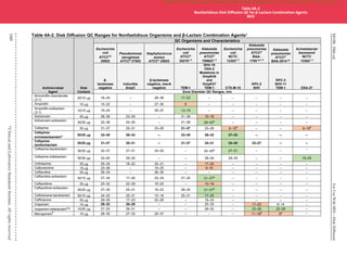
This document provides updated tables for antimicrobial susceptibility testing standards from CLSI documents M02, M07, and M11. It includes the most current information for drug selection, interpretation, and quality control using standardized procedures. Changes from the previous edition are indicated in bold. Laboratories should replace older tables with these new tables to ensure optimal testing and reporting of results.

![M100, 29th ed.
January 2019
Replaces M100, 28th ed.
Performance Standards for Antimicrobial Susceptibility Testing
Melvin P. Weinstein, MD
Jean B. Patel, PhD, D(ABMM)
April M. Bobenchik, PhD, D(ABMM)
Shelley Campeau, PhD, D(ABMM)
Sharon K. Cullen, BS, RAC
Marcelo F. Galas
Howard Gold, MD, FIDSA
Romney M. Humphries, PhD, D(ABMM)
Thomas J. Kirn, Jr., MD, PhD
James S. Lewis II, PharmD, FIDSA
Brandi Limbago, PhD
Amy J. Mathers, MD, D(ABMM)
Tony Mazzulli, MD, FACP, FRCP(C)
Sandra S. Richter, MD, D(ABMM), FCAP, FIDSA
Michael Satlin, MD, MS
Audrey N. Schuetz, MD, MPH, D(ABMM)
Jana M. Swenson, MMSc
Pranita D. Tamma, MD, MHS
Abstract
The data in the tables are valid only if the methodologies in CLSI documents M02,1
M07,2
and M113
are followed. These
standards contain information about disk diffusion (M021
) and dilution (M072
and M113
) test procedures for aerobic and
anaerobic bacteria. Clinicians depend heavily on information from the microbiology laboratory for treating their seriously ill
patients. The clinical importance of antimicrobial susceptibility test results demands that these tests be performed under optimal
conditions and that laboratories have the capability to provide results for the newest antimicrobial agents. The tables presented in
M100 represent the most current information for drug selection, interpretation, and quality control using the procedures
standardized in M02,1
M07,2
and M11.3
Users should replace previously published tables with these new tables. Changes in the
tables since the previous edition appear in boldface type.
Clinical and Laboratory Standards Institute (CLSI). Performance Standards for Antimicrobial Susceptibility Testing. 29th ed.
CLSI supplement M100 (ISBN 978-1-68440-032-4 [Print]; ISBN 978-1-68440-033-1 [Electronic]). Clinical and Laboratory
Standards Institute, 950 West Valley Road, Suite 2500, Wayne, Pennsylvania 19087 USA, 2019.
The Clinical and Laboratory Standards Institute consensus process, which is the mechanism for moving a document through
two or more levels of review by the health care community, is an ongoing process. Users should expect revised editions of any
given document. Because rapid changes in technology may affect the procedures, methods, and protocols in a standard or
guideline, users should replace outdated editions with the current editions of CLSI documents. Current editions are listed in
the CLSI catalog and posted on our website at www.clsi.org. If you or your organization is not a member and would like to
become one, or to request a copy of the catalog, contact us at: Telephone: +1.610.688.0100; Fax: +1.610.688.0700; E-Mail:
customerservice@clsi.org; Website: www.clsi.org.
M100, 30th ed.
January 2020
Replaces M100, 29th ed.
Performance Standards for Antimicrobial Susceptibility Testing
Melvin P. Weinstein, MD
James S. Lewis II, PharmD, FIDSA
April M. Bobenchik, PhD, D(ABMM)
Shelley Campeau, PhD, D(ABMM)
Sharon K. Cullen, BS, RAC
Marcelo F. Galas
Howard Gold, MD, FIDSA
Romney M. Humphries, PhD, D(ABMM)
Thomas J. Kirn, Jr., MD, PhD
Brandi Limbago, PhD
Amy J. Mathers, MD, D(ABMM)
Tony Mazzulli, MD, FACP, FRCP(C)
Michael Satlin, MD, MS
Audrey N. Schuetz, MD, MPH, D(ABMM)
Patricia J. Simner, PhD, D(ABMM)
Pranita D. Tamma, MD, MHS
Abstract
The data in the tables are valid only if the methodologies in CLSI documents M02,1
M07,2
and M113
are followed. These
standards contain information about disk diffusion (M021
) and dilution (M072
and M113
) test procedures for aerobic and
anaerobic bacteria. Clinicians depend heavily on information from the microbiology laboratory for treating their seriously ill
patients. The clinical importance of antimicrobial susceptibility test results demands that these tests be performed under optimal
conditions and that laboratories have the capability to provide results for the newest antimicrobial agents. The tables presented in
M100 represent the most current information for drug selection, interpretation, and quality control using the procedures
standardized in M02,1
M07,2
and M11.3
Users should replace previously published tables with these new tables. Changes in the
tables since the previous edition appear in boldface type.
Clinical and Laboratory Standards Institute (CLSI). Performance Standards for Antimicrobial Susceptibility Testing. 30th ed.
CLSI supplement M100 (ISBN 978-1-68440-066-9 [Print]; ISBN 978-1-68440-067-6 [Electronic]). Clinical and Laboratory
Standards Institute, 950 West Valley Road, Suite 2500, Wayne, Pennsylvania 19087 USA, 2020.
The Clinical and Laboratory Standards Institute consensus process, which is the mechanism for moving a document through
two or more levels of review by the health care community, is an ongoing process. Users should expect revised editions of any
given document. Because rapid changes in technology may affect the procedures, methods, and protocols in a standard or
guideline, users should replace outdated editions with the current editions of CLSI documents. Current editions are listed in
the CLSI catalog and posted on our website at www.clsi.org. If you or your organization is not a member and would like to
become one, or to request a copy of the catalog, contact us at: Telephone: +1.610.688.0100; Fax: +1.610.688.0700; E-Mail:
customerservice@clsi.org; Website: www.clsi.org.
CLSI
eCLIPSE
-
Dewanand
Mahto
-
BD
-
01/29/2020.
Unauthorized
duplication
or
network
sharing
is
not
allowed.](https://image.slidesharecdn.com/clsi2020-220831170657-a1139808/85/CLSI-2020-pdf-3-320.jpg)


![M100,
30th
ed.
xvi
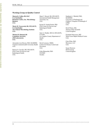
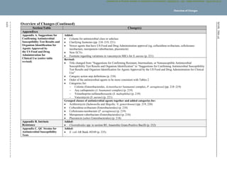
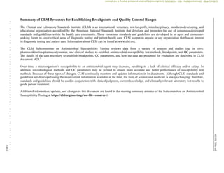
Overview of Changes (Continued)
Section/Table Change(s)
Instructions for Use of Tables (Continued)
VIII. Routine, Supplemental,
Screening, Surrogate Agent,
and Equivalent Agent Testing
to Determine Susceptibility
and Resistance to
Antimicrobial Agents
Supplemental Tests (Optional) table
Added:
Colistin agar test (p. 11)
Colistin broth disk elution (p. 11)
Supplemental Tests (Required and Optional) tables
Revised:
References to appropriate Tables 3 (pp. 10–11)
Screening Tests table
Revised:
References to appropriate Tables 3 (p. 12)
Surrogate Agent Tests table
Clarified:
Cefoxitin test description for specific Staphylococcus spp. (p. 12)
Revised:
Reference to appropriate Table 3 (p. 12)
Examples of Equivalent Agent Tests table
Added:
Colistin and polymyxin B for Enterobacterales, P. aeruginosa, and Acinetobacter baumannii complex (p. 13)
X. Abbreviations and
Acronyms
Added:
CAT (colistin agar test) (p. 14)
CBDE (colistin broth disk elution) (p. 14)
ICR (inducible clindamycin resistance) (p. 14)
MH-F agar (Mueller-Hinton fastidious agar) (p. 14)
Revised:
MRS (methicillin [oxacillin]-resistant staphylococci) (p. 15)
MRSA (methicillin [oxacillin]-resistant Staphylococcus aureus) (p. 15)
NAD (β-nicotinamide adenine dinucleotide) (p. 15)
Deleted:
CoNS (coagulase-negative staphylococci)
KPC (Klebsiella pneumoniae carbapenemase)
NDM (New Delhi metallo--lactamase)
Overview of Changes
CLSI
eCLIPSE
-
Dewanand
Mahto
-
BD
-
01/29/2020.
Unauthorized
duplication
or
network
sharing
is
not
allowed.](https://image.slidesharecdn.com/clsi2020-220831170657-a1139808/85/CLSI-2020-pdf-18-320.jpg)


![For
Use
With
M02
and
M07
M100,
30th
ed.
1
©
Clinical
and
Laboratory
Standards
Institute.
All
rights
reserved.
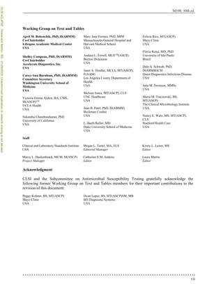
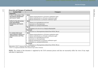
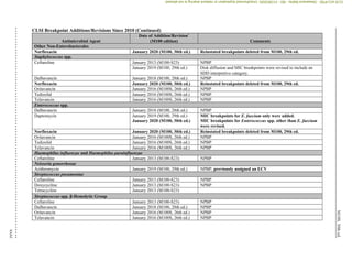
Instructions for Use of Tables
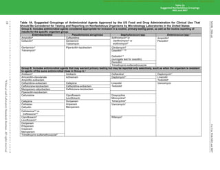
I. Selecting Antimicrobial Agents for Testing and Reporting
A. Selecting the most appropriate antimicrobial agents to test and report is a decision best made by each laboratory in consultation with the
infectious diseases and pharmacy practitioners, the pharmacy and therapeutics and infection prevention committees of the medical staff,
and the antimicrobial stewardship team. The recommendations for each organism group include agents of proven efficacy that show
acceptable in vitro test performance. Considerations in the assignment of agents to specific test/report groups include clinical efficacy,
prevalence of resistance, minimizing emergence of resistance, cost, FDA clinical indications for use, and current consensus
recommendations for first-choice and alternative drugs. Tests on selected agents may be useful for infection prevention purposes.
These instructions apply to:
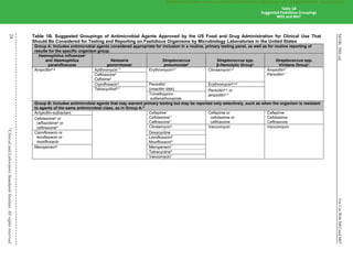
Tables 1A and 1B: suggested groupings of antimicrobial agents that should be considered for testing and reporting by microbiology
laboratories. These guidelines are based on antimicrobial agents approved by the US Food and Drug Administration (FDA) for clinical use
in the United States. In other countries, placement of antimicrobial agents in Tables 1A and 1B should be based on available drugs
approved for clinical use by relevant regulatory organizations.
Tables 2A through 2I: tables for each organism group that contain:
– Recommended testing conditions
– Routine QC recommendations (also see Chapter 4 in M021
and M072
)
– General comments for testing the organism group and specific comments for testing particular agent/organism combinations
– Suggested agents that should be considered for routine testing and reporting by medical microbiology laboratories, as specified in
Tables 1A and 1B (test/report groups A, B, C, U)
– Additional drugs that are appropriate for the respective organism group but would generally not warrant routine testing by a medical
microbiology laboratory in the United States (test/report group O for “other”; test/report group Inv. for “investigational” [not yet FDA
approved])
– Zone diameter and minimal inhibitory concentration (MIC) breakpoints
Tables 1C and 2J: tables containing specific recommendations for testing and reporting results on anaerobes and some of the information
listed in the bullets above
Tables 3A to 3J: tables describing tests to detect particular resistance types in specific organisms or organism groups
CLSI
eCLIPSE
-
Dewanand
Mahto
-
BD
-
01/29/2020.
Unauthorized
duplication
or
network
sharing
is
not
allowed.](https://image.slidesharecdn.com/clsi2020-220831170657-a1139808/85/CLSI-2020-pdf-37-320.jpg)


![M100,
30th
ed.
For
Use
With
M02
and
M07
8
©
Clinical
and
Laboratory
Standards
Institute.
All
rights
reserved
.
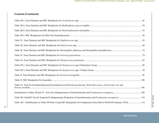
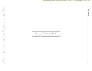
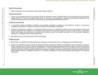
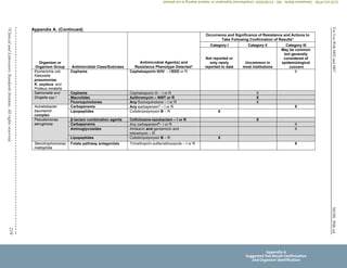
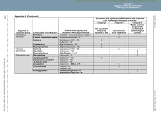
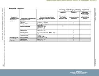
V. Confirmation of Patient Results
Multiple test parameters are monitored by following the QC recommendations described in M100. However, acceptable results derived
from testing QC strains do not guarantee accurate results when testing patient isolates. It is important to review all the results obtained
from all drugs tested on a patient’s isolate before reporting the results. This review should include but not be limited to ensuring that 1) the
AST results are consistent with the identification of the isolate; 2) the results from individual agents within a specific drug class follow the
established hierarchy of activity rules (eg, in general, third-generation cephems are more active than first- or second-generation cephems
against Enterobacterales); and 3) the isolate is susceptible to those agents for which resistance has not been documented (eg, vancomycin
and Streptococcus spp.) and for which only “susceptible” breakpoints exist in M100.
Unusual or inconsistent results should be confirmed by rechecking various testing parameters detailed in Appendix A. Each laboratory
must develop its own policies for confirming unusual or inconsistent antimicrobial susceptibility test results. The list provided in
Appendix A emphasizes results that are most likely to affect patient care.
VI. Development of Resistance and Testing of Repeat Isolates
Isolates that are initially susceptible may become intermediate or resistant after therapy is initiated. Therefore, subsequent isolates of the
same species from a similar anatomical site should be tested to detect resistance that may have developed. Development of resistance can
occur within as little as three to four days and has been noted most frequently in Enterobacter (including Klebsiella [formerly
Enterobacter] aerogenes), Citrobacter, and Serratia spp. with third-generation cephalosporins, in P. aeruginosa with all antimicrobial
agents, and in staphylococci with fluoroquinolones. For S. aureus, vancomycin-susceptible isolates may become vancomycin intermediate
during the course of prolonged therapy.
In certain circumstances, the decision to perform susceptibility tests on subsequent isolates necessitates knowledge of the specific situation
and the severity of the patient’s condition (eg, an isolate of E. cloacae from a blood culture on a premature infant or methicillin
(oxacillin)-resistant S. aureus [MRSA] from a patient with prolonged bacteremia). Laboratory guidelines on when to perform
susceptibility testing on repeat isolates should be determined after consultation with the medical staff.
CLSI
eCLIPSE
-
Dewanand
Mahto
-
BD
-
01/29/2020.
Unauthorized
duplication
or
network
sharing
is
not
allowed.](https://image.slidesharecdn.com/clsi2020-220831170657-a1139808/85/CLSI-2020-pdf-44-320.jpg)


![M100,
30th
ed.
For
Use
With
M02
and
M07
28
©
Clinical
and
Laboratory
Standards
Institute.
All
rights
reserved
.
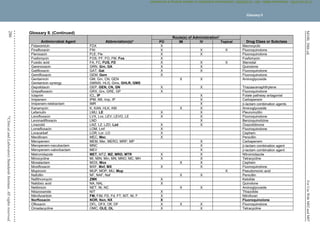
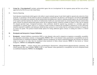
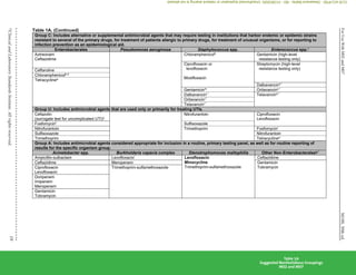
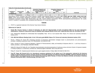
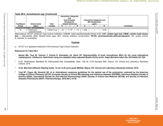
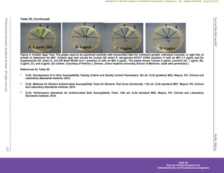
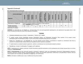
Table 1B. (Continued)
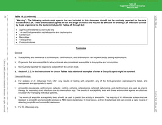
p. Rx: Recommendations for intrapartum prophylaxis for group B streptococci are penicillin or ampicillin. Although cefazolin is recommended for
penicillin-allergic women at low risk for anaphylaxis, those at high risk for anaphylaxis may receive clindamycin. Group B streptococci are
susceptible to ampicillin, penicillin, and cefazolin, but may be resistant to erythromycin and clindamycin. When group B Streptococcus is
isolated from a pregnant woman with severe penicillin allergy (high risk for anaphylaxis), erythromycin and clindamycin (including inducible
clindamycin resistance [ICR]) should be tested, and only clindamycin should be reported. Erythromycin, even when tested for
determination of ICR, should not be reported. See Table 3H.
q. For this table, the β-hemolytic group includes the large colony–forming pyogenic strains of streptococci with group A (S. pyogenes), C, or G
antigens and strains with group B (S. agalactiae) antigen. Small colony–forming β-hemolytic strains with group A, C, F, or G antigens
(Streptococcus anginosus group, previously termed “Streptococcus milleri”) are considered part of the viridans group, and breakpoints for the
viridans group should be used.
r. Daptomycin should not be reported for isolates from the respiratory tract.
s. For reporting against S. pyogenes and Streptococcus agalactiae only.
t. For reporting against S. anginosus group (includes S. anginosus, Streptococcus intermedius, and Streptococcus constellatus) only.
u. For reporting against S. pyogenes, S. agalactiae, Streptococcus dysgalactiae, and S. anginosus group.
NOTE 1: For information about the selection of appropriate antimicrobial agents; explanation of test/report groups A, B, C, and U; and explanation
of the listing of agents within boxes, including the meaning of “or” between agents, refer to the Instructions for Use of Tables that precede Table
1A.
NOTE 2: Information in boldface type is new or modified since the previous edition.
Reference for Table 1B
1
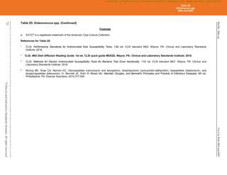
CLSI. Methods for Dilution Antimicrobial Susceptibility Tests for Bacteria That Grow Aerobically. 11th ed. CLSI standard M07. Wayne, PA:
Clinical and Laboratory Standards Institute; 2018.
Table 1B
Suggested Fastidious Groupings
M02 and M07
CLSI
eCLIPSE
-
Dewanand
Mahto
-
BD
-
01/29/2020.
Unauthorized
duplication
or
network
sharing
is
not
allowed.](https://image.slidesharecdn.com/clsi2020-220831170657-a1139808/85/CLSI-2020-pdf-64-320.jpg)


![M100,
30th
ed.
For
Use
With
M02
and
M07
59
©
Clinical
and
Laboratory
Standards
Institute.
All
rights
reserved.
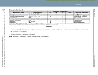
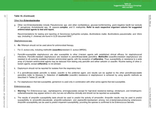
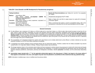
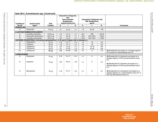
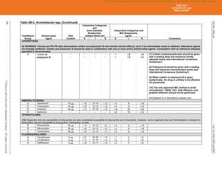
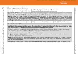
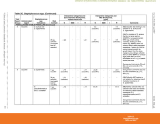
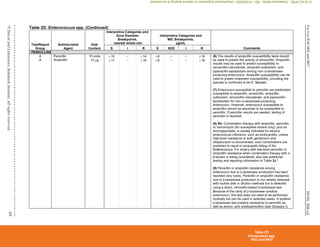
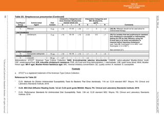
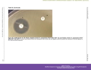
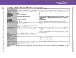
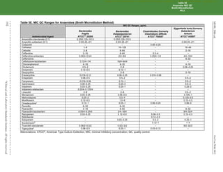
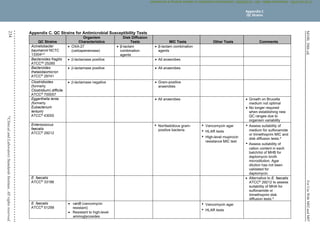
Table 2C. Staphylococcus spp. (Continued)
(4) Historically, resistance to the penicillinase-stable penicillins (see Glossary I) has been referred to as “methicillin resistance” or “oxacillin resistance.” MRSA
are strains of S. aureus that express mecA, mecC, or another mechanism of methicillin (oxacillin) resistance, such as changes in affinity of penicillin-binding
proteins for oxacillin (modified S. aureus strains).
(5) Most methicillin (oxacillin) resistance is mediated by mecA, encoding PBP2a (also called PBP2'). Isolates that test positive for mecA or PBP2a should be
reported as methicillin (oxacillin) resistant (see Appendix H).
Detection of methicillin (oxacillin) resistance in staphylococci is achieved by using specific methods as listed in Table 2C and further described in Table 3F.
Organism
Methods for Detection of Methicillin (Oxacillin)-Resistant Staphylococcus spp.
Cefoxitin MIC
Cefoxitin disk
diffusion Oxacillin MIC
Oxacillin disk
diffusion Oxacillin salt agar
S. aureus
Yes
(16–20 h)
Yes
(16–18 h)
Yes
(24 h)
No Yes
(24 h)
S. lugdunensis
Yes
(16–20 h)
Yes
(16–18 h)
Yes
(24 h)
No No
S. epidermidis
No Yes
(16–18 h)
Yes
(24 h)
Yes
(16–18 h)
No
S. pseudintermedius
No No Yes
(24 h)
Yes
(16–18 h)
No
S. schleiferi
No No Yes
(24 h)
Yes
(16–18 h)
No
Other Staphylococcus spp.
(not listed above)
No Yesa
(24 h)
Yesa
(24 h)
No No
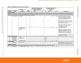
Abbreviations: h, hour(s); MIC, minimal inhibitory concentration; MRS, methicillin (oxacillin)-resistant staphylococci; PBP2a, penicillin-binding
protein 2a.
a
For isolates of “other Staphylococcus spp.” from serious infections for which the oxacillin MICs are 0.5–2 μg/mL, testing for mecA or PBP2a
should be considered (see comment [17]). Cefoxitin disk diffusion is not currently recommended.
Mechanisms of methicillin (oxacillin) resistance other than mecA are rare and include a novel mecA homologue, mecC.4
MICs for strains with mecC are
typically cefoxitin resistant and oxacillin susceptible; mecC resistance cannot be detected by tests directed at mecA or PBP2a.
(6) MRS, as defined by cefoxitin or oxacillin testing, as appropriate to the species, are considered resistant to other -lactam agents, ie, penicillins, -lactam
combination agents, cephems (with the exception of ceftaroline), and carbapenems. This is because most cases of documented MRS infections have
responded poorly to -lactam therapy or because convincing clinical data that document clinical efficacy for those agents have not been presented.
(7) For tests for β-lactamase production, methicillin (oxacillin) resistance and mecA-mediated methicillin (oxacillin) resistance using cefoxitin, reduced
susceptibility to vancomycin, ICR, and high-level mupirocin resistance (S. aureus only), refer to Tables 3E, 3F, 3G, 3H, and 3I, respectively.
NOTE: Information in boldface type is new or modified since the previous edition.
Table 2C
Staphylococcus spp.
M02 and M07
CLSI
eCLIPSE
-
Dewanand
Mahto
-
BD
-
01/29/2020.
Unauthorized
duplication
or
network
sharing
is
not
allowed.](https://image.slidesharecdn.com/clsi2020-220831170657-a1139808/85/CLSI-2020-pdf-95-320.jpg)


![M100,
30th
ed.
For
Use
With
M02
and
M07
74
©
Clinical
and
Laboratory
Standards
Institute.
All
rights
reserved
.
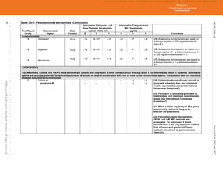
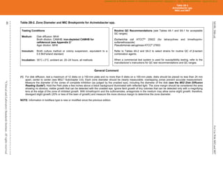
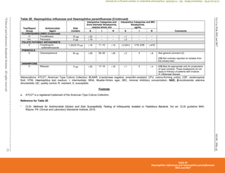
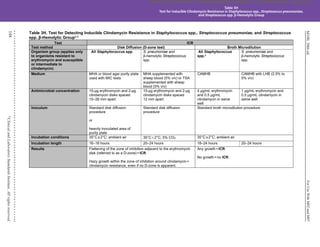
Table 2E. Zone Diameter and MIC Breakpoints for Haemophilus influenzae and Haemophilus parainfluenzae
Testing Conditions
Medium: Disk diffusion: HTM
Broth dilution: HTM broth
Inoculum: Colony suspension, equivalent to a 0.5 McFarland standard
prepared using colonies from an overnight (preferably 20- to
24-hour) chocolate agar plate (see comment [2])
Incubation: 35°C±2°C
Disk diffusion: 5% CO2; 16–18 hours
Broth dilution: ambient air; 20–24 hours
Routine QC Recommendations (see Tables 4A-1, 4B, 5A-1, and 5B for
acceptable QC ranges)
H. influenzae ATCC®a
49247
H. influenzae ATCC®
49766
Use either H. influenzae ATCC®
49247 or H. influenzae ATCC®
49766 or both
of these strains, based on the antimicrobial agents to be tested. Neither strain
has QC ranges for all agents that might be tested against H. influenzae or
H. parainfluenzae.
E. coli ATCC®
35218 (when testing amoxicillin-clavulanate)
When a commercial test system is used for susceptibility testing, refer to the
manufacturer’s instructions for QC test recommendations and QC ranges.
General Comments
(1) Haemophilus spp., as used in this table, includes only H. influenzae and H. parainfluenzae. See CLSI document M451
for testing and reporting
recommendations for other species of Haemophilus.
(2) The 0.5 McFarland suspension contains approximately 1 to 4 × 10⁸
CFU/mL. Use care in preparing this suspension, because higher inoculum concentrations
may lead to false-resistant results with some -lactam antimicrobial agents, particularly when -lactamase–producing strains of H. influenzae are tested.
(3) For disk diffusion, test a maximum of 9 disks on a 150-mm plate and 4 disks on a 100-mm plate. Measure the diameter of the zones of complete inhibition (as
judged by the unaided eye), including the diameter of the disk. Hold the Petri plate a few inches above a black background illuminated with reflected light. The
zone margin should be considered the area showing no obvious, visible growth that can be detected with the unaided eye. Ignore faint growth of tiny colonies
that can be detected only with a magnifying lens at the edge of the zone of inhibited growth. With trimethoprim and the sulfonamides, antagonists in the
medium may allow some slight growth; therefore, disregard slight growth (20% or less of the lawn of growth) and measure the more obvious margin to
determine the zone diameter.
(4) For isolates of H. influenzae from CSF, only results of testing with ampicillin, any of the 3rd-generation cephalosporins listed below, chloramphenicol, and
meropenem are appropriate to report.
(5) Amoxicillin-clavulanate, azithromycin, cefaclor, cefdinir, cefixime, cefpodoxime, cefprozil, cefuroxime, and clarithromycin are used as empiric therapy for
respiratory tract infections due to Haemophilus spp. The results of susceptibility tests with these antimicrobial agents are often not necessary for management
of individual patients.
Table 2E
Haemophilus influenzae and Haemophilus parainfluenzae
M02 and M07
CLSI
eCLIPSE
-
Dewanand
Mahto
-
BD
-
01/29/2020.
Unauthorized
duplication
or
network
sharing
is
not
allowed.](https://image.slidesharecdn.com/clsi2020-220831170657-a1139808/85/CLSI-2020-pdf-110-320.jpg)


![M100,
30th
ed.
For
Use
With
M02
and
M07
78
©
Clinical
and
Laboratory
Standards
Institute.
All
rights
reserved
.
Table 2F. Zone Diameter and MIC Breakpoints for Neisseria gonorrhoeae
Testing Conditions
Medium: Disk diffusion: GC agar base and 1% defined growth supplement. (The use of a
cysteine-free growth supplement is not required for disk diffusion testing.)
Agar dilution: GC agar base and 1% defined growth supplement. (The use of a
cysteine-free growth supplement is required for agar dilution tests with
carbapenems and clavulanate. Cysteine-containing defined growth supplement
does not significantly alter dilution test results with other drugs.)
Inoculum: Colony suspension, equivalent to a 0.5 McFarland standard prepared in MHB or
0.9% phosphate-buffered saline, pH 7, using colonies from an overnight (20- to
24-hour) chocolate agar plate incubated in 5% CO2
Incubation: 36°C±1°C (do not exceed 37°C); 5% CO2; all methods, 20–24 hours
Routine QC Recommendations (see Tables 4B and
5C for acceptable QC ranges)
N. gonorrhoeae ATCC®a
49226
When a commercial test system is used for
susceptibility testing, refer to the manufacturer’s
instructions for QC test recommendations and QC
ranges.
General Comments
(1) For disk diffusion, test a maximum of 9 disks on a 150-mm plate and 4 disks on a 100-mm plate. For some agents, eg, fluoroquinolones or cephalosporins,
only 2 to 3 disks may be tested per plate. Measure the diameter of the zones of complete inhibition (as judged by the unaided eye), including the diameter of
the disk. Hold the Petri plate a few inches above a black background illuminated with reflected light. The zone margin should be considered the area showing
no obvious, visible growth that can be detected with the unaided eye. Ignore faint growth of tiny colonies that can be detected only with a magnifying lens at
the edge of the zone of inhibited growth.
(2) The clinical effectiveness of cefotetan, cefoxitin, and spectinomycin for treating infections due to organisms that produce intermediate results with these
agents is unknown.
(3) For disk diffusion testing of N. gonorrhoeae, an intermediate result for an antimicrobial agent indicates either a technical problem that should be resolved by
repeat testing or a lack of clinical experience in treating infections due to organisms with these zones. Strains with intermediate zones to agents other than
cefotetan, cefoxitin, and spectinomycin have a documented lower clinical cure rate (85% to 95%) compared with >95% for susceptible strains.
(4) The recommended medium for testing N. gonorrhoeae consists of GC agar to which a 1% defined growth supplement (1.1 g L-cystine, 0.03 g guanine HCl,
0.003 g thiamine HCl, 0.013 g para-aminobenzoic acid, 0.01 g B12, 0.1 g cocarboxylase, 0.25 g NAD, 1 g adenine, 10 g L-glutamine, 100 g glucose, 0.02 g
ferric nitrate, 25.9 g L-cysteine HCl [in 1 L H2O]) is added after autoclaving.
NOTE: Information in boldface type is new or modified since the previous edition.
Table 2F
Neisseria gonorrhoeae
M02 and M07
CLSI
eCLIPSE
-
Dewanand
Mahto
-
BD
-
01/29/2020.
Unauthorized
duplication
or
network
sharing
is
not
allowed.](https://image.slidesharecdn.com/clsi2020-220831170657-a1139808/85/CLSI-2020-pdf-114-320.jpg)


![M100,
30th
ed.
For
Use
With
M02
and
M07
149
©
Clinical
and
Laboratory
Standards
Institute.
All
rights
reserved.
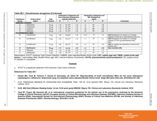
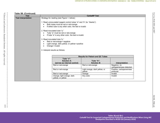
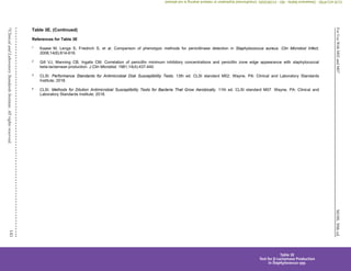
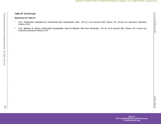
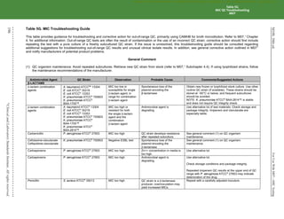
Table 3H. (Continued)
Test ICR
Test method Disk Diffusion (D-zone test) Broth Microdilution
Organism group (applies only
to organisms resistant to
erythromycin and susceptible
or intermediate to clindamycin)
All Staphylococcus spp. S. pneumoniae and
β-hemolytic Streptococcus
spp.
All Staphylococcus
spp.c
S. pneumoniae and β-hemolytic
Streptococcus spp.
Additional testing and
reporting
Report isolates with ICR as “clindamycin resistant.”
The following comment may be included with the report: “This isolate is presumed to be resistant based on detection of ICR,
as determined by testing clindamycin in combination with erythromycin.”
QC recommendations –
routinec
S. aureus ATCC®d
25923 for
routine QC of erythromycin
and clindamycin disks
S. pneumoniae ATCC
49619
for routine QC of erythromycin
and clindamycin disks
S. aureus ATCC®
BAA-976™ or
S. aureus ATCC®
29213 – no growth
S. pneumoniae ATCC
49619 or
S. aureus ATCC
BAA-976™ –
no growth
QC recommendations –
lot/shipmente
S. aureus ATCC
BAA-977™ – growth
QC recommendations –
supplementalf
S. aureus ATCC®
BAA-976™ (D-zone test negative)
S. aureus ATCC®
BAA-977™ (D-zone test positive)
Use of unsupplemented MHA is acceptable for these strains.
S. aureus ATCC®
BAA-976™ (no growth)
S. aureus ATCC®
BAA-977™ (growth)
Abbreviations: ATCC®
, American Type Culture Collection; CAMHB, cation-adjusted Mueller-Hinton broth; ICR, inducible clindamycin resistance; LHB, lysed
horse blood; MHA, Mueller-Hinton agar; MIC, minimal inhibitory concentration; QC, quality control; TSA, tryptic soy agar.
Footnotes
a. Antimicrobial susceptibility testing of β-hemolytic streptococci does not need to be performed routinely (see general comment [4] in Table 2H-1). When
susceptibility testing is clinically indicated, test for ICR in strains that are erythromycin resistant and clindamycin susceptible or intermediate.
b. In accordance with 2010 guidance from the Centers for Disease Control and Prevention, colonizing isolates of group B streptococci from penicillin-allergic
pregnant women should be tested for clindamycin (including ICR) (see comment [12] in Table 2H-1).1
For isolates that test susceptible to clindamycin
(with erythromycin induction), consider adding the following comment to the patient’s report: “This group B Streptococcus does not demonstrate
inducible clindamycin resistance as determined by testing clindamycin in combination with erythromycin.”
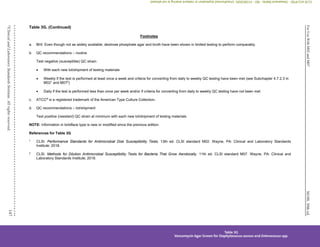
Table 3H
Test for Inducible Clindamycin Resistance in Staphylococcus spp., Streptococcus pneumoniae,
and Streptococcus spp. β-Hemolytic Group
CLSI
eCLIPSE
-
Dewanand
Mahto
-
BD
-
01/29/2020.
Unauthorized
duplication
or
network
sharing
is
not
allowed.](https://image.slidesharecdn.com/clsi2020-220831170657-a1139808/85/CLSI-2020-pdf-185-320.jpg)


![For
Use
With
M07—MIC
Testing
182
M100,
30th
ed.
©
Clinical
and
Laboratory
Standards
Institute.
All
rights
reserved
.
Table 5A-2. (Continued)
Footnotes
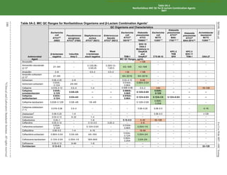
a. Unsupplemented Mueller-Hinton medium (cation-adjusted if broth). See Table 5A-1 for QC ranges for combination agents from other drug classes.
b. ATCC®
is a registered trademark of the American Type Culture Collection. Per ATCC®
convention, the trademark symbol is used after “BAA” in each catalog
number, in conjunction with the registered ATCC®
name.
c. Careful attention to organism maintenance (eg, minimal subcultures) and storage (eg, −60°C or below) is especially important for these QC strains because
spontaneous loss of the plasmid encoding the β-lactamase has been documented. If stored at temperatures above −60°C or if repeatedly subcultured, these
strains may lose their resistance characteristics and QC results may be outside the acceptable ranges.
d. To confirm the integrity of the QC strain, test one of the single β-lactam agents highlighted in orange by either a disk diffusion or MIC test method when the
strain is first subcultured from a frozen or lyophilized stock culture. In-range results for the single agent indicate the QC strain is reliable for QC of β-lactam
combination agents. It is not necessary to check the QC strain again with a single agent until a new frozen or lyophilized stock culture is put into use, providing
recommendations for handling QC strains as described in M021
and M072
are followed. If the highest concentration tested on a panel is lower than the QC
range listed for the particular antimicrobial agent and the MIC result obtained for the QC strain is interpreted as resistant, the QC strain can be considered
reliable for QC of β-lactam combination agents (eg, ampicillin panel concentrations 1–16 µg/mL; ampicillin Enterobacterales breakpoints [µg/mL]: ≤ 8 [S], 16
[I], ≥32 [R]; MIC of >16 µg/ml [R] would be acceptable for K. pneumoniae ATCC®
700603).
e. Either strain highlighted in green may be used for routine QC of this antimicrobial agent.
f. Not tested as a single agent routinely.
NOTE: Information in boldface type is new or modified since the previous edition.
References for Table 5A-2
1
CLSI. Performance Standards for Antimicrobial Disk Susceptibility Tests. 13th ed. CLSI standard M02. Wayne, PA: Clinical and Laboratory Standards
Institute; 2018.
2
CLSI. Methods for Dilution Antimicrobial Susceptibility Tests for Bacteria That Grow Aerobically. 11th ed. CLSI standard M07. Wayne, PA: Clinical and
Laboratory Standards Institute; 2018.
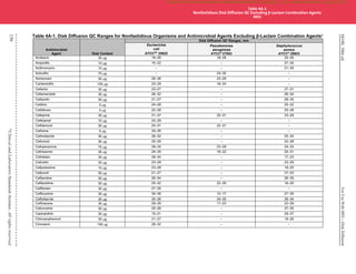
Table 5A-2
Nonfastidious MIC QC for β-Lactam Combination Agents
M07
CLSI
eCLIPSE
-
Dewanand
Mahto
-
BD
-
01/29/2020.
Unauthorized
duplication
or
network
sharing
is
not
allowed.](https://image.slidesharecdn.com/clsi2020-220831170657-a1139808/85/CLSI-2020-pdf-218-320.jpg)


![M100,
30th
ed.
For
Use
With
M07—MIC
Testing
206
©
Clinical
and
Laboratory
Standards
Institute.
All
rights
reserved
.
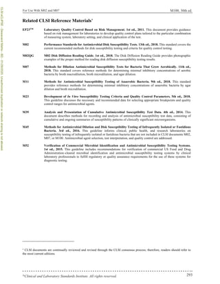
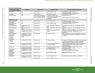
Table 6B. Preparing Stock Solutions for Antimicrobial Agents Provided With Activity Expressed as Units
Footnote
a. Do not use colistin methanesulfonate for in vitro antimicrobial susceptibility tests.
References for Table 6B
1
Geddes AM, Gould IM. Benzylpenicillin (penicillin G). In: Grayson ML, ed. Kucers’ The Use of Antibiotics: A Clinical Review of Antibacterial, Antifungal,
Antiparasitic and Antiviral Drugs. 6th ed. Boca Raton, FL: CRC Press, Taylor & Francis Group; 2010:5-58.
2
Polymyxins. In: Kucers A, Crowe SM, Grayson ML, Hoy JF, eds. The Use of Antibiotics: A Clinical Review of Antibacterial, Antifungal, Antiparasitic and
Antiviral Drugs. 5th ed. Oxford, UK: Butterworth-Heinemann; 1997:667-675.
3
United States Department of Agriculture, Food Safety and Inspection Service, Office of Public Health Science, Laboratory QA/QC Division. Bioassay for the
detection, identification and quantitation of antimicrobial residues in meat and poultry tissue. Microbiology Laboratory Guidebook (MLG) 34.03; 2011.
Antimicrobial
Agent
Pure Agent
(Reference) Calculation for µg/mg Example
Potassium
Penicillin G
0.625 µg/unit1
Multiply the activity expressed in units/mg by 0.625
µg/unit.
Activity units/mg•0.625 µg/unit=Activity µg/mg
(eg, 1592 units/mg•0.625 µg/unit=995 µg/mg)
Sodium
Penicillin G
0.6 µg/unit1
Multiply the activity expressed in units/mg by 0.6 µg/unit. Activity units/mg•0.6 µg/unit=Activity µg/mg
(eg, 1477 units/mg•0.6 µg/unit=886.2 µg/mg)
Polymyxin B 10 000 units/mg=
10 units/µg=
0.1 µg/unit2
Multiply the activity expressed in units/mg by 0.1 µg/unit. Activity units/mg•0.1 µg/unit=Activity µg/mg
(eg, 8120 units/mg•0.1 µg/unit=812 µg/mg)
Divide the activity expressed in units/mg by 10 units/µg. Activity units/mg/10 units/µg=Activity µg/mg
(eg, 8120 units/mg/10 units/mg=812 µg/mg)
Colistin sulfatea 30 000 units/mg=
30 units/µg=
0.03333 µg/unit2
Multiply the activity expressed in units/mg by 0.03333
µg/unit.
Activity units/mg•0.03333 µg/unit=Activity µg/mg
(eg, 20277 units/mg•0.03333 µg/unit=676 µg/mg)
Divide the activity expressed in units/mg by 30 units/mg. Activity units/mg /30 units/µg=Activity µg/mg
(eg, 20277 units/mg/30 units/µg=676 µg/mg)
Streptomycin 785 units/mg3
Divide the number of units given for the powder by 785.
This gives the percent purity of the powder. Multiply the
percent purity by 850, which is the amount in the purest
form of streptomycin. This result equals the activity factor
in µg/mg.
([Potency units/mg]/[785 units/mg])•(850 µg/mg)=Potency µg/mg
(eg, [751 units/mg/785 units/mg]•850 µg/mg=813 µg/mg)
If powder contains 2.8% water:
813•(1–0.028)=potency
813•0.972=790 µg/mg
Table 6B
Preparing Stock Solutions
M07
CLSI
eCLIPSE
-
Dewanand
Mahto
-
BD
-
01/29/2020.
Unauthorized
duplication
or
network
sharing
is
not
allowed.](https://image.slidesharecdn.com/clsi2020-220831170657-a1139808/85/CLSI-2020-pdf-242-320.jpg)


![M100,
30th
ed.
For
Use
With
M02
and
M07
251
©
Clinical
and
Laboratory
Standards
Institute.
All
rights
reserved.
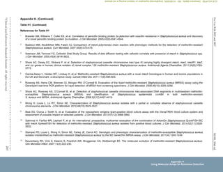
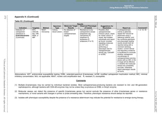
Appendix F. (Continued)
SDD is already well established for use in antifungal susceptibility testing.
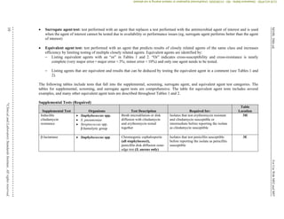
Antibiotic stewardship programs, which emphasize dosage regimen and duration of therapy options, are increasing awareness of appropriate use of
antibiotics. Personnel from these programs should be able to describe the significance to clinicians of an SDD result.
How should this change be implemented?
Meet with the appropriate practitioners at your institution (eg, members of the antimicrobial stewardship team, infectious diseases staff, pathology group,
pharmacy) to explain SDD and determine a plan for implementation, if appropriate.
Talk to the manufacturer of your antimicrobial susceptibility testing (AST) device to determine how to implement reporting SDD on your device.
– NOTE: Because the US Food and Drug Administration (FDA) does not yet recognize the SDD interpretive category and commercial manufacturers must
use FDA breakpoints, the manufacturer cannot adopt the CLSI SDD breakpoints. However, for most systems, you can manually change the breakpoints
and implement, following a verification study.
Work with your laboratory information system staff to report "SDD" or dose ("D") when MICs or zone diameters are in the SDD range. Some laboratory
information systems may handle only a single character and use of "D" for "dose" may be appropriate. Ideally, this could be translated to SDD on the final
patient report. Regardless of approach, make certain that SDD will be transmitted to the hospital information system and appropriately displayed on reports
viewed by clinicians.
Distribute user-specific educational materials to laboratory staff and clinicians receiving AST results from your laboratory. Examples of these materials can be
found on the CLSI Subcommittee on Antimicrobial Susceptibility Testing webpage at www.clsi.org.
Additional Questions and Answers:
1. Q: Does CLSI recommend a comment to be reported with the new SDD breakpoints?
A: If a laboratory chooses to report a comment explaining the SDD range, CLSI recommends the following: “The interpretive criterion for
susceptible is based on a dosage regimen of [dose] (refer to Appendix E). The interpretive criterion for SDD is based on dosage regimens that
result in higher antimicrobial exposure, either higher doses or more frequent doses, or both.”
2. Q: Will all intermediate ranges become SDD?
A: No, the SDD category will be implemented for drug and organism combinations only when there is sufficient evidence to suggest alternative
approved dosage regimens may be appropriate for organisms that have MICs or zone diameters between the susceptible and resistant
categories.
3. Q: Will SDD be applied to other antimicrobial agents?
A: CLSI will examine the SDD category possibility for additional drug and organism combinations for which multiple dosing options exist and have
been well studied.
Appendix F
Susceptible-Dose Dependent
Interpretive Category
CLSI
eCLIPSE
-
Dewanand
Mahto
-
BD
-
01/29/2020.
Unauthorized
duplication
or
network
sharing
is
not
allowed.](https://image.slidesharecdn.com/clsi2020-220831170657-a1139808/85/CLSI-2020-pdf-287-320.jpg)


![M100,
30th
ed.
For
Use
With
M02
and
M07
254
©
Clinical
and
Laboratory
Standards
Institute.
All
rights
reserved
.
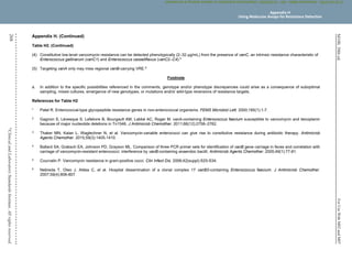
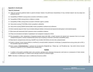
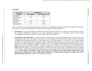
Appendix G. Epidemiological Cutoff Values
Abbreviations for Appendix G
ECV epidemiological cutoff value
MIC minimal inhibitory concentration
NWT non-wild-type
WT wild-type
G1 Defining Epidemiological Cutoff Values
G1.1 Definitions
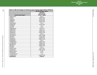
epidemiological cutoff value (ECV) – the minimal inhibitory concentration (MIC) or zone diameter value that separates microbial populations into those with and
without phenotypically detectable resistance (non-wild-type [NWT] or wild-type [WT], respectively). The ECV defines the highest MIC or smallest zone diameter for
the WT population of isolates.
EXAMPLE:
Interpretive Category
ECVs
MIC, μg/mL Zone Diameter, mm
Wild-type ≤ 4 ≥ 20
Non-wild-type ≥ 8 ≤ 19
wild-type (WT) – an interpretive category defined by an ECV that describes the microbial population with no phenotypically detectable
mechanisms of resistance or reduced susceptibility for the antimicrobial (antifungal) agent being evaluated.
non-wild-type (NWT) – an interpretive category defined by an ECV that describes the microbial population with phenotypically detectable
mechanisms of resistance and reduced susceptibility for the antimicrobial (antifungal) agent being evaluated.
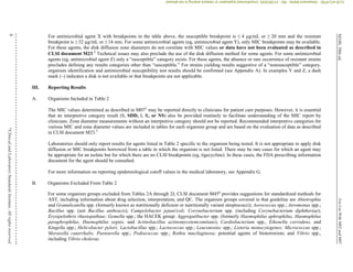
G1.2 Epidemiological Cutoff Values vs Clinical Breakpoints
ECVs are based on in vitro data only, using MIC or zone diameter distributions. ECVs are not clinical breakpoints, and the clinical relevance of ECVs for a
particular patient has not yet been identified or approved by CLSI or any regulatory agency.
By contrast, clinical breakpoints are established using MIC distributions, pharmacokinetic-pharmacodynamic data, and clinical outcome data, when available (as
described in CLSI document M231
).
“Caution”: Zone diameter (disk diffusion) and MIC values for which ECVs are defined are not to be interpreted or reported as susceptible,
intermediate, or resistant, but rather as WT or NWT. The ECVs should not be used as clinical breakpoints.
Appendix G
Epidemiological Cutoff Values
CLSI
eCLIPSE
-
Dewanand
Mahto
-
BD
-
01/29/2020.
Unauthorized
duplication
or
network
sharing
is
not
allowed.](https://image.slidesharecdn.com/clsi2020-220831170657-a1139808/85/CLSI-2020-pdf-290-320.jpg)


![M100,
30th
ed.
For
Use
With
M02
and
M07
260
©
Clinical
and
Laboratory
Standards
Institute.
All
rights
reserved
.
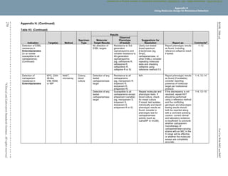
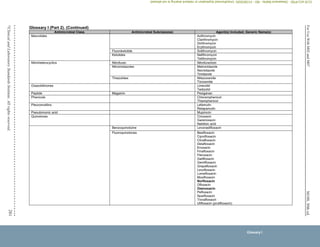
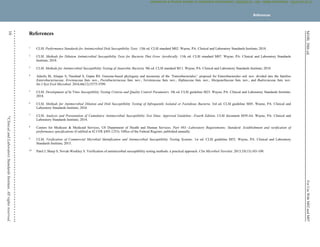
Appendix H. Using Molecular Assays for Resistance Detection
Abbreviations for Appendix H
AST antimicrobial susceptibility testing
ESBL extended-spectrum β-lactamase
MIC minimal inhibitory concentration
MRSA methicillin (oxacillin)-resistant Staphylococcus aureus
A not applicable
PBP2a penicillin-binding protein 2a
VRE vancomycin-resistant enterococci
Antimicrobial resistance and susceptibility are complex, and current in vitro methods have been developed to predict a microorganism’s response to antibacterial
therapy in vivo. Standardized phenotypic methods have evolved over many decades, but faster and potentially more reliable nucleic acid– and protein-based
methods have been recently developed to detect antimicrobial resistance. The current challenge for medical laboratories is to integrate molecular assays for
antimicrobial resistance determinants with conventional antimicrobial susceptibility testing (AST) procedures, sometimes despite an incomplete understanding of
test limitations.
The tables in this appendix provide a practical approach for testing and reporting results among medical laboratories that routinely use molecular techniques (with
or without a phenotypic test) for detecting antimicrobial resistance. Antimicrobial resistance is genetically complex and based on available data. Molecular methods
are often used as a screening tool (eg, methicillin (oxacillin)-resistant Staphylococcus aureus [MRSA] from nasal swabs) or as a rapid adjunct to traditional
phenotypic methods (eg, KPC from instrument-flagged blood culture bottles). Interpretation necessitates critical thinking and an understanding of the dynamics
between detecting “resistance” determinants and testing phenotypic “susceptibility.” Detecting a resistance marker does not necessarily predict therapeutic failure
of antimicrobial agents. The gene may be nonfunctional or expressed at clinically insignificant levels. Conversely, the absence of the genetic marker does not
necessarily indicate susceptibility, because technical issues may interfere with detection (eg, inhibition of amplification, emergence of genetic variants). In some
cases, a molecular approach may be superior to traditional phenotypic methods, such as in the case of low in vitro expression, heteroresistance, or poor growth
masking higher minimal inhibitory concentrations (MICs). Overall, laboratorians should attempt to apply a consistent approach to molecular-based methods and
aim to resolve discordant results with repeat or supplementary testing, by referral to a reference laboratory or by reporting both results in accordance with
institutional policies.
As understanding of the molecular mechanisms of antimicrobial resistance continues to develop, more sophisticated approaches to molecular detection of
antimicrobial resistance in the medical microbiology laboratory will undoubtedly emerge. The following tables will be updated as needed to ensure the provision of
relevant guidance as methods evolve.
Appendix H
Using Molecular Assays for Resistance Detection
CLSI
eCLIPSE
-
Dewanand
Mahto
-
BD
-
01/29/2020.
Unauthorized
duplication
or
network
sharing
is
not
allowed.](https://image.slidesharecdn.com/clsi2020-220831170657-a1139808/85/CLSI-2020-pdf-296-320.jpg)
![M100,
30th
ed.
For
Use
With
M02
and
M07
261
©
Clinical
and
Laboratory
Standards
Institute.
All
rights
reserved.
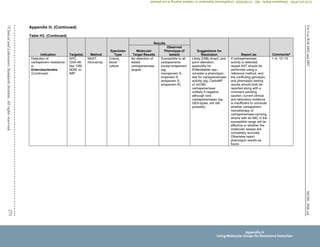
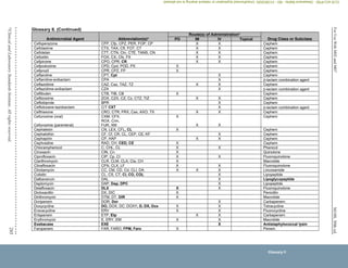
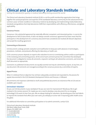
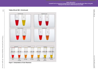
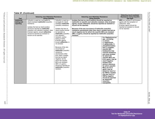
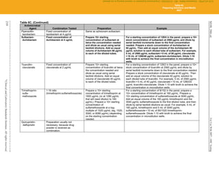
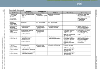
Appendix H. (Continued)
Table H1. Strategies for Reporting Methicillin (Oxacillin) Results When Using Molecular and Phenotypic AST Methods for S. aureus
Indication Target(s) Method
Specimen
Type
Results
Suggestions for Resolution
Consider
reporting asa
: Commentsb
Genotype or
Predicted
Phenotype
Observed
Colony
Phenotype
(if tested)
Detecting
methicillin
(oxacillin)
resistance in
S. aureus
PBP2a Latex agglutination,
immuno-
chromatography
Colony PBP2a positive Cefoxitin R N/A Methicillin (oxacillin) R 1
PBP2a negative Cefoxitin S N/A Methicillin (oxacillin) S 1
PBP2a positive Cefoxitin S Confirm isolate identification,
repeat latex agglutination and
AST, and consider mecA
colony NAAT, if available.
If discrepancy is not
resolved by suggested
testing, report as
methicillin (oxacillin)
R.
1–2
PBP2a negative Cefoxitin R Confirm isolate identification,
repeat latex agglutination and
AST, and consider mecA
colony NAAT, if available.
If discrepancy is not
resolved by suggested
testing, report as
methicillin (oxacillin)
R.
1
mecA NAAT, microarray
hybridization, ISH
Colony, blood
culture broth,
surveillance
specimen
mecA detected Cefoxitin R N/A If tested, report
phenotypic result as
found (methicillin
[oxacillin] R) and
consider reporting
molecular result per
institutional protocol.
3–6
mecA not
detected
Cefoxitin S N/A If tested, report
phenotypic result as
found (methicillin
[oxacillin] S) and
consider reporting
molecular result per
institutional protocol.
3–6
mecA detected Cefoxitin S Confirm isolate identification,
repeat AST, and repeat or
perform mecA colony NAAT,
if available. If mixed
specimen, test isolates
individually.
If discrepancy is not
resolved by suggested
testing, report as
methicillin (oxacillin)
R.
2–5, 8–9
mecA not
detected
Cefoxitin R Confirm isolate identification,
repeat AST, and repeat or
perform mecA colony NAAT,
if available. If mixed
specimen, test isolates
individually.
If discrepancy is not
resolved by suggested
testing, report as
methicillin (oxacillin)
R.
3, 7
Appendix H
Using Molecular Assays for Resistance Detection
CLSI
eCLIPSE
-
Dewanand
Mahto
-
BD
-
01/29/2020.
Unauthorized
duplication
or
network
sharing
is
not
allowed.](https://image.slidesharecdn.com/clsi2020-220831170657-a1139808/85/CLSI-2020-pdf-297-320.jpg)
![M100,
30th
ed.
For
Use
With
M02
and
M07
262
©
Clinical
and
Laboratory
Standards
Institute.
All
rights
reserved
.
Appendix H. (Continued)
Table H1. (Continued)
Indication Target(s) Method
Specimen
Type
Results
Suggestions for Resolution
Consider reporting
asa
: Commentsb
Genotype or
Predicted
Phenotype
Observed
Colony
Phenotype
(if tested)
Detecting
methicillin
(oxacillin)
resistance in
S. aureus
(Continued)
SCCmec-
orfX
functional
regions
only
NAAT Blood
culture
broth,
surveillance
specimen
SCCmec
detected
Cefoxitin R N/A If tested, report
phenotypic result as
found (methicillin
[oxacillin] R) and
consider reporting
molecular result per
institutional protocol.
3–6
SCCmec not
detected
Cefoxitin S N/A If tested, report
phenotypic result as
found (methicillin
[oxacillin] S) and
consider reporting
molecular result per
institutional protocol.
3–6
SCCmec
detected
Cefoxitin S Confirm isolate identification,
repeat AST and consider mecA
colony NAAT, if available. If
mixed culture, test isolates
individually.
If discrepancy is not
resolved by suggested
testing, report as
methicillin (oxacillin) R.
2, 10
SCCmec not
detected
Cefoxitin R Confirm isolate identification,
repeat AST and consider mecA
colony NAAT, if available. If
mixed culture, test isolates
individually.
If discrepancy is not
resolved by suggested
testing, report as
methicillin (oxacillin) R.
7, 12
Appendix H
Using Molecular Assays for Resistance Detection
CLSI
eCLIPSE
-
Dewanand
Mahto
-
BD
-
01/29/2020.
Unauthorized
duplication
or
network
sharing
is
not
allowed.](https://image.slidesharecdn.com/clsi2020-220831170657-a1139808/85/CLSI-2020-pdf-298-320.jpg)
![M100,
30th
ed.
For
Use
With
M02
and
M07
263
©
Clinical
and
Laboratory
Standards
Institute.
All
rights
reserved.
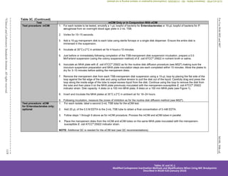
Appendix H. (Continued)
Table H1. (Continued)
Abbreviations: AST, antimicrobial susceptibility testing; ISH, in situ hybridization; MSSA, methicillin (oxacillin)-susceptible Staphylococcus aureus; MRSA,
methicillin (oxacillin)-resistant S. aureus; N/A, not applicable; NAAT, nucleic acid amplification test; PBP2a, penicillin-binding protein 2a; PCR, polymerase chain
reaction; R, resistant; S, susceptible.
Indication Target(s) Method
Specimen
Type
Results
Suggestions for
Resolution Consider reporting asa
: Commentsb
Genotype or
Predicted
Phenotype
Observed Colony
Phenotype
(if tested)
Detection of
methicillin
resistance in
S. aureus
(Continued)
SCCmec-
orfX
junctional
regions
and mecA
and/or
other
targets
NAAT Blood culture
broth,
surveillance
specimen
SCCmec AND
mecA or
other target
detected
Cefoxitin R N/A If tested, report phenotypic
result as found (methicillin
[oxacillin] R) and consider
reporting molecular result
per institutional protocol.
3–6
SCCmec AND
mecA or
other target not
detected
Cefoxitin S N/A If tested, report phenotypic
result as found (methicillin
[oxacillin] S) and consider
reporting molecular result
per institutional protocol.
3–6
SCCmec AND
mecA or
other target
detected
Cefoxitin S Confirm isolate
identification, repeat AST
and consider mecA
colony NAAT if available.
If mixed culture, test
isolates individually
If discrepancy is not
resolved by suggested
testing, report as methicillin
(oxacillin) R.
2
SCCmec AND
mecA or other
target not
detected
Cefoxitin R Confirm isolate
identification, repeat AST
and consider mecA
colony NAAT if available.
If mixed culture, test
isolates individually
If discrepancy is not
resolved by suggested
testing, report as methicillin
(oxacillin) R.
3, 11
Appendix H
Using Molecular Assays for Resistance Detection
CLSI
eCLIPSE
-
Dewanand
Mahto
-
BD
-
01/29/2020.
Unauthorized
duplication
or
network
sharing
is
not
allowed.](https://image.slidesharecdn.com/clsi2020-220831170657-a1139808/85/CLSI-2020-pdf-299-320.jpg)