Clinical Digital Photography in Orthodontics Shadi Samawi - JDJ; Vol18-2012
•
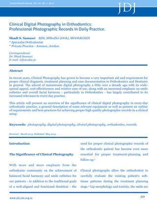
11 likes•3,916 views

Clinical digital photography is an important tool in orthodontics for diagnosis, treatment planning, and documentation. The advent of digital photography has increased its relevance due to lower costs and ease of use compared to film. Proper clinical photographs require a digital SLR camera, macro lens, ring flash, and cheek retractors. Extraoral photos should include full face relaxed, full face smiling, right profile relaxed, and 3/4 profile smiling views. Intraoral photos include upper occlusal, lower occlusal, right buccal, left buccal, and frontal smiling views. Clinical photos provide important soft tissue and dental information to evaluate patients and monitor their treatment.

Recommended
More Related Content
What's hot
What's hot (20)
Viewers also liked
Viewers also liked (19)
Similar to Clinical Digital Photography in Orthodontics Shadi Samawi - JDJ; Vol18-2012
Similar to Clinical Digital Photography in Orthodontics Shadi Samawi - JDJ; Vol18-2012 (20)
Recently uploaded
Recently uploaded (20)
Clinical Digital Photography in Orthodontics Shadi Samawi - JDJ; Vol18-2012
- 1. JDJ Jordan Dental Journal, Vol. 18, No. 1, 2012 Clinical Digital Photography in Orthodontics: Professional Photographic Records in Daily Practice. Shadi S. Samawi BDS, MMedSci (Orth), MOrthRCSED * Specialist Orthodontist * Private Practice - Amman, Jordan. Correspondence: Dr. Shadi Samawi E-mail: info@sdoc.jo Abstract In recent years, Clinical Photography has grown to become a very important aid and requirement for proper clinical diagnosis, treatment planning and case documentation in Orthodontics and Dentistry in general. The advent of mainstream digital photography a little over a decade ago with its widespread appeal, cost-effectiveness and relative ease of use, along with an increased emphasis on smile esthetics and overall facial harmony - particularly in Orthodontics - has largely contributed to its increased relevance to every-day practice. This article will present an overview of the significance of clinical digital photography in every-day orthodontic practice, a general description of some relevant equipment as well as present an outline of requirements and best-practices for achieving proper high quality photographic records in a clinical setup. Keywords: photography, digital photography, clinical photography, orthodontics, records. Received: March 2013, Published: May 2013 Introduction need for proper clinical photographic records of the orthodontic patient has become even more The Significance of Clinical Photography essential for proper treatment-planning and follow-up.1 With more and more emphasis from the orthodontic community on the achievement of Clinical photographs allow the orthodontist to balanced facial harmony and smile esthetics for carefully evaluate the existing patient's soft- our patients - in addition to the traditional goals tissue patterns during the treatment planning of a well-aligned and functional dentition - the stage.2 Lip morphology and tonicity, the smile arc www. jd j. jd a. o rg. j o 20
- 2. JDJ Jordan Dental Journal, Vol. 18, No. 1, 2012 and the degree of incisal show upon smiling and suitable digital camera setup, one or two high overall smile esthetics can all be assessed from capacity memory cards, and a computer with a various angles at any time. Thus, they allow us to reasonably-sized hard disk setup for processing, study the patient in a so called “social” setting, storage and back-up purposes. and all without the patient ever being present. Such information greatly aids the orthodontist in One of the main advantages of digital formulating the best possible treatment plan for photography is the ability to enhance, or “post- each patient, and monitoring any changes in process” your images. Even if some images are subsequent follow-ups. still not suitably aligned, rotated, or the color, brightness or saturation etc. is not up to Needless to say, the need for photographic standard, it is usually relatively easy to adjust records for purposes of research and publication, those using a suitable image-editing software on lecturing and for teaching presentations has your computer, before saving the images in the always been present. In addition, the growing patient’s file. 1,5 importance for the need for such records for medico-legal reasons cannot be ignored.2,3 However, it is vital not to overlook the ethical issues raised by such enhancement procedures Going for a digital camera is the obvious choice and as such, any photo manipulation beyond the in this digital age. One of the major reasons is the basic general improvement of the properties of relative ease of use of such cameras, along with the original image as described in this article is the ability to repeat or delete unsuitable images not advised and is considered - by all means - on the spot. There is no need to wait until the ethically and professionally unacceptable, and film is developed to check your photos. Any may ultimately lead to legal ramifications. problems can be easily rectified immediately. Another important advantage is the issue of Recommended Equipment “running cost”; Digital camera setups are costeffective; eliminating the cost of photographic There are several types of digital cameras film, developing costs and hassles, as well as available on the market today. Most compact solving the issue of “physical storage” of slides cameras i.e “Point and Shoot” cameras available and photographs of large numbers of patients.3,4 today have sufficient image quality and All that is required is a one-time investment in a sophistication to produce reasonable clinical www. jd j. jd a. o rg. j o 21
- 3. JDJ Jordan Dental Journal, Vol. 18, No. 1, 2012 dental photographs, although at the expense of build quality and thus would be a solid long-term proper illumination of the scene due to investment for the practice. Many DSLRs now limitations of their built-in Single-point Flash come with High Definition (HD) video recording units. However, their results are often capabilities as well, that may prove useful in inconsistent and require too much time and trial certain clinical situations. An entry-level DSLR and error to get the desired outcome. The ideal camera is recommended for clinical photography, camera setup that is best-suited and as it will produce the highest quality results while recommended for clinical photography is a being a more cost-effective option than other Digital Single Lens Reflex camera setup (DSLR) high-end DSLRs. with a suitable Ring Flash unit.3,6,7 The minimum accepted image resolution that would provide us B. The Lens with high quality photos for screen presentation or 4x6 prints - if desired - would be around 4 Although zoom lenses can generally be used for Megapixels. However, current cameras can clinical photography, the recommended lens to produce much higher-resolution photos than this use is a dedicated Macro lens e.g. a 100mm focal minimum requirement, therefore it can be length Macro lens. generally determined that any current camera with a resolution of 8 Megapixels or higher Macro Lens vs Macro Function would be more than adequate for orthodontic record-taking purposes. This setup is described Macro photography refers to close-up in more detail in the following sections. photography; the classical definition that the image projected on the "film plane" (i.e film or a A. The Digital Camera digital sensor) is the same size as the subject. Most Point-&-Shoot digital cameras have a built- A DSLR provides full manual control over all in Macro function that is actually very good for exposure parameters in photography, in addition dental photography purposes. However, a to the usual automatic and semi-automatic dedicated Macro lens attached to a DSLR camera programs available on most cameras today. provides even better close-up photos usually with Therefore, they allow maximum flexibility and higher definition and better focus, and is by far customization for producing the highest possible the superior choice.8 quality of digital images.7 They are of a superior www. jd j. jd a. o rg. j o 22
- 4. Jordan Dental Journal, Vol. 18, No. 1, 2012 JDJ C. The Flash Ring Flash vs Point Flash The Single Point Flash built into most compact digital cameras and some DSLR may occasionally produce fairly good light distribution when used for clinical photographs, but the results are very inconsistent and largely depend on camera orientation and pre-existing lighting conditions in the clinic. Dark distracting shadows, which Figure 1 Canon MR-14EX TTL Macro Ring Lite Flash (Canon USA) may obstruct important details frequently occur. These are often irreparable even by using image editing software, and will detract from the final quality of the image, and possibly the information gained from it. In contrast, A dedicated Ring Flash - eliminates almost all shadows by providing a more even distribution of light during extra and intra-oral photographs (Figure 1), and thus the quality of the image is enhanced due to overall better illumination. Ring Flash for orthodontic photography.1,3,4,7 Figure 2 Large and Small Double-ended Cheek Retractors and Medium-sized, Front-silver coated Dental Mirror. D. Retractors There are two sets of double-ended retractors; Therefore, it is highly recommended to use a one set with a regular and small size either end. The recommended cheek retractors to be used These are mainly used for intra-oral occlusal for best results in clinical photography are two shots (mirror shots). The other set has a narrow pairs of variable-size double-ended retractors end and a wide end on the other. These are used shown (Figure 2).1,3 for intra-oral frontal and buccal shots. www. jd j. jd a. o rg. j o 23
- 5. JDJ Jordan Dental Journal, Vol. 18, No. 1, 2012 Although other types of retractors are available resulting in haziness or a “Double-Image”. Also, on the market, it is accepted by many that this light reflection is not on par with the front- selection presents an ideal set to be used for silvered mirrors, leading to a dimmed, darkened clinical photography, as they greatly facilitate image as an end-result. Therefore front-coated taking almost any kind of intra-oral photographs silvered mirrors are highly recommended over with clarity and with the largest possible field of other types (Figure 2).1,3 vision. Smaller one-piece orthodontic bonding retractors are generally not a good choice for It is generally preferred to use long-handled orthodontic purposes, especially for buccal and mirrors (see Image) as they allow better control occlusal shots, as their retraction potential is very and handling by the clinician during the occlusal limited, and it can often prove to be a “painful shots. Different sizes can be found for use with experience” for the patient.9 different patients depending on age and mouthopening size, with medium-sized mirrors It is a wise long-term investment to buy good generally being fit for use with most patients. quality retractors to ensure durability and reliability, with recurrent disinfection Mirrors with no handles may also be used procedures. successfully but are trickier to handle, especially when juggling an expensive digital camera in the E. Dental Photography Mirrors other hand. Many types of mirrors may be used for clinical It is important to mention that the previously photography, ranging from front-silvered mentioned equipment are not - by any means - mirrors to highly polished Stainless Steel mirrors the only recommended equipment for the of various shapes and sizes. Many authorities in purposes of clinical photography, nor are they the field recommend front-coated silvered necessarily the best for all clinicians, and should mirrors as they offer the best image quality and be considered as a basic recommendation that - light distribution over other types of mirrors. in the author’s personal opinion and experience - With front-silvered mirrors, no “ghost” image, or represents the simplest, most cost-effective and “double-layering” occurs. In contrast, when using easy-to-use setup that will produce the highest glass or rear-coated silvered mirrors, “ghosting” quality professional results in a consistent can severely affect the quality of the image, fashion. www. jd j. jd a. o rg. j o 24
- 6. Jordan Dental Journal, Vol. 18, No. 1, 2012 How Many Photographs Do We Need? JDJ exposure as it determines the other variables automatically. The aperture (F number) is best There is no “standard” set of clinical photographs set to f22 or higher; this will ensure a high depth that is universally approved as a rule of thumb. of field and focus throughout the various extra However, it can generally be accepted - based on and intra-oral photographs.1,2,3 The use of a Ring many practitioners’ opinions - that a complete flash is essential in this case to ensure proper “Clinical Photographic Set” for any orthodontic lighting and illumination of the scene. Clinicians patient at any stage of treatment, that would with a good understanding of photographic enable the clinician to obtain maximum benefit exposure principles may choose to use the Full and information, should include a minimum of Manual setting (M) and control all the exposure nine photographs; four extra-oral, and five variables themselves as they see fit.8 intraoral photographs per set, with a minimum of two sets; Pre and Post-treatment sets. It is important to note that these are only general recommendations and some trial and error may However, a clinician may also choose to take additional views, as needed to document the still be required depending on the existing clinical lighting and situation. entire case in further detail. Extra-oral Clinical Photographs Clinical Photographic Technique They are usually the first and simplest General Camera Settings photographs to take. They require proper positioning of the patient and clinician in As mentioned earlier, DSLRs allow maximum control and customization of camera settings to achieve the best possible results. In most cases, it is recommended to set the camera dial to Aperture Priority Mode (A or Av, depending on front of a suitable plain background; in addition to the digital camera setup itself. It is recommended to use a plain-white or darkblue background or alternatively a large wall- camera brand). Aperture Priority mode allows mounted light box behind the patient’s head, the photographer to control the aperture in order to maintain the best definition of the dimension, through which light enters the lens, soft tissue profile of the patient with no while leaving the camera to balance the best distractions in the background.3,7 www. jd j. jd a. o rg. j o 25
- 7. JDJ Jordan Dental Journal, Vol. 18, No. 1, 2012 Standardizing extra-oral photographs by head in the Natural Head Position, eyes maintaining a fixed distance between the looking straight at the camera and the teeth patient and the camera is preferred as it and lips relaxed in the rest position. The would provide a consistent magnification whole of the patient’s face should be clearly factor and add a professional appearance to visible. Ensuring that the inter-pupillary line the photos. This may be done by using a is horizontally leveled is vital (Figure 3). tripod set at a fixed marked distance from the background and marking a line on the floor The camera should be held in the vertical at a certain distance from the background portrait orientation for this and all extra-oral where patients should consistently stand photographs and the photograph taken at 90 while being photographed. degrees to the facial midline for best results, while ensuring proper framing of the entire Extra-oral photos consist of the following head and neck. four photographs, taken in the following sequence: 2. Face-Frontal (Smiling) 1. Face-Frontal (lips relaxed). The same principles apply as in the first 2. Face-Frontal (Smiling). photograph with the important exception 3. Profile (Lips relaxed). that the patient should be smiling in a natural 4. (45 °) Profile (also known as 3/4 Profile - relaxed manner with the anterior teeth Smiling). clearly visible, in order to properly showcase overall smile esthetics from the front (Figure These four shots provide the clinician with 4). maximum possible information about the patient’s facial and soft tissue features, 3. Profile (Lips relaxed) proportions, and overall smile esthetics. The profile view has an extremely high diagnostic value for the orthodontist. The 1. Face-Frontal (lips relaxed) patient is required to turn to bodily to their The patient should stand at the marked left thus having their right profile visible to distance form the background with their the clinician. Ideally, the whole of the right www. jd j. jd a. o rg. j o 26
- 8. JDJ Jordan Dental Journal, Vol. 18, No. 1, 2012 Figure 3: Full-face Frontal - Lips at Rest. Figure 4: Full-face Frontal Smiling. Figure 5: Right Profile View - Lips at Rest. Figure 6: 3/4 Profile Smiling View. side of the face should be clearly visible with here is particularly important, as it will no obstructions such as hair, hats or scarfs. eliminate any shadowing of the border of the The head should be in the Natural Head patient’s profile onto the background, which Position, with their eyes fixed horizontally - can very distracting and considerably preferably at a specific point at eye-level, or compromise the quality of the photo and the at the reflection of their own pupils in a information gained from it. mirror (Figure 5). The use of a Ring flash www. jd j. jd a. o rg. j o 27
- 9. JDJ Jordan Dental Journal, Vol. 18, No. 1, 2012 4. (45 °) Profile (also known as 3/4 There are five essential intra-oral Profile - Smiling) photographs - taken in the following From the Profile photo position, the patient sequence: is asked to turn their heads slightly to their 1. Frontal - in occlusion right - about 3/4 of the way, hence the name - 2. Right Buccal (in occlusion) while keeping their body still in the previous 3. Left Buccal (in occlusion) “Profile Shot” position i.e. Facing forward. 4. Upper Occlusal (mirror) They are then instructed to look into the 5. Lower Occlusal (mirror) camera, and then smile. It is essential that the patient’s teeth show clearly when smiling, 1. Frontal (in occlusion) otherwise the photograph would be of The patient is seated in the dental chair with minimum benefit (Figure 6). head raised to the clinician’s elbow level. The dental assistant stands behind the patient This shot conveys the patient as if in “social and uses the larger set of retractors from interaction”, and can provide valuable their wider ends to retract the patient's lips information regarding the smile esthetics’ sideways and outward, away from the teeth changes pre and post-treatment. and gingivae. Maximum field of view is required for visualization of all teeth and Intra-oral Clinical Photographs associated tissues. The photo should be taken 90° to the facial mid-line & central incisors. Intra-oral photos require the proper cheek With the occlusal plane properly leveled retractors, dental photography mirrors, as according to the patient’s existing occlusion. well as a well-trained assistant if possible - in The Ring-Flash will greatly aid in producing a addition to the camera setup. They require quality photograph by ensuring the best more attention to detail to produce excellent possible illumination with no shadows, results. especially in the deeper parts of the oral cavity and buccal vestibules (Figure 7). www. jd j. jd a. o rg. j o 28
- 10. JDJ Jordan Dental Journal, Vol. 18, No. 1, 2012 2. Right Buccal (in occlusion) molar is visible if possible, while the assistant Here, the assistant flips the right retractor to maintains hold of the left retractor, without the narrower side, while the left retractor undue stretching. Again, the shot is taken 90° remains in place as for the previous frontal to the canine-premolar area for best shot. The patient is asked to turn their head visualization of the buccal segment to their left so their right side will be facing relationship, as this is very important in the clinician (Figure 8). orthodontic assessment. A useful tip would be for the clinician to fully stretch the right The clinician holds the right retractor and retractor just before taking the shot to stretches it to the extent that the last present minimize any discomfort for the patient, and Figure 7 Figure 8 Frontal View - In Occlusion. Right Buccal View - In Occlusion. www. jd j. jd a. o rg. j o Figure 9 Left Buccal View - In Occlusion. 29
- 11. JDJ Jordan Dental Journal, Vol. 18, No. 1, 2012 Figure 11 Figure 10 Upper Occlusal View - Mirror. achieve maximum visibility of the last present molar, if possible. 3. Left Buccal (in occlusion) The assistant now switches the retractors with the narrow end on the photo side (patient’s left) and the wide end on the other (patient’s right). The same principles as for the right buccal shot apply. The clinician should move their body slightly to the right while holding the retractor on the photo side, to ensure the photo is taken at 90 degrees to the canine-premolar area, while the patient turns their head all the way to their right (Figure 9). Lower Occlusal View - Mirror. 4. Upper Occlusal (mirror) The assistant now switches to the smaller retractor set and with the patient’s mouth held open, the retractors are inserted in a “V” shape to retract the upper lips sideways and away from the teeth. The clinician inserts the mirror with its wider end inwards to capture maximum width of the arch posteriorly, and pulls it slightly downwards so that the whole upper arch is visible to the last present molar (Figure 10). The shot must be taken 90° to the plane of the mirror for best visibility and no visual distortion. The mid-palatal raphe is used as a guide to level the palate in the photo. Minimal retractor visibility in the image is recommended, and no fingers should be visible in the final photo, if possible. www. jd j. jd a. o rg. j o 30
- 12. JDJ Jordan Dental Journal, Vol. 18, No. 1, 2012 5. Lower Occlusal (mirror) esthetics, or a close-up view of the overjet The assistant now lowers the smaller from one side may be taken to demonstrate retractors into a Reverse “V” shape to retract the amount of incisor overjet and overbite the lower lips sideways and away from the present. Close-ups focusing on certain teeth. The clinician would now lift the mirror aspects of existing appliances, brackets or upwards so he/she may visualize the arch-wires and associated auxiliaries may be reflection of the lower arch, while the patient individually taken as needed, although many is be asked to “lift their chin up” slightly. clinicians prefer to crop the necessary details Ideally, the shot should be taken 90° to the out of the complete photographic set plane of the mirror, with the last molar described earlier, provided that they are present visible. An important issue here taken using a high resolution setting, to avoid would be the tongue position of the patient any deterioration in image quality upon while taking the photo. It is best to ask the cropping.10 patient to “roll back” their tongue behind the mirror so that it won’t interfere with the Helpful Tips for Best Results visibility of any teeth, particularly in the posterior area (Figure 11). It is recommended that all photographic records be taken before impression-taking, to Additional Photographic Views eliminate the possibility of impression material being stuck between the teeth or on There are several other views that may be the face during photographic record-taking.3,7 taken by the clinician to fully document the case at hand, depending on his or her needs. Wetting the retractors just before insertion Functional Occlusion views may be taken by eases their proper positioning in the mouth the orthodontist to demonstrate canine with minimum patient discomfort. guidance or Group Function of the occlusion to supplement existing records. A close-up of The direction of pull of the retractors is the lips and mouth upon smiling may be always sideways and slightly forward and taken for closer examination of overall smile outward, away from the gingival tissues. www. jd j. jd a. o rg. j o 31
- 13. JDJ Jordan Dental Journal, Vol. 18, No. 1, 2012 This maximizes the field of view and References minimizes patient discomfort. 1. Sandler J, Murray A; Digital Photography in When taking occlusal views with the mirror, Orthodontics. Journal of Orthodontics, 2001, 28:197-201 slightly warming the mirror in warm water 2. Mizrahi E; Orthodontic Pearls: A Selection of prior to insertion in the mouth helps prevent Practical Tips and Clinical Expertise; 2004, Taylor & “Fogging” of the mirrors which would Francis Group. (Sandler J, Murray A; Ch 4: prevent a clear image from being obtained.1,9 Orthodontic Photography) 3. Sandler J, Murray A; Current Products and Practice: Clinical Photographs - The Gold Standard. With some patients, profuse salivary flow and Journal of Orthodontics, 2002, 29:158-67 “frothing” can affect the quality of the image 4. Sandler J, Murray A; Recent Developments in being taken, thus a saliva ejector can be used Clinical Photography. British Journal Of Orthodontics, 1999, 26: 269-72 to eliminate saliva prior to taking each 5. Mah J, Ritto K; The Cutting Edge. Journal of photograph.3 Clinical Orthodontics, 2002, 36: 11; 619-625 6. Palomo J, Wolf G, Hans M; Use of Digital Post-Processing Photography in the Case Orthodontic Clinic. Am J Orthod Dentofacial Orthop 2004; 126: 381-5 Once all photographic records are obtained, 7. Terry D.A, Snow S.R, Mclaren E.A; Contemporary they should be downloaded to the computer Dental Photography: Selection and Application. and an initial back-up of the original files Functional Esthetics & Restorative Dentistry, 2008; performed onto a separate computer, hard Series 1, No. 1. 8. Mastering Digital SLR Photography: The Serious disk or suitable removable media prior to Photographer’s Guide to High Quality Digital SLR post-processing the photos for final Photography, Busch D, 2005, Thomson Publishing. archiving, to ensure the originals are always 9. Mckeown H.F, Sandler P.J, Murray A.M; How to available in case of a computer or hard disk avoid common errors in clinical photography. Journal of Orthodontics, 2005, 32: 43-54 failure or if something goes wrong during 10. Aperture: Digital Photography Fundamentals. post-processing.10 This will be the subject of 2005. Apple Inc. another article. www. jd j. jd a. o rg. j o 32