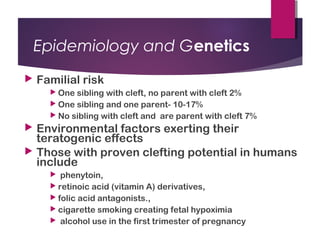
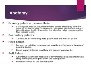
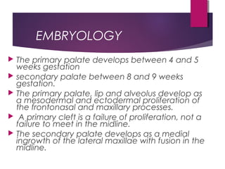
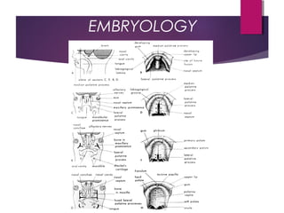
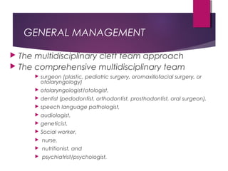
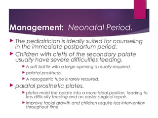
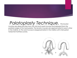
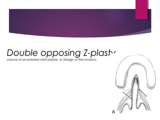
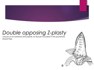
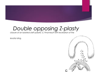
Cleft Lip and Palate - Presentation.
Cleft Lip and Palate is the 2nd most common Congenital Anomaly after Clubfoot. This presentation goes in depth about the Presentation, eitiology, Genetics, Medical management, Nasoalveolar Moulding, Surgical management of Cleft Lip & Palate
Cleft lip and Cleft palate embryology, features, and management Augustine raj
cleft lip and Cleft palate is one of the most common congenital anomalies encountered in ENT and Pediatrics practice. It is important to be familiar with the clinical features and complications, Surgical procedures, timing of surgery and complications associated with the surgeries. this presentation will give you a simple approach towards the same.
At the end of this lecture, students should be able to:
Develop an understanding terms Cleft lip & Palate
Develop an understanding of incidence of the condition
Describe the etiology and pathogenesis
Describe classification and dental implications
Cleft lip and palate is the most common developmental anomaly of the craniofacial region, and they have been depicted throughout in the past civilizations.
Management of Orofacial Cleft Dr. Sunil (2).pptx Management of Orofacial Clef...ssuser12303b
Management of Orofacial Cleft Dr. Sunil (2).pptxManagement of Orofacial Cleft Dr. Sunil (2).pptxManagement of Orofacial Cleft Dr. Sunil (2).pptxManagement of Orofacial Cleft Dr. Sunil (2).pptx
The Indian Dental Academy is the Leader in continuing dental education , training dentists in all aspects of dentistry and
offering a wide range of dental certified courses in different formats.for more details please visit
www.indiandentalacademy.com
- Video recording of this lecture in English language: https://youtu.be/lK81BzxMqdo
- Video recording of this lecture in Arabic language: https://youtu.be/Ve4P0COk9OI
- Link to download the book free: https://nephrotube.blogspot.com/p/nephrotube-nephrology-books.html
- Link to NephroTube website: www.NephroTube.com
- Link to NephroTube social media accounts: https://nephrotube.blogspot.com/p/join-nephrotube-on-social-media.html
New Directions in Targeted Therapeutic Approaches for Older Adults With Mantl...i3 Health
i3 Health is pleased to make the speaker slides from this activity available for use as a non-accredited self-study or teaching resource.
This slide deck presented by Dr. Kami Maddocks, Professor-Clinical in the Division of Hematology and
Associate Division Director for Ambulatory Operations
The Ohio State University Comprehensive Cancer Center, will provide insight into new directions in targeted therapeutic approaches for older adults with mantle cell lymphoma.
STATEMENT OF NEED
Mantle cell lymphoma (MCL) is a rare, aggressive B-cell non-Hodgkin lymphoma (NHL) accounting for 5% to 7% of all lymphomas. Its prognosis ranges from indolent disease that does not require treatment for years to very aggressive disease, which is associated with poor survival (Silkenstedt et al, 2021). Typically, MCL is diagnosed at advanced stage and in older patients who cannot tolerate intensive therapy (NCCN, 2022). Although recent advances have slightly increased remission rates, recurrence and relapse remain very common, leading to a median overall survival between 3 and 6 years (LLS, 2021). Though there are several effective options, progress is still needed towards establishing an accepted frontline approach for MCL (Castellino et al, 2022). Treatment selection and management of MCL are complicated by the heterogeneity of prognosis, advanced age and comorbidities of patients, and lack of an established standard approach for treatment, making it vital that clinicians be familiar with the latest research and advances in this area. In this activity chaired by Michael Wang, MD, Professor in the Department of Lymphoma & Myeloma at MD Anderson Cancer Center, expert faculty will discuss prognostic factors informing treatment, the promising results of recent trials in new therapeutic approaches, and the implications of treatment resistance in therapeutic selection for MCL.
Target Audience
Hematology/oncology fellows, attending faculty, and other health care professionals involved in the treatment of patients with mantle cell lymphoma (MCL).
Learning Objectives
1.) Identify clinical and biological prognostic factors that can guide treatment decision making for older adults with MCL
2.) Evaluate emerging data on targeted therapeutic approaches for treatment-naive and relapsed/refractory MCL and their applicability to older adults
3.) Assess mechanisms of resistance to targeted therapies for MCL and their implications for treatment selection
Prix Galien International 2024 Forum ProgramLevi Shapiro
June 20, 2024, Prix Galien International and Jerusalem Ethics Forum in ROME. Detailed agenda including panels:
- ADVANCES IN CARDIOLOGY: A NEW PARADIGM IS COMING
- WOMEN’S HEALTH: FERTILITY PRESERVATION
- WHAT’S NEW IN THE TREATMENT OF INFECTIOUS,
ONCOLOGICAL AND INFLAMMATORY SKIN DISEASES?
- ARTIFICIAL INTELLIGENCE AND ETHICS
- GENE THERAPY
- BEYOND BORDERS: GLOBAL INITIATIVES FOR DEMOCRATIZING LIFE SCIENCE TECHNOLOGIES AND PROMOTING ACCESS TO HEALTHCARE
- ETHICAL CHALLENGES IN LIFE SCIENCES
- Prix Galien International Awards Ceremony
New Drug Discovery and Development .....NEHA GUPTA
The "New Drug Discovery and Development" process involves the identification, design, testing, and manufacturing of novel pharmaceutical compounds with the aim of introducing new and improved treatments for various medical conditions. This comprehensive endeavor encompasses various stages, including target identification, preclinical studies, clinical trials, regulatory approval, and post-market surveillance. It involves multidisciplinary collaboration among scientists, researchers, clinicians, regulatory experts, and pharmaceutical companies to bring innovative therapies to market and address unmet medical needs.
These lecture slides, by Dr Sidra Arshad, offer a quick overview of physiological basis of a normal electrocardiogram.
Learning objectives:
1. Define an electrocardiogram (ECG) and electrocardiography
2. Describe how dipoles generated by the heart produce the waveforms of the ECG
3. Describe the components of a normal electrocardiogram of a typical bipolar leads (limb II)
4. Differentiate between intervals and segments
5. Enlist some common indications for obtaining an ECG
Study Resources:
1. Chapter 11, Guyton and Hall Textbook of Medical Physiology, 14th edition
2. Chapter 9, Human Physiology - From Cells to Systems, Lauralee Sherwood, 9th edition
3. Chapter 29, Ganong’s Review of Medical Physiology, 26th edition
4. Electrocardiogram, StatPearls - https://www.ncbi.nlm.nih.gov/books/NBK549803/
5. ECG in Medical Practice by ABM Abdullah, 4th edition
6. ECG Basics, http://www.nataliescasebook.com/tag/e-c-g-basics
micro teaching on communication m.sc nursing.pdfAnurag Sharma
Microteaching is a unique model of practice teaching. It is a viable instrument for the. desired change in the teaching behavior or the behavior potential which, in specified types of real. classroom situations, tends to facilitate the achievement of specified types of objectives.
Tom Selleck Health: A Comprehensive Look at the Iconic Actor’s Wellness Journeygreendigital
Tom Selleck, an enduring figure in Hollywood. has captivated audiences for decades with his rugged charm, iconic moustache. and memorable roles in television and film. From his breakout role as Thomas Magnum in Magnum P.I. to his current portrayal of Frank Reagan in Blue Bloods. Selleck's career has spanned over 50 years. But beyond his professional achievements. fans have often been curious about Tom Selleck Health. especially as he has aged in the public eye.
Follow us on: Pinterest
Introduction
Many have been interested in Tom Selleck health. not only because of his enduring presence on screen but also because of the challenges. and lifestyle choices he has faced and made over the years. This article delves into the various aspects of Tom Selleck health. exploring his fitness regimen, diet, mental health. and the challenges he has encountered as he ages. We'll look at how he maintains his well-being. the health issues he has faced, and his approach to ageing .
Early Life and Career
Childhood and Athletic Beginnings
Tom Selleck was born on January 29, 1945, in Detroit, Michigan, and grew up in Sherman Oaks, California. From an early age, he was involved in sports, particularly basketball. which played a significant role in his physical development. His athletic pursuits continued into college. where he attended the University of Southern California (USC) on a basketball scholarship. This early involvement in sports laid a strong foundation for his physical health and disciplined lifestyle.
Transition to Acting
Selleck's transition from an athlete to an actor came with its physical demands. His first significant role in "Magnum P.I." required him to perform various stunts and maintain a fit appearance. This role, which he played from 1980 to 1988. necessitated a rigorous fitness routine to meet the show's demands. setting the stage for his long-term commitment to health and wellness.
Fitness Regimen
Workout Routine
Tom Selleck health and fitness regimen has evolved. adapting to his changing roles and age. During his "Magnum, P.I." days. Selleck's workouts were intense and focused on building and maintaining muscle mass. His routine included weightlifting, cardiovascular exercises. and specific training for the stunts he performed on the show.
Selleck adjusted his fitness routine as he aged to suit his body's needs. Today, his workouts focus on maintaining flexibility, strength, and cardiovascular health. He incorporates low-impact exercises such as swimming, walking, and light weightlifting. This balanced approach helps him stay fit without putting undue strain on his joints and muscles.
Importance of Flexibility and Mobility
In recent years, Selleck has emphasized the importance of flexibility and mobility in his fitness regimen. Understanding the natural decline in muscle mass and joint flexibility with age. he includes stretching and yoga in his routine. These practices help prevent injuries, improve posture, and maintain mobilit
TEST BANK for Operations Management, 14th Edition by William J. Stevenson, Ve...kevinkariuki227
TEST BANK for Operations Management, 14th Edition by William J. Stevenson, Verified Chapters 1 - 19, Complete Newest Version.pdf
2. INTRODUCTION
common congenital disorder
have numerous associated problems,
including
basic anatomic deformity,
deficient facial growth,
dental problems
speech problems
otologic problems
psychologic disorders, and
additional congenital anomalies.
3. History
400BC Hippocrates and 150AD Galen mentioned cleft lip
500 AD 1st Operation on palate due to inflammation of the uvula
1564 Pare described the use of obturators for palatal perforations
1556Cleft palate recognised as a congenital disorder by Franco.
1764 LeMonnier, a French dentist performed 1st successful
repair of a cleft velum.
1834Dieffennbach performed the 1st closure of the hard palate
1861Von Langenbeck described the use of mucoperiosteal flaps in
cleft palate closure
1930Kilner and Wardill independently developed the ‘pushback’
procedure.
1944 Schweckendiek began closing cleft defects in young patients.
4. Epidemiology and Genetics
The incidence of cleft palate only is half the incidence of
CL/P (more than 1 : 2000 live births).
There is usually little difference between racial groups.
The incidence is much higher (about 4 : 1) in females
than in males.
About 20% to 30% of human cleft palate only are
syndromic.
Pierre-Robin Sequence,
Treacher-Collins Malformation,
Trisomies 13 and 18,
Apert’s Syndrome,
Stickler’s Syndrome and
Waardenburg’s Syndrome.
5. Epidemiology and Genetics
Familial risk
One sibling with cleft, no parent with cleft 2%
One sibling and one parent- 10-17%
No sibling with cleft and are parent with cleft 7%
Environmental factors exerting their
teratogenic effects
Those with proven clefting potential in humans
include
phenytoin,
retinoic acid (vitamin A) derivatives,
folic acid antagonists.,
cigarette smoking creating fetal hypoximia
alcohol use in the first trimester of pregnancy
6. Anatomy
Primary palate or premaxilla is
a triangular area of the anterior hard palate extending from the
anterior to the incisive foramen and to a point just lateral to each
lateral incisor tooth. It includes the alveolar ridge containing the
four incisor teeth.
Secondary palate:-
Consist of all remaining hard palate and are the soft palate.
Hard palate-
Formed by palatine processes of maxilla and horizontal lamina of
palatine bones.
Blood supply internal maxillary art- greater palative Ar.
Soft Palate
A fibromuscular shelf made up of several muscles attached like a
sling to the posterior portion of the hard palate
Function- close off the nasopharynx
7. EMBRYOLOGY
The primary palate develops between 4 and 5
weeks gestation
secondary palate between 8 and 9 weeks
gestation.
The primary palate, lip and alveolus develop as
a mesodermal and ectodermal proliferation of
the frontonasal and maxillary processes.
A primary cleft is a failure of proliferation, not a
failure to meet in the midline.
The secondary palate develops as a medial
ingrowth of the lateral maxillae with fusion in the
midline.
9. CLASSIFICATION
no standard classification is universally
accepted.
Cleft palate
A. Location in reference to incisive foramen
1. Primary—anterior
2. Secondary—posterior
B. Unilateral or bilateral
C. Complete or incomplete
Submucous cleft palate
10. Classification (Iowa system)
Group I - cleft of lip only
Group II - Cleft of palate only
Group III - Clefts of lip alveolus
Group IV - Cleft of the lip and alveolus+
Palate
Group V - Miscellaneous
11. GENERAL MANAGEMENT
The multidisciplinary cleft team approach
The comprehensive multidisciplinary team
surgeon (plastic, pediatric surgery, oromaxillofacial surgery, or
otolaryngology)
otolaryngologist/otologist,
dentist (pedodontist, orthodontist, prosthodontist, oral surgeon),
speech language pathologist,
audiologist,
geneticist,
Social worker,
nurse,
nutritionist, and
psychiatrist/psychologist.
12. Management: Neonatal Period.
The pediatrician is ideally suited for counseling
in the immediate postpartum period.
Children with clefts of the secondary palate
usually have severe difficulties feeding.
A soft bottle with a large opening is usually required.
palatal prosthesis.
A nasogastric tube is rarely required.
palatal prosthetic plates.
plates mold the palate into a more ideal position, leading to
less difficulty feeding and an easier surgical repair.
improve facial growth and children require less intervention
throughout time
13. Management:
The timing of the repair is controversial
Palatal repair is performed anywhere from
6 months onward.
delay in palatal repair results in improved midfacial
growth
earlier repair results in better speech
14. Management: Toddler Years.
The most important process in the toddler
years is the development of adequate
speech
requires speech therapy
The child is trained to speak slowly and forcefully in order to
maximize palatal elevation and minimize hypernasality
up to 20% of patients still require
pharyngoplasty to reduce VPI
recurrent or chronic ear infections
growth hormone deficiency is 40 times more
common in this population
15. Management: School Years
orthodontic management
psychological growth
psychotherapist is essential to ensure
adequate social development
high rate of depression and poor self esteem
Child abuse is also more common
16. Management: Teenage Years.
The teenage growth spurt is marked by
midface retrusion, altering appearance.
Palatal repair is likely responsible
ideal timing for palatal repair so that midface retrusion is minimized is
currently an area of intense research, with many conflicting studies in
the literature
The treatment requires maxillary
advancement through a combination of
LeForte I, II and III osteotomies.
The treatment of midfacial retrusion requires maxillary
advancement through a combination of LeForte I, II and
III osteotomies.
usually performed in young adulthood, after growth is complete
Rhinoplasty, when required, is usually the last surgery
performed.
17. Surgical Techniques
The child with CLAP usually undergoes
numerous surgical procedures.
The first procedure is repair of the lip, which occurs around
10 weeks.
The palate is usually repaired between 6 and 18 months,
with newer studies advocating earlier repair
Pharyngeal flaps, if required, are performed around age 4.
Alveolar bone grafting occurs between ages 9 and 11,
definitive orthodontics at age 12-13.
Maxillary advancement and rhinoplasty are delayed until
the later teenage years.
18. Surgical Techniques
Palatoplasty
The basic surgical techniques of palate repair
include Schweckendiek’s procedure or primary
veloplasty,
Von Langenbeck’s palatoplasty,
V-Ypush-back
double reversing Z-plasty (Furlow’s palatoplasty),
and two-flap palatoplasty.
A combination of palatoplasty and pharyngeal flap
may be used for the repair of extremely large
clefts15 and occasionally for primary palate repair.
19. • Pre-Surgical
Orthopedics allowing
gingivoperiosteoplasty.
A: Wide unilateral cleft lip with separation of
the arch and collapse of the lesser segment.
B: Improved arch alignment and nasal shape
after presurgical orthopedic treatment.
C: Results after rotation-advancement lip
repair, nasal repair, and
gingivoperiosteoplasty, all performed at the
first operation, when the child is
approximately 3 months of age.
20. Palatoplasty Technique. A: Cleft palate closure after healing of
gingivoperiosteoplasty at 11–12 months of age. Bilateral, unipedicle
mucoperiosteal flaps based on the greater palatine arteries are elevated. B:
Anteriorly, the nasal floor is repaired by suturing the vomerine mucosa to the nasal
mucosa on the cleft side.
21. Palatoplasty Technique. The levator
muscles are dissected free from the oral and nasal mucosa and released from the
posterior edge of the hard palate. The levator muscles are approximated to each other
in the midline. D: The oral mucosa is reapproximated in the midline with interrupted
horizontal mattress sutures.
25. SUBMUCOUS CLEFT PALATE
The submucous cleft palate is traditionally defined by a triad of
deformities:
a bifid uvula,
a notched posterior hard palate, and
muscular diastasis of the velum.
A submucous cleft palate can vary considerably, however, and
muscular diastasis can occur in the absence of a bifid uvula.
The majority of patients with are asymptomatic, although
approximately 15% will develop velopharyngeal insufficiency.
Velopharyngeal incompetence (VPI) correlates with short palatal
length, limited mobility, and easy fatigability of the palate.
Since the majority of patients remain asymptomatic, a
nonoperative approach is recommended until speech can be
26. Otologic Disease
Children with cleft palate suffer from a greater
incidence of middle ear disease and hearing loss,
the primary pathology being eustachian tube dysfunction due to
abnormal insertion of the palatal musculature
Robinson et al performed a prospective study of 150
cleft palate children whose palates were repaired
between 2 and 18 months.
They found 92% of these children developed chronic otitis media
with effusion which required tympanostomy tubes
Muntz et al,
who found 96% of cleft palate children developed chronic middle
ear effusions
decreasing incidence of middle ear disease with age.
27. Otologic Disease
An aggressive approach is required in the
management of middle ear disease including
frequent myringotomy and
tympanostomy tube placement
the goal of the management of ear disease in
cleft palate patients is to
provide adequate hearing,
maintain ossicular continuity,
Maintain adequate middle ear space, and
prevent deterioration of the tympanic membrane until the
patient develops adequate eustachian tube function.
28. Otologic Disease
Indications for myringotomy and
insertion of ventilating tubes
include
persistent middle ear effusion with a
conductive hearing loss despite
adequate medical management,
recurrent acute otitis media failing
prophylaxis,
a combination of both, or
presence of a retraction pocket
29. Otologic Disease
Abnormal insertion of the levator and tensor veli palatini
muscles
into the posterior margin of the hard palate and the muscular
hypoplasia appears to be the primary cause of eustachain tube
dysfunction in cleft patients.
Recent evidence that the nature of middle ear effusions in
cleft patients is different from that in noncleft patients.
Hutton and others recently published a study on the characterization
of mucin from the effusions of cleft patients.
When compared with mucoid and serous effusions from
noncleft patients, the mucin in the middle ears of cleft
patients, contained larger glycopeptides with a larger
hydrodynamic size.
This suggests that infections in cleft patients may be
different from those in noncleft patients.