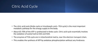
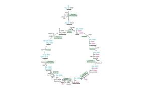
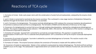
Citric acid cycle
Dr. Kayeen Vadakkan
Citric Acid Cycle
• The citric acid cycle (Krebs cycle or tricarboxylic acid—TCA cycle) is the most important
metabolic pathway for the energy supply to the body.
• About 65-70% of the ATP is synthesized in Krebs cycle. Citric acid cycle essentially involves
the oxidation of acetyl CoA to CO2 and H2O.
• The enzymes of TCA cycle are in mitochondrial matrix, near the electron transport chain.
• This enables the synthesis of ATP by oxidative phosphorylation without any hindrance.
Reactions of TCA cycle
1. Formation of citrate : Krebs cycle proper starts with the condensation of acetyl CoA and oxaloacetate, catalysed by the enzyme citrate
synthase.
2. and 3. Citrate is isomerized to isocitrate by the enzyme aconitase. This is achieved in a two-stage reaction of dehydration followed by
hydration through the formation of an intermediate—cis-aconitate.
4. and 5. Formation of α-ketoglutarate : The enzyme isocitrate dehydrogenase (ICD) catalyses the conversion (oxidative decarboxylation)
of isocitrate to oxalosuccinate and then to α-ketoglutarate. The formation of NADH and the liberation of CO2 occur at this stage.
6. Conversion of α-ketoglutarate to succinyl CoA occurs through oxidative decarboxylation, catalysed by α-ketoglutarate dehydrogenase
complex. This enzyme is dependent on five cofactors—TPP, lipoamide, NAD+, FAD and CoA. The mechanism of the reaction is analogous to
the conversion of pyruvate to acetyl CoA
7. Formation of succinate : Succinyl CoA is converted to succinate by succinate thiokinase. This reaction is coupled with the
phosphorylation of GDP to GTP. This is a substrate level phosphorylation. GTP is converted to ATP by the enzyme nucleoside diphosphate
kinase.
8. Conversion of succinate to fumarate : Succinate is oxidized by succinate dehydrogenase to fumarate. This reaction results in the
production of FADH2 and not NADH.
9. Formation of malate : The enzyme fumarase catalyses the conversion of fumarate to malate with the addition of H2O.
10. Conversion of malate to oxaloacetate : Malate is then oxidized to oxaloacetate by malate dehydrogenase. The third and final synthesis
of NADH occurs at this stage. The oxaloacetate is regenerated which can combine with another molecule of acetyl CoA, and continue the
cycle.
Pyruvate
dehydrogenase
complex (PDH),
• Pyruvate is converted to acetyl CoA by oxidative decarboxylation.
• This is an irreversible reaction, catalysed by a multienzyme complex, known as pyruvate dehydrogenase complex
(PDH), which is found only in the mitochondria.
• In mammals, the PDH complex has an approximate molecular weight of 9×10⁶.
• The enzyme PDH requires five cofactors (coenzymes), namely—TPP, lipoamide, FAD, coenzyme A and NAD+
(lipoamide contains lipoic acid linked to ε-amino group of lysine).
• Structure- pyruvate dehydrogenase complex (PDH) is composed of three subunits in eukaryotes
1. Pyruvate dehydrogenase (E1)
The E1 subunit, called the pyruvate dehydrogenase subunit, has a structure that consists of two chains (an “ɑ” and
“ꞵ” chain).
A magnesium ion forms a 4-coordinate complex with three, polar amino acid residues (Asp, Asn, and Tyr) located on
the alpha chain, and the thiamine diphosphate (TPP) cofactor directly involved in decarboxylation of the pyruvate
2. Dihydrolipoyl transacetylase (E2)
The E2 subunit, or dihydrolipoyl acetyltransferase, for both prokaryotes and eukaryotes, is generally composed of
three domains.
The N-terminal domain (the lipoyl domain), consists of 1-3 lipoyl groups of approximately 80 amino acids each.
The peripheral subunit binding domain (PBSD), serves as a selective binding site for other domains of the E1 and E3
subunits.
Finally, the C-terminal (catalytic) domain catalyzes the transfer of acetyl groups and acetyl-CoA synthesis.
3. Dihydrolipoyl dehydrogenase (E3)
The E3 subunit, called the dihydrolipoyl dehydrogenase enzyme, is characterized as a homodimer protein wherein
two cysteine residues, engaged in disulfide bonding, and the FAD cofactor in the active site facilitate its main
purpose as an oxidizing catalyst.
One example of E3 structure, found in Pseudomonas putida, is formed such that each individual homodimer subunit
contains two binding domains responsible for FAD binding and NAD binding, as well as a central domain and an
interface domain.
Dihydrolipoyl dehydrogenase Binding protein (E3BP) - An auxiliary protein unique to most eukaryotes is the E3
binding protein (E3BP), which serves to bind the E3 subunit to the PDC complex. In the case of human E3BP,
hydrophobic proline and leucine residues in the BP interact with the surface recognition site formed by the binding
of two identical E3 monomers
α-ketoglutarate
dehydrogenase
complex
• Three classes of these multienzyme complexes have been
characterized: one specific for pyruvate, a second specific for 2-
oxoglutarate, and a third specific for branched-chain α-keto acids.
The oxoglutarate dehydrogenase complex has the same subunit
structure and thus uses the same coenzymes as the pyruvate
dehydrogenase complex and the branched-chain alpha-keto acid
dehydrogenase complex.
• This enzyme forms a complex composed of three components
Unit Name Cofactor
E1 oxoglutarate dehydrogenase
thiamine
pyrophosphate(TPP)
E2 dihydrolipoyl succinyltransferase lipoic acid, Coenzyme A
E3 dihydrolipoyl dehydrogenase FAD, NAD

Citric acid cycle

  • 1.
    Citric acid cycle Dr.Kayeen Vadakkan
  • 2.
    Citric Acid Cycle •The citric acid cycle (Krebs cycle or tricarboxylic acid—TCA cycle) is the most important metabolic pathway for the energy supply to the body. • About 65-70% of the ATP is synthesized in Krebs cycle. Citric acid cycle essentially involves the oxidation of acetyl CoA to CO2 and H2O. • The enzymes of TCA cycle are in mitochondrial matrix, near the electron transport chain. • This enables the synthesis of ATP by oxidative phosphorylation without any hindrance.
  • 4.
    Reactions of TCAcycle 1. Formation of citrate : Krebs cycle proper starts with the condensation of acetyl CoA and oxaloacetate, catalysed by the enzyme citrate synthase. 2. and 3. Citrate is isomerized to isocitrate by the enzyme aconitase. This is achieved in a two-stage reaction of dehydration followed by hydration through the formation of an intermediate—cis-aconitate. 4. and 5. Formation of α-ketoglutarate : The enzyme isocitrate dehydrogenase (ICD) catalyses the conversion (oxidative decarboxylation) of isocitrate to oxalosuccinate and then to α-ketoglutarate. The formation of NADH and the liberation of CO2 occur at this stage. 6. Conversion of α-ketoglutarate to succinyl CoA occurs through oxidative decarboxylation, catalysed by α-ketoglutarate dehydrogenase complex. This enzyme is dependent on five cofactors—TPP, lipoamide, NAD+, FAD and CoA. The mechanism of the reaction is analogous to the conversion of pyruvate to acetyl CoA 7. Formation of succinate : Succinyl CoA is converted to succinate by succinate thiokinase. This reaction is coupled with the phosphorylation of GDP to GTP. This is a substrate level phosphorylation. GTP is converted to ATP by the enzyme nucleoside diphosphate kinase. 8. Conversion of succinate to fumarate : Succinate is oxidized by succinate dehydrogenase to fumarate. This reaction results in the production of FADH2 and not NADH. 9. Formation of malate : The enzyme fumarase catalyses the conversion of fumarate to malate with the addition of H2O. 10. Conversion of malate to oxaloacetate : Malate is then oxidized to oxaloacetate by malate dehydrogenase. The third and final synthesis of NADH occurs at this stage. The oxaloacetate is regenerated which can combine with another molecule of acetyl CoA, and continue the cycle.
  • 5.
    Pyruvate dehydrogenase complex (PDH), • Pyruvateis converted to acetyl CoA by oxidative decarboxylation. • This is an irreversible reaction, catalysed by a multienzyme complex, known as pyruvate dehydrogenase complex (PDH), which is found only in the mitochondria. • In mammals, the PDH complex has an approximate molecular weight of 9×10⁶. • The enzyme PDH requires five cofactors (coenzymes), namely—TPP, lipoamide, FAD, coenzyme A and NAD+ (lipoamide contains lipoic acid linked to ε-amino group of lysine). • Structure- pyruvate dehydrogenase complex (PDH) is composed of three subunits in eukaryotes 1. Pyruvate dehydrogenase (E1) The E1 subunit, called the pyruvate dehydrogenase subunit, has a structure that consists of two chains (an “ɑ” and “ꞵ” chain). A magnesium ion forms a 4-coordinate complex with three, polar amino acid residues (Asp, Asn, and Tyr) located on the alpha chain, and the thiamine diphosphate (TPP) cofactor directly involved in decarboxylation of the pyruvate 2. Dihydrolipoyl transacetylase (E2) The E2 subunit, or dihydrolipoyl acetyltransferase, for both prokaryotes and eukaryotes, is generally composed of three domains. The N-terminal domain (the lipoyl domain), consists of 1-3 lipoyl groups of approximately 80 amino acids each. The peripheral subunit binding domain (PBSD), serves as a selective binding site for other domains of the E1 and E3 subunits. Finally, the C-terminal (catalytic) domain catalyzes the transfer of acetyl groups and acetyl-CoA synthesis. 3. Dihydrolipoyl dehydrogenase (E3) The E3 subunit, called the dihydrolipoyl dehydrogenase enzyme, is characterized as a homodimer protein wherein two cysteine residues, engaged in disulfide bonding, and the FAD cofactor in the active site facilitate its main purpose as an oxidizing catalyst. One example of E3 structure, found in Pseudomonas putida, is formed such that each individual homodimer subunit contains two binding domains responsible for FAD binding and NAD binding, as well as a central domain and an interface domain. Dihydrolipoyl dehydrogenase Binding protein (E3BP) - An auxiliary protein unique to most eukaryotes is the E3 binding protein (E3BP), which serves to bind the E3 subunit to the PDC complex. In the case of human E3BP, hydrophobic proline and leucine residues in the BP interact with the surface recognition site formed by the binding of two identical E3 monomers
  • 6.
    α-ketoglutarate dehydrogenase complex • Three classesof these multienzyme complexes have been characterized: one specific for pyruvate, a second specific for 2- oxoglutarate, and a third specific for branched-chain α-keto acids. The oxoglutarate dehydrogenase complex has the same subunit structure and thus uses the same coenzymes as the pyruvate dehydrogenase complex and the branched-chain alpha-keto acid dehydrogenase complex. • This enzyme forms a complex composed of three components Unit Name Cofactor E1 oxoglutarate dehydrogenase thiamine pyrophosphate(TPP) E2 dihydrolipoyl succinyltransferase lipoic acid, Coenzyme A E3 dihydrolipoyl dehydrogenase FAD, NAD