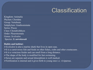
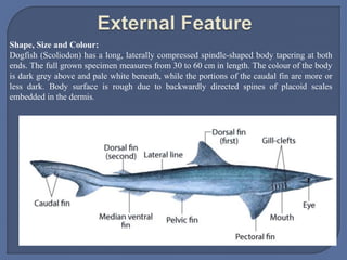
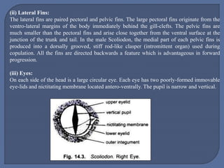
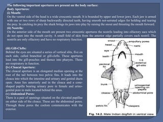
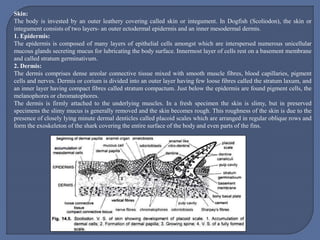
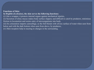
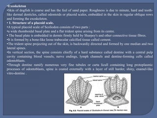
Dr. Arun Gaware
Assistant Professor,
Department of Zoology,
Shri Shivaji College, Motala, Dist. Buldhana
Kingdom: Animalia
Phylum: Chordata
Group: Vertebrata
Subphylum: Gnathostomata
Series: Pisces
Class: Chondrichthyes
Order: Pleurotremata
Genus: Scoliodon
Species: S. sarrokawah
Habits and habitat:
Scoliodon is also a marine shark that lives in open seas.
It is a carnivorous fish and feeds on other fishes, crabs and other crustaceans .
It is a voracious feeder and can smell from a long distance.
The shape of the body is modified for fast swimming.
Sexes are separate and sexual dimorphism is well marked.
Fertilization is internal and it gives birth to young ones i.e. viviparous.
Shape, Size and Colour:
Dogfish (Scoliodon) has a long, laterally compressed spindle-shaped body tapering at both
ends. The full grown specimen measures from 30 to 60 cm in length. The colour of the body
is dark grey above and pale white beneath, while the portions of the caudal fin are more or
less dark. Body surface is rough due to backwardly directed spines of placoid scales
embedded in the dermis.
Division of Body:
The body is divisible into head, trunk and tail, though there are no distinct boundaries between these
regions.
(i) Head:The head is strongly compressed dorso-ventrally and is produced in front into a wedge-
shaped snout or rostrum.
(ii) Trunk:The trunk is almost elliptical in transverse section. Its thickest part lying in front of the
middle of the body. The trunk gradually tapers behind into the tail.
(iii) Tail:The tail is laterally compressed and is bent upwards at a small angle and fringed with a caudal
fin. Such a tail is known as heterocercal tail.
Fins:Dogfish (Scoliodon) is provided with two sets of fins which are flattened expansions of the skin
supported by cartilaginous rods and horny fin rays- these are unpaired or median fins and paired lateral
fins.
(i) Median Fins:
 The median fins are two dorsal fins, a ventral or anal fin and a caudal fin. The first dorsal fin is
large and triangular in shape and is situated a little in front of the middle of the body.
 It has a basal lobe. The second dorsal fin is also triangular in outline but is very small and is
situated midway between the first dorsal and the tip of the tail.
 The caudal fin extends along the dorsal and ventral surfaces of the tail in the median line and forms
a dorsal and ventral lobe.
 The dorsal lobe is very much reduced and forms only a low ridge along the greater part of the upper
surface but the ventral lobe is well developed and is divided into two parts, the anterior part being
much larger and more extensive than the posterior.
 The ventral or anal fin is situated in the mid-ventral line about 5.0 cm in front of the caudal fin,
more or less opposite the second dorsal. Each of the two dorsals and the ventral fin are produced
behind into long and narrow fleshy processes called basal lobes.
(ii) Lateral Fins:
The lateral fins are paired pectoral and pelvic fins. The large pectoral fins originate from the
ventro-lateral margins of the body immediately behind the gill-clefts. The pelvic fins are
much smaller than the pectoral fins and arise close together from the ventral surface at the
junction of the trunk and tail. In the male Scoliodon, the medial part of each pelvic fins is
produced into a dorsally grooved, stiff rod-like clasper (intromittent organ) used during
copulation. All the fins are directed backwards a feature which is advantageous in forward
progression.
(iii) Eyes:
On each side of the head is a large circular eye. Each eye has two poorly-formed immovable
eye-lids and nictitating membrane located antero-ventrally. The pupil is narrow and vertical.
The following important apertures are present on the body surface:
Body Apertures:
(i) Mouth:
On the ventral side of the head is a wide crescentic mouth. It is bounded by upper and lower jaws. Each jaw is armed
with one or two rows of sharp backwardly directed teeth, having smooth not-serrated edges for holding and tearing
the prey. In catching its prey the shark brings its jaws into play by raising the snout and thrusting the mouth forward.
(ii) Nostrils:
On the anterior side of the mouth are present two crescentic apertures the nostrils leading into olfactory sacs which
do not open into the mouth cavity. A small fold of skin from the anterior edge partially covers each nostril. The
nostrils are only olfactory and have no respiratory function.
(iii) Gill-Clefts:
Behind the eyes are situated a series of vertical slits, five on
each side, called branchial or gill-clefts. These apertures
lead into the gill-pouches and thence into pharynx. These
are respiratory in function.
(iv) Cloacal Aperture:
The cloacal aperture is an elongated median opening at the
root of the tail between two pelvic fins. It leads into the
cloaca into which the intestine and urinary and genital ducts
open. Anus lies anteriorly and in the cloaca and a cone-
shaped papilla bearing urinary pore in female and urino-
genital pore in male located behind the anus.
(v) Abdominal Pores:
There is a pair of openings situated on the elevated papillae
on either side of the cloaca. These are the abdominal pores.
Through these pores the coelom communicates with the
exterior.
(vi) Caudal Pit:
At the root of the tail, just in front of the caudal fins, is a small dorsal and a ventral shallow
depressions, the caudal pits. The caudal pits are the characteristic feature of the genus Dogfish
(Scoliodon).
(vii) Lateral Line and Pores:
A faint line runs on either side of the body extending from the head to the posterior end of the
tail, this is called the lateral line. It marks the position of an underlying canal which runs along
each side of the body and contains special receptor organs. The lateral line canal extends
anteriorly into the head where it branches into several canals; at intervals these canals open to
the exterior through minute pores.
(viii) Ampullary Pores:
On the head and snout open several groups of minute ampullary pores of the receptors called
ampullae of Lorenzini. When pressed they exude mucus.
Sexual Dimorphism:
In the male the inner margins of the pelvic fins bear a pair of rod-shaped copulatory organs
called claspers or myxopterygia, they are modified parts of the pelvic fins. Each clasper is a
stiff rod-like appendage having a groove on its dorsal surface which leads into a cavity, the
siphon, beginning at the base of the clasper.
Skin:
The body is invested by an outer leathery covering called skin or integument. In Dogfish (Scoliodon), the skin or
integument consists of two layers- an outer ectodermal epidermis and an inner mesodermal dermis.
1. Epidermis:
The epidermis is composed of many layers of epithelial cells amongst which are interspersed numerous unicellular
mucous glands secreting mucus for lubricating the body surface. Innermost layer of cells rest on a basement membrane
and called stratum germinativum.
2. Dermis:
The dermis comprises dense areolar connective tissue mixed with smooth muscle fibres, blood capillaries, pigment
cells and nerves. Dermis or corium is divided into an outer layer having few loose fibres called the stratum laxum, and
an inner layer having compact fibres called stratum compactum. Just below the epidermis are found pigment cells, the
melanophores or chromatophores.
The dermis is firmly attached to the underlying muscles. In a fresh specimen the skin is slimy, but in preserved
specimens the slimy mucus is generally removed and the skin becomes rough. This roughness of the skin is due to the
presence of closely lying minute dermal denticles called placoid scales which are arranged in regular oblique rows and
form the exoskeleton of the shark covering the entire surface of the body and even parts of the fins.
Functions of Skin:
In Dogfish (Scoliodon), the skin serves the following functions:
(i) Like a wrapper, it protects internal organs against mechanical injuries,
(ii) Secretion of slimy mucus makes body surface slippery and difficult to catch by predators, minimises
friction in locomotion and resists entry of microorganisms into body.
(iii) Its colouration imparts camouflage, as the fish blends with silvery surface of water when seen from
below and with the dark bottom when seen from above, by predators,
(iv) Skin receptors help in reacting to changes in the surrounding.
•Exoskeleton
•Skin of dogfish is coarse and has the feel of sand paper. Roughness is due to minute, hard and tooth-
like dermal denticles, called odontoids or placoid scales, embedded in the skin in regular oblique rows
and forming the exoskeleton.
• 1. Structure of a placoid scale.
•A typical placoid scale of Scoliodon consists of two parts :
•a wide rhomboidal basal plate and a flat trident spine arising from its centre.
• The basal plate is embedded in dermis firmly held by Sharpey's and other connective tissue fibres.
•It is formed by a bone-like loose trabecular calcified tissue called cement.
•The trident spine projecting out of the skin, is backwardiy directed and formed by one median and two
lateral spines.
•In vertical section, the spine consists chiefly of a hard substance called dentine with a central pulp
cavity containing blood vessels, nerve endings, lymph channels and dentine-forming cells called
odontoblasts.
•Through dentine ramify numerous very fine tubules or carta licull containing long protoplasmic
processes of odontoblasts, spine is coated externally with a layer of still harder, shiny, enamel-like
vitro-dentine .
2. Development of a placoid scale.
 The first sign of a developing scale is an aggregation of mesodermal cells of dermis,
lying just beneath epidermis and forming a dermal papilla.
 As the epidermis is pushed up, the outermost cells of dermal papilla become odontoblasts
(or scleroblasts) and deposit dentine between themselves and epidermis.
 Meanwhile, the overlying epidermal cells of stratum germinativum or Malpighian layer,
now called ameloblasts, form the so-called enamel organ which deposits vitrodentine
over dentine. Cement (basal plate) is formed from surrounding cells of dermis.
 In a growing scale, the dermal papilla forms nutritive pulp occupying the pulp cavity. In a
fully formed scale, epidermis wears off around the spine which projects above the skin,
while the basal plate remains embedded.
Scoliodon Morphology.pptx

Scoliodon Morphology.pptx

  • 1.
    Dr. Arun Gaware AssistantProfessor, Department of Zoology, Shri Shivaji College, Motala, Dist. Buldhana
  • 2.
    Kingdom: Animalia Phylum: Chordata Group:Vertebrata Subphylum: Gnathostomata Series: Pisces Class: Chondrichthyes Order: Pleurotremata Genus: Scoliodon Species: S. sarrokawah Habits and habitat: Scoliodon is also a marine shark that lives in open seas. It is a carnivorous fish and feeds on other fishes, crabs and other crustaceans . It is a voracious feeder and can smell from a long distance. The shape of the body is modified for fast swimming. Sexes are separate and sexual dimorphism is well marked. Fertilization is internal and it gives birth to young ones i.e. viviparous.
  • 3.
    Shape, Size andColour: Dogfish (Scoliodon) has a long, laterally compressed spindle-shaped body tapering at both ends. The full grown specimen measures from 30 to 60 cm in length. The colour of the body is dark grey above and pale white beneath, while the portions of the caudal fin are more or less dark. Body surface is rough due to backwardly directed spines of placoid scales embedded in the dermis.
  • 4.
    Division of Body: Thebody is divisible into head, trunk and tail, though there are no distinct boundaries between these regions. (i) Head:The head is strongly compressed dorso-ventrally and is produced in front into a wedge- shaped snout or rostrum. (ii) Trunk:The trunk is almost elliptical in transverse section. Its thickest part lying in front of the middle of the body. The trunk gradually tapers behind into the tail. (iii) Tail:The tail is laterally compressed and is bent upwards at a small angle and fringed with a caudal fin. Such a tail is known as heterocercal tail. Fins:Dogfish (Scoliodon) is provided with two sets of fins which are flattened expansions of the skin supported by cartilaginous rods and horny fin rays- these are unpaired or median fins and paired lateral fins. (i) Median Fins:  The median fins are two dorsal fins, a ventral or anal fin and a caudal fin. The first dorsal fin is large and triangular in shape and is situated a little in front of the middle of the body.  It has a basal lobe. The second dorsal fin is also triangular in outline but is very small and is situated midway between the first dorsal and the tip of the tail.  The caudal fin extends along the dorsal and ventral surfaces of the tail in the median line and forms a dorsal and ventral lobe.  The dorsal lobe is very much reduced and forms only a low ridge along the greater part of the upper surface but the ventral lobe is well developed and is divided into two parts, the anterior part being much larger and more extensive than the posterior.  The ventral or anal fin is situated in the mid-ventral line about 5.0 cm in front of the caudal fin, more or less opposite the second dorsal. Each of the two dorsals and the ventral fin are produced behind into long and narrow fleshy processes called basal lobes.
  • 5.
    (ii) Lateral Fins: Thelateral fins are paired pectoral and pelvic fins. The large pectoral fins originate from the ventro-lateral margins of the body immediately behind the gill-clefts. The pelvic fins are much smaller than the pectoral fins and arise close together from the ventral surface at the junction of the trunk and tail. In the male Scoliodon, the medial part of each pelvic fins is produced into a dorsally grooved, stiff rod-like clasper (intromittent organ) used during copulation. All the fins are directed backwards a feature which is advantageous in forward progression. (iii) Eyes: On each side of the head is a large circular eye. Each eye has two poorly-formed immovable eye-lids and nictitating membrane located antero-ventrally. The pupil is narrow and vertical.
  • 6.
    The following importantapertures are present on the body surface: Body Apertures: (i) Mouth: On the ventral side of the head is a wide crescentic mouth. It is bounded by upper and lower jaws. Each jaw is armed with one or two rows of sharp backwardly directed teeth, having smooth not-serrated edges for holding and tearing the prey. In catching its prey the shark brings its jaws into play by raising the snout and thrusting the mouth forward. (ii) Nostrils: On the anterior side of the mouth are present two crescentic apertures the nostrils leading into olfactory sacs which do not open into the mouth cavity. A small fold of skin from the anterior edge partially covers each nostril. The nostrils are only olfactory and have no respiratory function. (iii) Gill-Clefts: Behind the eyes are situated a series of vertical slits, five on each side, called branchial or gill-clefts. These apertures lead into the gill-pouches and thence into pharynx. These are respiratory in function. (iv) Cloacal Aperture: The cloacal aperture is an elongated median opening at the root of the tail between two pelvic fins. It leads into the cloaca into which the intestine and urinary and genital ducts open. Anus lies anteriorly and in the cloaca and a cone- shaped papilla bearing urinary pore in female and urino- genital pore in male located behind the anus. (v) Abdominal Pores: There is a pair of openings situated on the elevated papillae on either side of the cloaca. These are the abdominal pores. Through these pores the coelom communicates with the exterior.
  • 7.
    (vi) Caudal Pit: Atthe root of the tail, just in front of the caudal fins, is a small dorsal and a ventral shallow depressions, the caudal pits. The caudal pits are the characteristic feature of the genus Dogfish (Scoliodon). (vii) Lateral Line and Pores: A faint line runs on either side of the body extending from the head to the posterior end of the tail, this is called the lateral line. It marks the position of an underlying canal which runs along each side of the body and contains special receptor organs. The lateral line canal extends anteriorly into the head where it branches into several canals; at intervals these canals open to the exterior through minute pores. (viii) Ampullary Pores: On the head and snout open several groups of minute ampullary pores of the receptors called ampullae of Lorenzini. When pressed they exude mucus. Sexual Dimorphism: In the male the inner margins of the pelvic fins bear a pair of rod-shaped copulatory organs called claspers or myxopterygia, they are modified parts of the pelvic fins. Each clasper is a stiff rod-like appendage having a groove on its dorsal surface which leads into a cavity, the siphon, beginning at the base of the clasper.
  • 8.
    Skin: The body isinvested by an outer leathery covering called skin or integument. In Dogfish (Scoliodon), the skin or integument consists of two layers- an outer ectodermal epidermis and an inner mesodermal dermis. 1. Epidermis: The epidermis is composed of many layers of epithelial cells amongst which are interspersed numerous unicellular mucous glands secreting mucus for lubricating the body surface. Innermost layer of cells rest on a basement membrane and called stratum germinativum. 2. Dermis: The dermis comprises dense areolar connective tissue mixed with smooth muscle fibres, blood capillaries, pigment cells and nerves. Dermis or corium is divided into an outer layer having few loose fibres called the stratum laxum, and an inner layer having compact fibres called stratum compactum. Just below the epidermis are found pigment cells, the melanophores or chromatophores. The dermis is firmly attached to the underlying muscles. In a fresh specimen the skin is slimy, but in preserved specimens the slimy mucus is generally removed and the skin becomes rough. This roughness of the skin is due to the presence of closely lying minute dermal denticles called placoid scales which are arranged in regular oblique rows and form the exoskeleton of the shark covering the entire surface of the body and even parts of the fins.
  • 9.
    Functions of Skin: InDogfish (Scoliodon), the skin serves the following functions: (i) Like a wrapper, it protects internal organs against mechanical injuries, (ii) Secretion of slimy mucus makes body surface slippery and difficult to catch by predators, minimises friction in locomotion and resists entry of microorganisms into body. (iii) Its colouration imparts camouflage, as the fish blends with silvery surface of water when seen from below and with the dark bottom when seen from above, by predators, (iv) Skin receptors help in reacting to changes in the surrounding.
  • 10.
    •Exoskeleton •Skin of dogfishis coarse and has the feel of sand paper. Roughness is due to minute, hard and tooth- like dermal denticles, called odontoids or placoid scales, embedded in the skin in regular oblique rows and forming the exoskeleton. • 1. Structure of a placoid scale. •A typical placoid scale of Scoliodon consists of two parts : •a wide rhomboidal basal plate and a flat trident spine arising from its centre. • The basal plate is embedded in dermis firmly held by Sharpey's and other connective tissue fibres. •It is formed by a bone-like loose trabecular calcified tissue called cement. •The trident spine projecting out of the skin, is backwardiy directed and formed by one median and two lateral spines. •In vertical section, the spine consists chiefly of a hard substance called dentine with a central pulp cavity containing blood vessels, nerve endings, lymph channels and dentine-forming cells called odontoblasts. •Through dentine ramify numerous very fine tubules or carta licull containing long protoplasmic processes of odontoblasts, spine is coated externally with a layer of still harder, shiny, enamel-like vitro-dentine .
  • 11.
    2. Development ofa placoid scale.  The first sign of a developing scale is an aggregation of mesodermal cells of dermis, lying just beneath epidermis and forming a dermal papilla.  As the epidermis is pushed up, the outermost cells of dermal papilla become odontoblasts (or scleroblasts) and deposit dentine between themselves and epidermis.  Meanwhile, the overlying epidermal cells of stratum germinativum or Malpighian layer, now called ameloblasts, form the so-called enamel organ which deposits vitrodentine over dentine. Cement (basal plate) is formed from surrounding cells of dermis.  In a growing scale, the dermal papilla forms nutritive pulp occupying the pulp cavity. In a fully formed scale, epidermis wears off around the spine which projects above the skin, while the basal plate remains embedded.