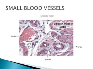
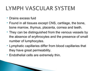
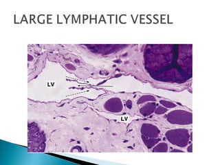
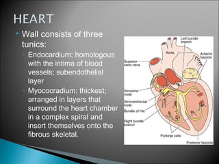
MUSCULAR ARTERY HAS THICK MEDIA
 Drains excess fuid
 Found in all tissues except CNS, cartilage, the bone,
bone marrow, thymus, placenta, cornea and teeth.
 They can be distinguished from the venous vessels by
the absence of erythrocytes and the presence of small
number of lumphocytes.
 Lymphatic capillaries differ from blood capillaries that
they have great permeability.
 Endothelial cells are extremely thin.
 Basement membrane is absent
 There are no pericytes.
 Fine collagenous filaments known as anchoring
filaments link the endothelium to the surrounding
supportive tissue preventing collapse.
 Wall consists of three
tunics:
◦ Endocardium: homologous
with the intima of blood
vessels; subendothelial
layer
◦ Myococradium: thickest;
arranged in layers that
surround the heart chamber
in a complex spiral and
insert themselves onto the
fibrous skeletal.
 Epicardium: a subepicardial layer of loose
connective tissue contains veins, nerves and
nerve ganglia and edipose tissue; it corresponds
to the visceral layer of pericardium.
 Fibrous skeletal is composed of dense connective
tissue. Its components are trigona fibrosa and
annuli fibrosi. These structures are composed of
dense connective tissue with thick collagen fibers.
They also contain nodules of fibrous cartilage.
 Two nodes located in
the atrium are SA node,
AV node and the AV
bundle.
 The cells of the impulse
conducting system are
functionally integrated
by gap junctions.
The SA node is a mass of modified cardiac muscle
cells that is fusiform smaller than the atrial muscle
cells and has fewer myofibrils.
 The cells of AV node are similar to that of the SA
node but their cytoplasmic projections branch in
various directions to form a network.
 The AV bundle is formed by similar cells of the AV
node but they are larger than ordinary cardiac
muscle cells and acquire a special apperance so
called purkinje cells.
 They have one or two central nucleis.
 Mitochondria and glycogen rich cytolplasm.
 Few myofibrils which are restricted to the
periphery of the cytoplasm.
 After travelling the subendocardial layer, they
penetrate the ventricle and become
intramyocardiac. This arrangement allows the
stimulus to get into the inner most layer of the
ventricular muscles.

Circulatory system

  • 9.
  • 17.
     Drains excessfuid  Found in all tissues except CNS, cartilage, the bone, bone marrow, thymus, placenta, cornea and teeth.  They can be distinguished from the venous vessels by the absence of erythrocytes and the presence of small number of lumphocytes.  Lymphatic capillaries differ from blood capillaries that they have great permeability.  Endothelial cells are extremely thin.
  • 18.
     Basement membraneis absent  There are no pericytes.  Fine collagenous filaments known as anchoring filaments link the endothelium to the surrounding supportive tissue preventing collapse.
  • 21.
     Wall consistsof three tunics: ◦ Endocardium: homologous with the intima of blood vessels; subendothelial layer ◦ Myococradium: thickest; arranged in layers that surround the heart chamber in a complex spiral and insert themselves onto the fibrous skeletal.
  • 22.
     Epicardium: asubepicardial layer of loose connective tissue contains veins, nerves and nerve ganglia and edipose tissue; it corresponds to the visceral layer of pericardium.  Fibrous skeletal is composed of dense connective tissue. Its components are trigona fibrosa and annuli fibrosi. These structures are composed of dense connective tissue with thick collagen fibers. They also contain nodules of fibrous cartilage.
  • 23.
     Two nodeslocated in the atrium are SA node, AV node and the AV bundle.  The cells of the impulse conducting system are functionally integrated by gap junctions.
  • 24.
    The SA nodeis a mass of modified cardiac muscle cells that is fusiform smaller than the atrial muscle cells and has fewer myofibrils.  The cells of AV node are similar to that of the SA node but their cytoplasmic projections branch in various directions to form a network.  The AV bundle is formed by similar cells of the AV node but they are larger than ordinary cardiac muscle cells and acquire a special apperance so called purkinje cells.
  • 25.
     They haveone or two central nucleis.  Mitochondria and glycogen rich cytolplasm.  Few myofibrils which are restricted to the periphery of the cytoplasm.  After travelling the subendocardial layer, they penetrate the ventricle and become intramyocardiac. This arrangement allows the stimulus to get into the inner most layer of the ventricular muscles.