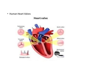
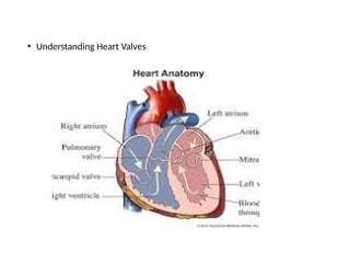
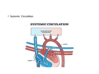
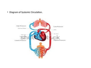
Introduction to cardiovascular system
Dr. E. Muralinath, Dr. C. Kalyan chakravrthy, Dr. K.
Sri Devi Dr. D. Kusumalatha, Dr. K. Sravani Pragna,
Dr. P. Manjari, Dr. V. Yaswanth Sai and Dr. T. Nikhil,
• Cardiovascular system incorporates heart and blood vessels. Heart pumps blood into the blood vessels. Blood
vessels circulate the blood throughout the body. Blood transports especially nutrients and oxygen to the tissues
and eliminates carbon dioxide and waste products from the tissues.
• Heart
• Heart is a muscular organ that pumps blood throughout the circulatory system. It is located in
between two lungs in the mediastinum. It consists of four chambers, two atria and two ventricles.
The musculature of ventricles is thicker than that of atria. Force of contraction of heart is associated
with the muscles
• RIGHT SIDE OF THE HEART
• Right side of the heart comprises two chambers, right atrium and right ventricle. Right atrium is a thin
walled and low pressure chamber. It contains the pacemaker termed as sinoatrial node that generates
cardiac impulses and Atrioventricular node that conducts the impulses to the ventricles.
• Right atrium gains venous (deoxygenated) blood via two large veins:
• Superior vena cava that returns venous blood from the head, neck and upper limbs
• Inferior vena cava that returns venous blood from lower parts of the body.
• Right atrium communicates with right ventricle With the help of tricuspid valve. Wall of
right ventricle is thick.
• Venous blood from the right atrium enters the right ventricle through this valve.
• From the right ventricle, pulmonary artery originates. It carries the venous blood from
right ventricle to lungs. In the lungs, the deoxygenated blood is changed into oxygenated
blood.
• LEFT SIDE OF THE HEART
• Left side of the heart consists of two chambers, left atrium and left ventricle. Left atrium is
a thin walled and low pressure chamber. It obtains oxygenated blood from the lungs via
pulmonary veins. This is the only exception in the body, where an artery carries
venous blood and vein carries the arterial blood
• Blood from left atrium gains an entry into the left ventricle through mitral valve (bicuspid
valve). Wall of the left ventricle is very thick. Left ventricle pumps the arterial blood to
different parts of the body through systemic aorta.
• SEPTA OF THE HEART
• Right and left atria are separated from one another by a fibrous septum termed as interatrial septum.
Right and left ventricles are separated from one another by interventricular septum. The upper part
of this septum is a membranous structure, whereas the lower part of itis muscular in nature.
• LAYERS OF WALL OF THE HEART
• Heart comprises three layers of tissues:
• Outer pericardium
• Middle myocardium
• Inner endocardium
• PERICARDIUM
• Pericardium is the outer covering of the heart. It comprises two layers:
• Outer parietal pericardium
• Inner visceral pericardium
• The space between the two layers is known as pericardial cavity or pericardial space and it consists of a
thin film of fluid.
• Outer Parietal Pericardium Parietal pericardium forms a strong protective sac for the heart. It assists
also to anchor the heart especially within the mediastinum.
• Parietal pericardium consists of two layers:
• Outer fibrous layer
• Inner serous layer.
• Fibrous layer
• The formation of ffibrous layer of the parietal pericardium happens by thick fibrous connective
tissue. It is connected to the diaphragm and it is continuous with tunica adventitia (outer wall) of the
blood vessels, entering and leaving the heart. It is attached with diaphragm below. Because of the
fibrous nature, it provides the p[protection to the heart from over stretching.
Serous layer
• The formation of serous layer occurs by mesothelium, together with a small amount of
connective tissue. Mesothelium consists of squamous epithelial cells which secrete a small
amount of fluid, which lines the pericardial space. This fluid prevents friction and permits free
movement of heart along within pericardium, if it contracts and relaxes. The total volume of
this fluid is only about 25 to 35 mL.
• Inner Visceral Pericardium
• Inner visceral pericardium lines the surface of myocardium. It comprises flattened epithelial
cells. This layer is also termed as epicardium
• MYOCARDIUM
• Myocardium is the middle layer of wall of t he heart and its formation takes place by cardiac
muscle fibers or cardiac myocytes. Myocardium leads to the formation of the bulk of the heart
and Plays an important role regarding pumping action of the heart. Unlike skeletal muscle
fibers, the cardiac muscle fibers are involuntary in nature.
• Myocardium consists of three types of muscle fibers:
• Muscle fibers which form contractile unit of Heart.
• Muscle fibers which form pacemaker
• Muscle fibers which form conductive system
• Muscle Fibers which Form Contractile Unit of Heart
• These cardiac muscle fibers are striated and resemble the skeletal muscle
fibers regarding structure. Cardiac muscle fiber is bound by sarcolemma.
It comprises a centrally placed nucleus. Myofibrils are embedded in the
sarcoplasm. Sarcomere of the cardiac muscle consists of all the
contractile proteins, namely actin, myosin, troponin and tropomyosin.
Sarcotubular system in cardiac muscle is similar to that of skeletal muscle.
Important difference between skeletal muscle and cardiac muscle is that
the cardiac muscle fiber is branched and the skeletal muscle is not
branched.
• Intercalated disk
• Intercalated disk is a tough double membranous structure, located at the
junction between the branches of neighboring cardiac muscle fibers. It is
formed by the fusion of the membrane of the cardiac muscle branches
• Intercalated disks generate adherent junctions, which play an important role
regarding the contraction of cardiac muscle as a single unit.
• Syncytium
• Syncytium means tissue along with cytoplasmic continuity between
adjacent cells. Whatever it may be, cardiac muscle is like a physiological
syncytium, since there is no continuity of the cytoplasm and the muscle
fibers are separated from each other due to cell membrane. At the sides,
the membranes of the adjacent muscle fibers fuse together and result in
the formation of gap junctions. Gap junction is permeable to ions and it
promotes the rapid conduction of action potential from one fiber to
another. Due to this, all the cardiac muscle fibers behave like a single unit,
which is also otherwise known as syncytium.
• Syncytium in human heart consists of two portions, syncytium of atria and the
syncytium of ventricles. Both the portions of syncytium are connected by a thick
non-conducting fibrous ring termed as the atrioventricular ring.
• ii. Muscle Fibers which Form the Pacemaker
• Some of the muscle fibers of heart are modified into a specialized structure
termed as pacemaker. These muscle fibers forming the pacemaker have less
striation.
• Pacemaker
• Pacemaker is structure in the heart that develops the impulses for heart beat.
The formation of pacemaker happens by pacemaker cells termed as P cells.
Sinoatrial (SA) node forms the pacemaker in human heart.
• iii. Muscle Fibers which Form Conductive System
• Conductive system of the heart is formed by modified cardiac muscle fibers. Impulses
from SA node are transmitted to the atria in a direct manner. Whatever it may be,
thee impulses are transmitted to ventricles through various components of
conducting system
• ENDOCARDIUM
• Endocardium is the inner most layer of heart wall. It is a thin, smooth and glistening
membrane. The formation of endocardium occurs by a single layer of endothelial
cells, lining the inner surface of the heart.
• Endocardium continues as endothelium of the blood vessels.
• VALVES OF THE HEART
• There are four valves in human heart. Two valves are in between atria and the ventricles
termed as Atrioventricular valves. Other two are the semilunar valves, situated at the
opening of blood vessels arising from ventricles, namely systemic aorta and pulmonary
artery. Valves of the heart permit the flow of blood through heart in only one direction.
• Atrioventricular Valves
• Left Atrioventricular valve is also termed as mitral valve or bicuspid valve. The formation of
left atrio-ventricular valve happened by two valvular cusps or flaps. Right Atrioventricular
valve is termed as tricuspid valve and it is formed by three cusps.
• Brim of the atrioventricular valves is connected to atrioventricular ring, which is the fibrous
connection between the atria and ventricles. Cusps of the valves are connected to papillary
muscles with the help of chordae tendineae.
• Papillary muscles arise from inner surface of the ventricles. Papillary muscles play an important
role regarding closure of the cusps and prevention of the back flow of blood from ventricle to
atria especially during ventricular contraction.
• Atrioventricular valves open only towards ventricles and inhibit the backflow of blood into atria.
• Semilunar Valves
• Semilunar valves are located at the openings of systemic aorta and pulmonary artery and
are termed as aortic valve and pulmonary valve respectively. Due to the half moon shape,
these two valves are also termed as semilunar valves. Semilunar valves are made up of
three flaps. Semilunar valves open only towards the aorta and pulmonary artery and inhibit
the backflow of blood into the ventricles
• ACTIONS OF THE HEART
• Actions of the heart are categorized into four types:
• Chronotropic action
• Inotropic action
• Dromotropic action
• Bathmotropic action.
• CHRONOTROPIC ACTION
• Chronotropic action is the frequency of heart beat or heart rate. It is of two types:
• Tachycardia or enhancement of heart rate
• Bradycardia or reduction of heart rate
• IONOTROPIC ACTION
• Force of contraction of heart is termed as inotropic action. It is of two types:
• Positive inotropic action or enhancement regarding the force of contraction
• Negative inotropic action or reduction regarding the force of contraction
• DROMOTROPIC ACTION
• Dromotropic action is the conduction of impulse through heart. It is of two types:
• Positive dromotropic action or enhancement rewgarding the velocity of conduction
• Negative dromotropic action or reduction regarding the velocity of conduction.
• BATHMOTROPIC ACTION
• Bathmotropic action is the excitability of cardiac muscle. It is also of two types:
• Positive bathmotropic action orenhancement regarding the excitability of cardiac muscle
• Negative bathmotropic action or reduction regarding the excitability of cardiac muscle
• Regulation of Actions of Heart
• All the actions of heart are regulated in a continuous manner. It is essential for the heart to
cope up with the requirements of the body. All the actions are altered by activation of nerves
supplying the heart or some hormones or hormonal substances secreted in the body.
• BLOOD VESSELS
• Vessels of circulatory system are the aorta, arteries, arterioles, capillaries, venules, veins and venae
cava
• ARTERIAL SYSTEM
• Arterial system consists of the aorta, arteries and arterioles. The formation of walls of the aorta
and arteries occurs by three layers:
• Outer tunica adventitia, which is made up of connective tissue layer. It is the continuation of
fibrous layer of parietal pericardium.
• Middle tunica media, which is formed by smooth muscles
• Inner tunica intima, which is made up of endothelium. It is the continuation of endocardium
• Aorta, arteries and arterioles consist of two lamina of elastic tissues:
• External elastic lamina between tunica adventitia and tunica media ii. Internal elastic lamina
between tunica media and tunica intima.
• Aorta and arteries contain more elastic tissues and the arterioles have more smooth muscles.
Arterial branches become narrower and their walls become thinner while reaching the periphery.
Aorta consists of the maximum diameter of about 25 mm.
• Diameterof the arteries is reduced in a gradual manner and at the end arteries, it is about
4 mm. It further reduces to 30 μ in the arterioles and ends up with 10 μ in the terminal
arterioles. Resistance (peripheral resistance) is offered to blood flow in the arterioles and
so these vessels are termed as resistant vessels.
• Arterioles are continued as capillaries, which are small, thin walled vessels along with a
diameter of about 5 to 8 μ. Capillaries are very important regarding functionality because,
the exchange of materials between the blood and the tissues happens through these
vessels.
• VENOUS SYSTEM
• From the capillaries, venous system begins and it includes venules, veins and venae
cavae. Capillaries end in venules, which are the smaller vessels along with thin
muscular wall than the arterioles. Diameter of the venules is about 20 μ. At a time, a
large quantity of blood is held in venules and hence the venules are otherwise
known as capacitance vessels. Venules are continued as veins, which have the
diameter of 5 mm. Veins form superior and inferior venae cava, which have a
diameter of about 30 mm
• Blood vessel Diameter Thickness of the wall Elastic tissue Smooth muscle fibers Fibrous tissue
• Aorta 25 mm 2 mm More Less More, Artery 4 mm 1 mm More More Moderate
• Arteriole 30 μ 6 μ Moderate More Moderate, Terminal arteriole 10 μ 2 μ Less More Moderate, Capillary 8 μ
0.5 μ Absent Absent Moderate Venule 20 μ 1 μ Absent Absent Less, Vein 5 mm 0.5 mm Less More Moderate,
Vena cava524 Section 8 t Cardiovascular System, Walls of the veins and venae cavae are made up of inner
endothelium, elastic tissues, smooth muscles and outer connective tissue layer. In the veins and venae
cavae, the elastic tissue is less but the smooth muscle fibers are more.
• COMPLICATIONS IN BLOOD VESSELS
• Aorta and Arteries
• Arterial blood vessels are highly susceptible for arteriosclerosis and atherosclerosis. Arteriosclerosis is
the disease of the arteries, related to hardening, thickening and loss of elasticity in the wall of the
vessels. Atherosclerosis is the disease manifested by the narrowing of lumen of arterial vessel because
of the deposition of cholesterol.
• Arterioles
• When the tone of the smooth muscles in the arterioles enhances, hypertension happens.
• Capillaries
• Permeability of the capillary membrane may enhance Leading to shock or edema because of leakage
of fluid, proteins and other substances from blood
• Veins
• Inflammation of the wall of veins results in the formation of intravascular clot called thrombosis. The
clot gets dislodged, as thrombus. The thrombus travels through blood and leads to embolism.
Embolism leads to the obstruction of the blood flow to vital organs namely brain, heart and lungs,
leading to many complications
• DIVISIONS OF CIRCULATION
• Blood flows through two divisions of circulatory system:
• Systemic circulation
• Pulmonary circulation.
• SYSTEMIC CIRCULATION
• Systemic circulation is also termed as greater circulation . Blood pumped from left
ventricle passes through a series of blood vessels, arterial system and attains the
tissues. Exchange of various substances between blood and the tissues happens
at the capillaries. After exchange of materials, blood enters the venous system
and returns to right atrium of the heart. From right atrium, blood enters the right
ventricle. Thus, through systemic circulation, oxygenated blood is supplied from
heart to the tissues and venous blood returns to the heart from tissues
• PULMONARY CIRCULATION
• Pulmonary circulation is termed as lesser circulation. Blood is pumped from right
ventricle to lungs through pulmonary artery. Exchange of gases happens between
blood and alveoli of the lungs at pulmonary capillaries. Oxygenated blood returns
to left atrium through the pulmonary veins. Thus, left side of the heart consists of
oxygenated or arterial blood and the right side of the heart cconsists of
deoxygenated or venous blood.
• Colour coded anatomy of heart
• Human Heart Valves
• Understanding Heart Valves
• Systemic Circulation
• Diagram of Systemic Circulation.
• References
• 1.
• Polak-Iwaniuk A, Harasim-Symbor E, Gołaszewska K, Chabowski A. How Hypertension Affects
Heart Metabolism. Front Physiol. 2019;10:435. [PMC free article] [PubMed]
• 2.
• Huang Y, Hu D, Huang C, Nichols CG. Genetic Discovery of ATP-Sensitive K+
Channels in
Cardiovascular Diseases. Circ Arrhythm Electrophysiol. 2019 May;12(5):e007322. [
PMC free article] [PubMed]
• 3.
• Tsibulnikov SY, Maslov LN, Gorbunov AS, Voronkov NS, Boshchenko AA, Popov SV, Prokudina
ES, Singh N, Downey JM. A Review of Humoral Factors in Remote Preconditioning of the
Heart. J Cardiovasc Pharmacol Ther. 2019 Sep;24(5):403-421. [PubMed]
• 4.
• Gruzdeva OV, Borodkina DA, Belik EV, Akbasheva OE, Palicheva EI, Barbarash OL. [Ghrelin
Physiology and Pathophysiology: Focus on the Cardiovascular System]. Kardiologiia. 2019 Apr
13;59(3):60-67. [PubMed]
• 5.
• Seo DY, Kwak HB, Kim AH, Park SH, Heo JW, Kim HK, Ko JR, Lee SJ, Bang HS, Sim JW,
Kim M, Han J. Cardiac adaptation to exercise training in health and disease.
Pflugers Arch. 2020 Feb;472(2):155-168. [PubMed]
• 6.
• Park S, Nguyen NB, Pezhouman A, Ardehali R. Cardiac fibrosis: potential
therapeutic targets. Transl Res. 2019 Jul;209:121-137. [PMC free article] [PubMed]
• 7.
• Rossignol P, Hernandez AF, Solomon SD, Zannad F. Heart failure drug treatment.
Lancet. 2019 Mar 09;393(10175):1034-1044. [PubMed]
• 8.
• Elkin HK, Winter A. Grounding Patients With Hypertension Improves Blood
Pressure: A Case History Series Study. Altern Ther Health Med. 2018 Nov;24(6):46-
50. [PubMed]

Introduction to Cardiovascular system_structure and functions-1

  • 1.
    Introduction to cardiovascularsystem Dr. E. Muralinath, Dr. C. Kalyan chakravrthy, Dr. K. Sri Devi Dr. D. Kusumalatha, Dr. K. Sravani Pragna, Dr. P. Manjari, Dr. V. Yaswanth Sai and Dr. T. Nikhil,
  • 2.
    • Cardiovascular systemincorporates heart and blood vessels. Heart pumps blood into the blood vessels. Blood vessels circulate the blood throughout the body. Blood transports especially nutrients and oxygen to the tissues and eliminates carbon dioxide and waste products from the tissues. • Heart • Heart is a muscular organ that pumps blood throughout the circulatory system. It is located in between two lungs in the mediastinum. It consists of four chambers, two atria and two ventricles. The musculature of ventricles is thicker than that of atria. Force of contraction of heart is associated with the muscles • RIGHT SIDE OF THE HEART • Right side of the heart comprises two chambers, right atrium and right ventricle. Right atrium is a thin walled and low pressure chamber. It contains the pacemaker termed as sinoatrial node that generates cardiac impulses and Atrioventricular node that conducts the impulses to the ventricles. • Right atrium gains venous (deoxygenated) blood via two large veins: • Superior vena cava that returns venous blood from the head, neck and upper limbs • Inferior vena cava that returns venous blood from lower parts of the body.
  • 3.
    • Right atriumcommunicates with right ventricle With the help of tricuspid valve. Wall of right ventricle is thick. • Venous blood from the right atrium enters the right ventricle through this valve. • From the right ventricle, pulmonary artery originates. It carries the venous blood from right ventricle to lungs. In the lungs, the deoxygenated blood is changed into oxygenated blood. • LEFT SIDE OF THE HEART • Left side of the heart consists of two chambers, left atrium and left ventricle. Left atrium is a thin walled and low pressure chamber. It obtains oxygenated blood from the lungs via pulmonary veins. This is the only exception in the body, where an artery carries venous blood and vein carries the arterial blood • Blood from left atrium gains an entry into the left ventricle through mitral valve (bicuspid valve). Wall of the left ventricle is very thick. Left ventricle pumps the arterial blood to different parts of the body through systemic aorta.
  • 4.
    • SEPTA OFTHE HEART • Right and left atria are separated from one another by a fibrous septum termed as interatrial septum. Right and left ventricles are separated from one another by interventricular septum. The upper part of this septum is a membranous structure, whereas the lower part of itis muscular in nature. • LAYERS OF WALL OF THE HEART • Heart comprises three layers of tissues: • Outer pericardium • Middle myocardium • Inner endocardium • PERICARDIUM • Pericardium is the outer covering of the heart. It comprises two layers: • Outer parietal pericardium • Inner visceral pericardium • The space between the two layers is known as pericardial cavity or pericardial space and it consists of a thin film of fluid. • Outer Parietal Pericardium Parietal pericardium forms a strong protective sac for the heart. It assists also to anchor the heart especially within the mediastinum.
  • 5.
    • Parietal pericardiumconsists of two layers: • Outer fibrous layer • Inner serous layer. • Fibrous layer • The formation of ffibrous layer of the parietal pericardium happens by thick fibrous connective tissue. It is connected to the diaphragm and it is continuous with tunica adventitia (outer wall) of the blood vessels, entering and leaving the heart. It is attached with diaphragm below. Because of the fibrous nature, it provides the p[protection to the heart from over stretching. Serous layer • The formation of serous layer occurs by mesothelium, together with a small amount of connective tissue. Mesothelium consists of squamous epithelial cells which secrete a small amount of fluid, which lines the pericardial space. This fluid prevents friction and permits free movement of heart along within pericardium, if it contracts and relaxes. The total volume of this fluid is only about 25 to 35 mL. • Inner Visceral Pericardium • Inner visceral pericardium lines the surface of myocardium. It comprises flattened epithelial cells. This layer is also termed as epicardium • MYOCARDIUM • Myocardium is the middle layer of wall of t he heart and its formation takes place by cardiac muscle fibers or cardiac myocytes. Myocardium leads to the formation of the bulk of the heart and Plays an important role regarding pumping action of the heart. Unlike skeletal muscle fibers, the cardiac muscle fibers are involuntary in nature. • Myocardium consists of three types of muscle fibers: • Muscle fibers which form contractile unit of Heart. • Muscle fibers which form pacemaker • Muscle fibers which form conductive system
  • 6.
    • Muscle Fiberswhich Form Contractile Unit of Heart • These cardiac muscle fibers are striated and resemble the skeletal muscle fibers regarding structure. Cardiac muscle fiber is bound by sarcolemma. It comprises a centrally placed nucleus. Myofibrils are embedded in the sarcoplasm. Sarcomere of the cardiac muscle consists of all the contractile proteins, namely actin, myosin, troponin and tropomyosin. Sarcotubular system in cardiac muscle is similar to that of skeletal muscle. Important difference between skeletal muscle and cardiac muscle is that the cardiac muscle fiber is branched and the skeletal muscle is not branched. • Intercalated disk • Intercalated disk is a tough double membranous structure, located at the junction between the branches of neighboring cardiac muscle fibers. It is formed by the fusion of the membrane of the cardiac muscle branches
  • 7.
    • Intercalated disksgenerate adherent junctions, which play an important role regarding the contraction of cardiac muscle as a single unit. • Syncytium • Syncytium means tissue along with cytoplasmic continuity between adjacent cells. Whatever it may be, cardiac muscle is like a physiological syncytium, since there is no continuity of the cytoplasm and the muscle fibers are separated from each other due to cell membrane. At the sides, the membranes of the adjacent muscle fibers fuse together and result in the formation of gap junctions. Gap junction is permeable to ions and it promotes the rapid conduction of action potential from one fiber to another. Due to this, all the cardiac muscle fibers behave like a single unit, which is also otherwise known as syncytium.
  • 8.
    • Syncytium inhuman heart consists of two portions, syncytium of atria and the syncytium of ventricles. Both the portions of syncytium are connected by a thick non-conducting fibrous ring termed as the atrioventricular ring. • ii. Muscle Fibers which Form the Pacemaker • Some of the muscle fibers of heart are modified into a specialized structure termed as pacemaker. These muscle fibers forming the pacemaker have less striation. • Pacemaker • Pacemaker is structure in the heart that develops the impulses for heart beat. The formation of pacemaker happens by pacemaker cells termed as P cells. Sinoatrial (SA) node forms the pacemaker in human heart. • iii. Muscle Fibers which Form Conductive System • Conductive system of the heart is formed by modified cardiac muscle fibers. Impulses from SA node are transmitted to the atria in a direct manner. Whatever it may be, thee impulses are transmitted to ventricles through various components of conducting system • ENDOCARDIUM • Endocardium is the inner most layer of heart wall. It is a thin, smooth and glistening membrane. The formation of endocardium occurs by a single layer of endothelial cells, lining the inner surface of the heart. • Endocardium continues as endothelium of the blood vessels.
  • 9.
    • VALVES OFTHE HEART • There are four valves in human heart. Two valves are in between atria and the ventricles termed as Atrioventricular valves. Other two are the semilunar valves, situated at the opening of blood vessels arising from ventricles, namely systemic aorta and pulmonary artery. Valves of the heart permit the flow of blood through heart in only one direction. • Atrioventricular Valves • Left Atrioventricular valve is also termed as mitral valve or bicuspid valve. The formation of left atrio-ventricular valve happened by two valvular cusps or flaps. Right Atrioventricular valve is termed as tricuspid valve and it is formed by three cusps. • Brim of the atrioventricular valves is connected to atrioventricular ring, which is the fibrous connection between the atria and ventricles. Cusps of the valves are connected to papillary muscles with the help of chordae tendineae. • Papillary muscles arise from inner surface of the ventricles. Papillary muscles play an important role regarding closure of the cusps and prevention of the back flow of blood from ventricle to atria especially during ventricular contraction. • Atrioventricular valves open only towards ventricles and inhibit the backflow of blood into atria. • Semilunar Valves • Semilunar valves are located at the openings of systemic aorta and pulmonary artery and are termed as aortic valve and pulmonary valve respectively. Due to the half moon shape, these two valves are also termed as semilunar valves. Semilunar valves are made up of three flaps. Semilunar valves open only towards the aorta and pulmonary artery and inhibit the backflow of blood into the ventricles
  • 10.
    • ACTIONS OFTHE HEART • Actions of the heart are categorized into four types: • Chronotropic action • Inotropic action • Dromotropic action • Bathmotropic action. • CHRONOTROPIC ACTION • Chronotropic action is the frequency of heart beat or heart rate. It is of two types: • Tachycardia or enhancement of heart rate • Bradycardia or reduction of heart rate • IONOTROPIC ACTION • Force of contraction of heart is termed as inotropic action. It is of two types: • Positive inotropic action or enhancement regarding the force of contraction • Negative inotropic action or reduction regarding the force of contraction • DROMOTROPIC ACTION • Dromotropic action is the conduction of impulse through heart. It is of two types: • Positive dromotropic action or enhancement rewgarding the velocity of conduction • Negative dromotropic action or reduction regarding the velocity of conduction. • BATHMOTROPIC ACTION • Bathmotropic action is the excitability of cardiac muscle. It is also of two types: • Positive bathmotropic action orenhancement regarding the excitability of cardiac muscle • Negative bathmotropic action or reduction regarding the excitability of cardiac muscle
  • 11.
    • Regulation ofActions of Heart • All the actions of heart are regulated in a continuous manner. It is essential for the heart to cope up with the requirements of the body. All the actions are altered by activation of nerves supplying the heart or some hormones or hormonal substances secreted in the body. • BLOOD VESSELS • Vessels of circulatory system are the aorta, arteries, arterioles, capillaries, venules, veins and venae cava • ARTERIAL SYSTEM • Arterial system consists of the aorta, arteries and arterioles. The formation of walls of the aorta and arteries occurs by three layers: • Outer tunica adventitia, which is made up of connective tissue layer. It is the continuation of fibrous layer of parietal pericardium. • Middle tunica media, which is formed by smooth muscles • Inner tunica intima, which is made up of endothelium. It is the continuation of endocardium • Aorta, arteries and arterioles consist of two lamina of elastic tissues: • External elastic lamina between tunica adventitia and tunica media ii. Internal elastic lamina between tunica media and tunica intima. • Aorta and arteries contain more elastic tissues and the arterioles have more smooth muscles. Arterial branches become narrower and their walls become thinner while reaching the periphery. Aorta consists of the maximum diameter of about 25 mm.
  • 12.
    • Diameterof thearteries is reduced in a gradual manner and at the end arteries, it is about 4 mm. It further reduces to 30 μ in the arterioles and ends up with 10 μ in the terminal arterioles. Resistance (peripheral resistance) is offered to blood flow in the arterioles and so these vessels are termed as resistant vessels. • Arterioles are continued as capillaries, which are small, thin walled vessels along with a diameter of about 5 to 8 μ. Capillaries are very important regarding functionality because, the exchange of materials between the blood and the tissues happens through these vessels. • VENOUS SYSTEM • From the capillaries, venous system begins and it includes venules, veins and venae cavae. Capillaries end in venules, which are the smaller vessels along with thin muscular wall than the arterioles. Diameter of the venules is about 20 μ. At a time, a large quantity of blood is held in venules and hence the venules are otherwise known as capacitance vessels. Venules are continued as veins, which have the diameter of 5 mm. Veins form superior and inferior venae cava, which have a diameter of about 30 mm
  • 13.
    • Blood vesselDiameter Thickness of the wall Elastic tissue Smooth muscle fibers Fibrous tissue • Aorta 25 mm 2 mm More Less More, Artery 4 mm 1 mm More More Moderate • Arteriole 30 μ 6 μ Moderate More Moderate, Terminal arteriole 10 μ 2 μ Less More Moderate, Capillary 8 μ 0.5 μ Absent Absent Moderate Venule 20 μ 1 μ Absent Absent Less, Vein 5 mm 0.5 mm Less More Moderate, Vena cava524 Section 8 t Cardiovascular System, Walls of the veins and venae cavae are made up of inner endothelium, elastic tissues, smooth muscles and outer connective tissue layer. In the veins and venae cavae, the elastic tissue is less but the smooth muscle fibers are more. • COMPLICATIONS IN BLOOD VESSELS • Aorta and Arteries • Arterial blood vessels are highly susceptible for arteriosclerosis and atherosclerosis. Arteriosclerosis is the disease of the arteries, related to hardening, thickening and loss of elasticity in the wall of the vessels. Atherosclerosis is the disease manifested by the narrowing of lumen of arterial vessel because of the deposition of cholesterol. • Arterioles • When the tone of the smooth muscles in the arterioles enhances, hypertension happens. • Capillaries • Permeability of the capillary membrane may enhance Leading to shock or edema because of leakage of fluid, proteins and other substances from blood • Veins • Inflammation of the wall of veins results in the formation of intravascular clot called thrombosis. The clot gets dislodged, as thrombus. The thrombus travels through blood and leads to embolism. Embolism leads to the obstruction of the blood flow to vital organs namely brain, heart and lungs, leading to many complications
  • 14.
    • DIVISIONS OFCIRCULATION • Blood flows through two divisions of circulatory system: • Systemic circulation • Pulmonary circulation. • SYSTEMIC CIRCULATION • Systemic circulation is also termed as greater circulation . Blood pumped from left ventricle passes through a series of blood vessels, arterial system and attains the tissues. Exchange of various substances between blood and the tissues happens at the capillaries. After exchange of materials, blood enters the venous system and returns to right atrium of the heart. From right atrium, blood enters the right ventricle. Thus, through systemic circulation, oxygenated blood is supplied from heart to the tissues and venous blood returns to the heart from tissues • PULMONARY CIRCULATION • Pulmonary circulation is termed as lesser circulation. Blood is pumped from right ventricle to lungs through pulmonary artery. Exchange of gases happens between blood and alveoli of the lungs at pulmonary capillaries. Oxygenated blood returns to left atrium through the pulmonary veins. Thus, left side of the heart consists of oxygenated or arterial blood and the right side of the heart cconsists of deoxygenated or venous blood.
  • 16.
    • Colour codedanatomy of heart
  • 17.
  • 18.
  • 19.
  • 20.
    • Diagram ofSystemic Circulation.
  • 21.
    • References • 1. •Polak-Iwaniuk A, Harasim-Symbor E, Gołaszewska K, Chabowski A. How Hypertension Affects Heart Metabolism. Front Physiol. 2019;10:435. [PMC free article] [PubMed] • 2. • Huang Y, Hu D, Huang C, Nichols CG. Genetic Discovery of ATP-Sensitive K+ Channels in Cardiovascular Diseases. Circ Arrhythm Electrophysiol. 2019 May;12(5):e007322. [ PMC free article] [PubMed] • 3. • Tsibulnikov SY, Maslov LN, Gorbunov AS, Voronkov NS, Boshchenko AA, Popov SV, Prokudina ES, Singh N, Downey JM. A Review of Humoral Factors in Remote Preconditioning of the Heart. J Cardiovasc Pharmacol Ther. 2019 Sep;24(5):403-421. [PubMed] • 4. • Gruzdeva OV, Borodkina DA, Belik EV, Akbasheva OE, Palicheva EI, Barbarash OL. [Ghrelin Physiology and Pathophysiology: Focus on the Cardiovascular System]. Kardiologiia. 2019 Apr 13;59(3):60-67. [PubMed]
  • 22.
    • 5. • SeoDY, Kwak HB, Kim AH, Park SH, Heo JW, Kim HK, Ko JR, Lee SJ, Bang HS, Sim JW, Kim M, Han J. Cardiac adaptation to exercise training in health and disease. Pflugers Arch. 2020 Feb;472(2):155-168. [PubMed] • 6. • Park S, Nguyen NB, Pezhouman A, Ardehali R. Cardiac fibrosis: potential therapeutic targets. Transl Res. 2019 Jul;209:121-137. [PMC free article] [PubMed] • 7. • Rossignol P, Hernandez AF, Solomon SD, Zannad F. Heart failure drug treatment. Lancet. 2019 Mar 09;393(10175):1034-1044. [PubMed] • 8. • Elkin HK, Winter A. Grounding Patients With Hypertension Improves Blood Pressure: A Case History Series Study. Altern Ther Health Med. 2018 Nov;24(6):46- 50. [PubMed]