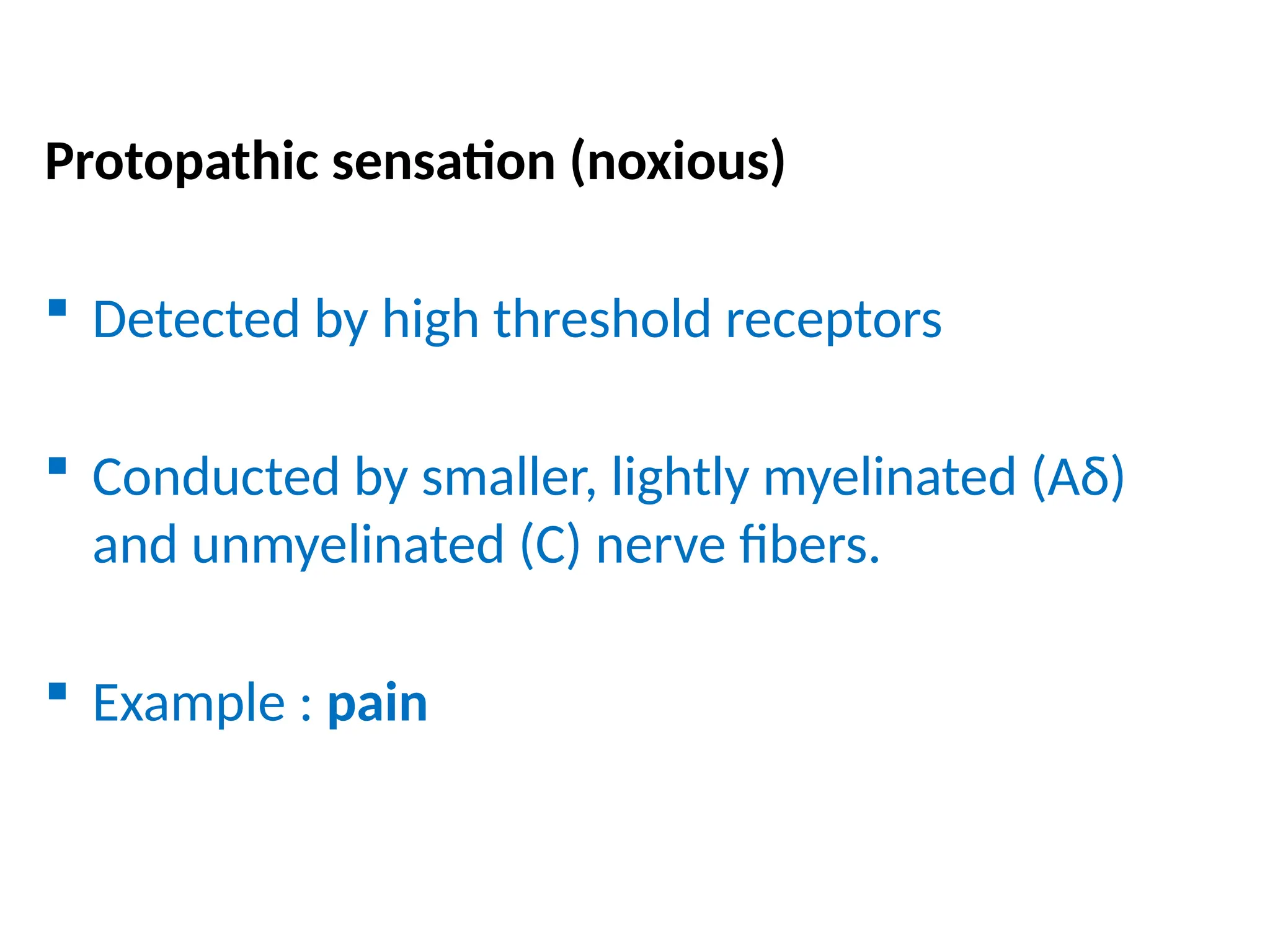
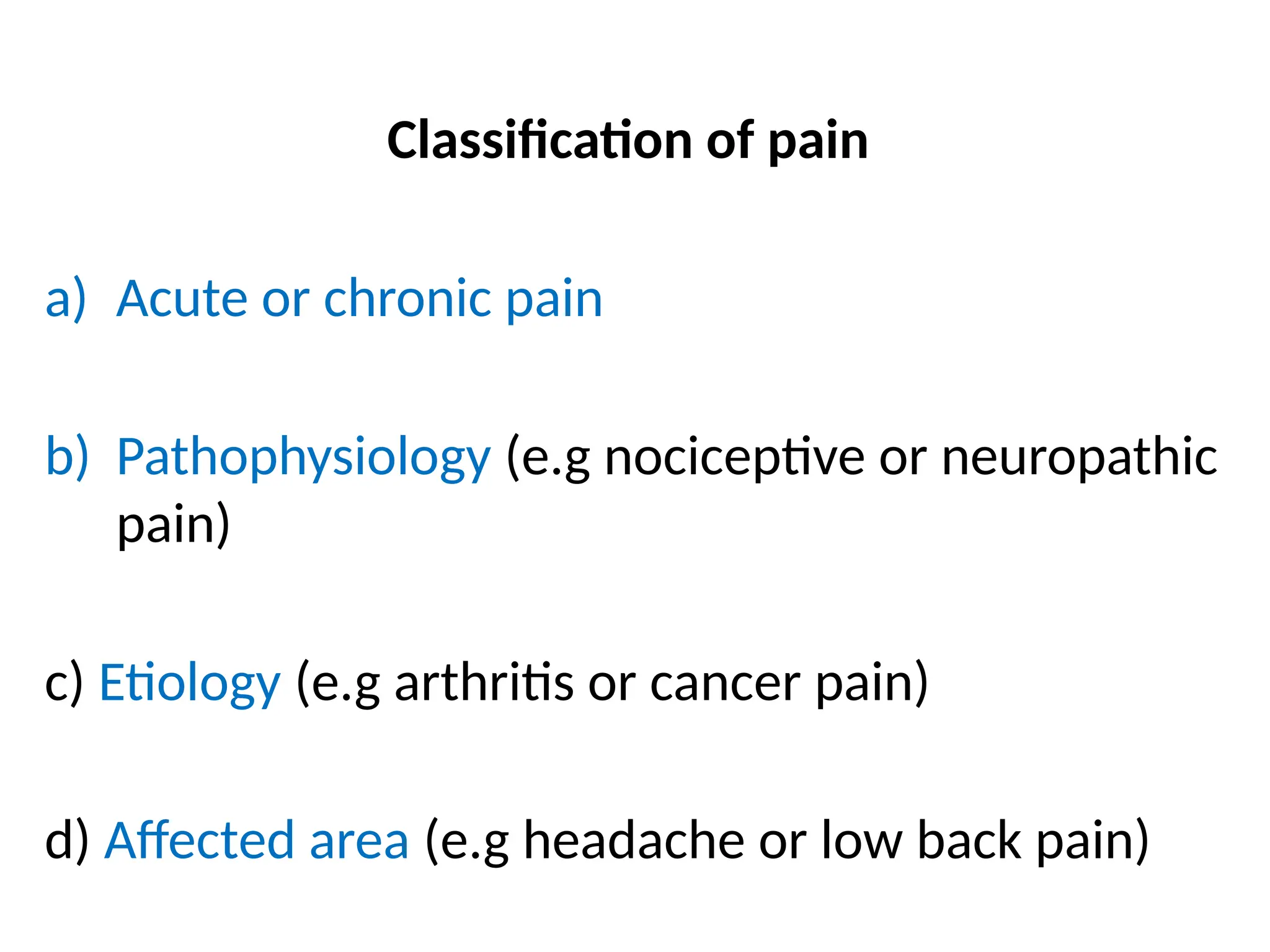
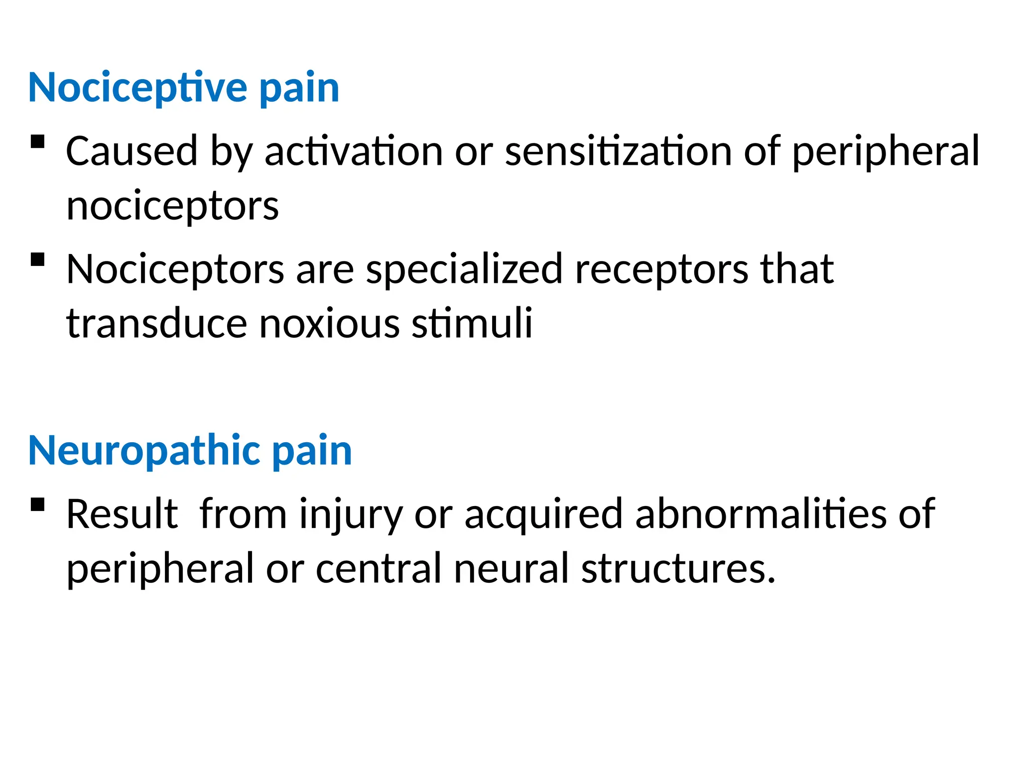
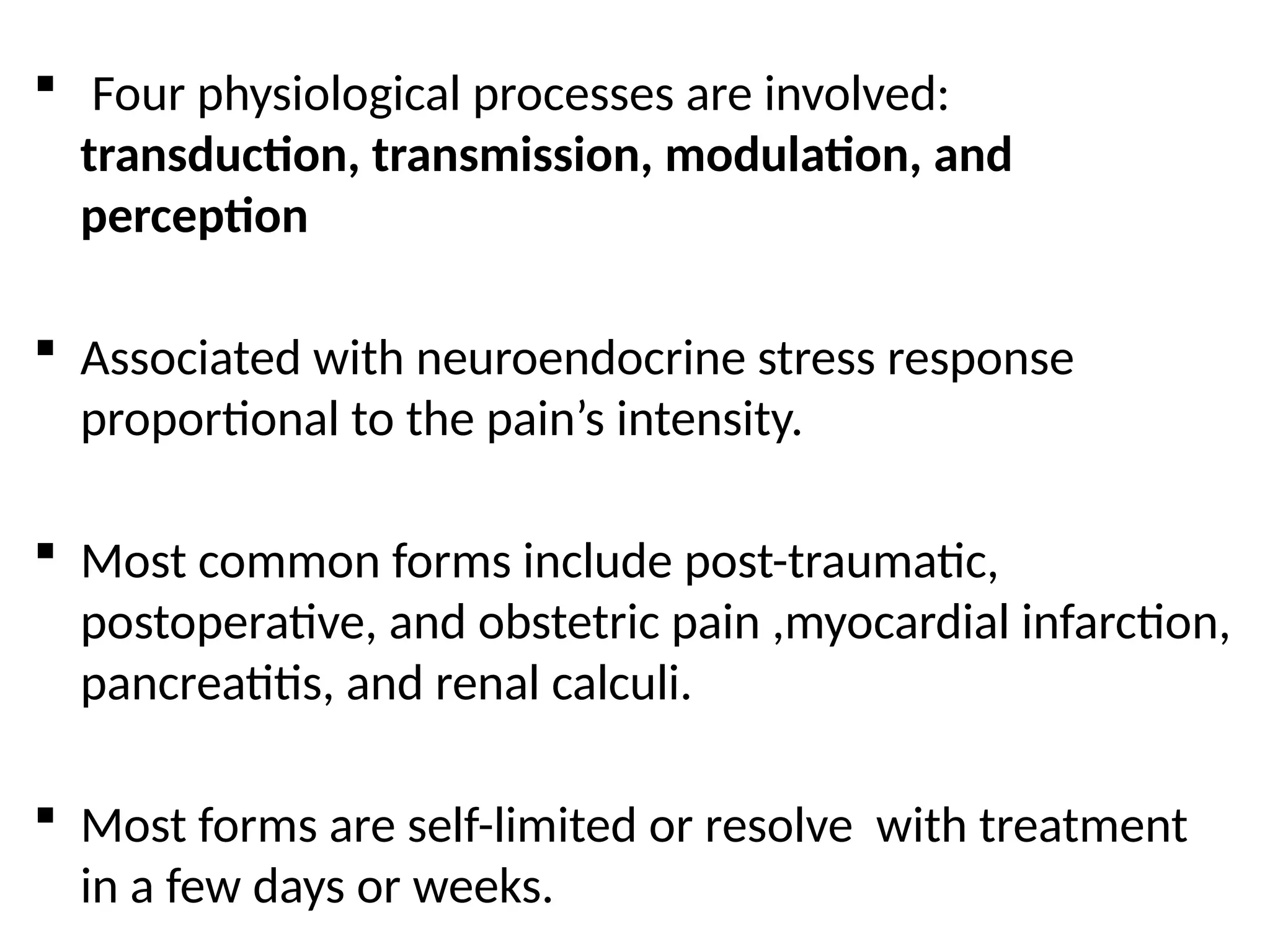
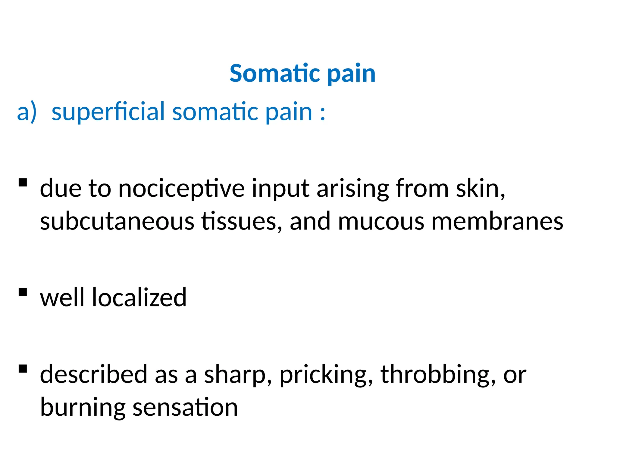
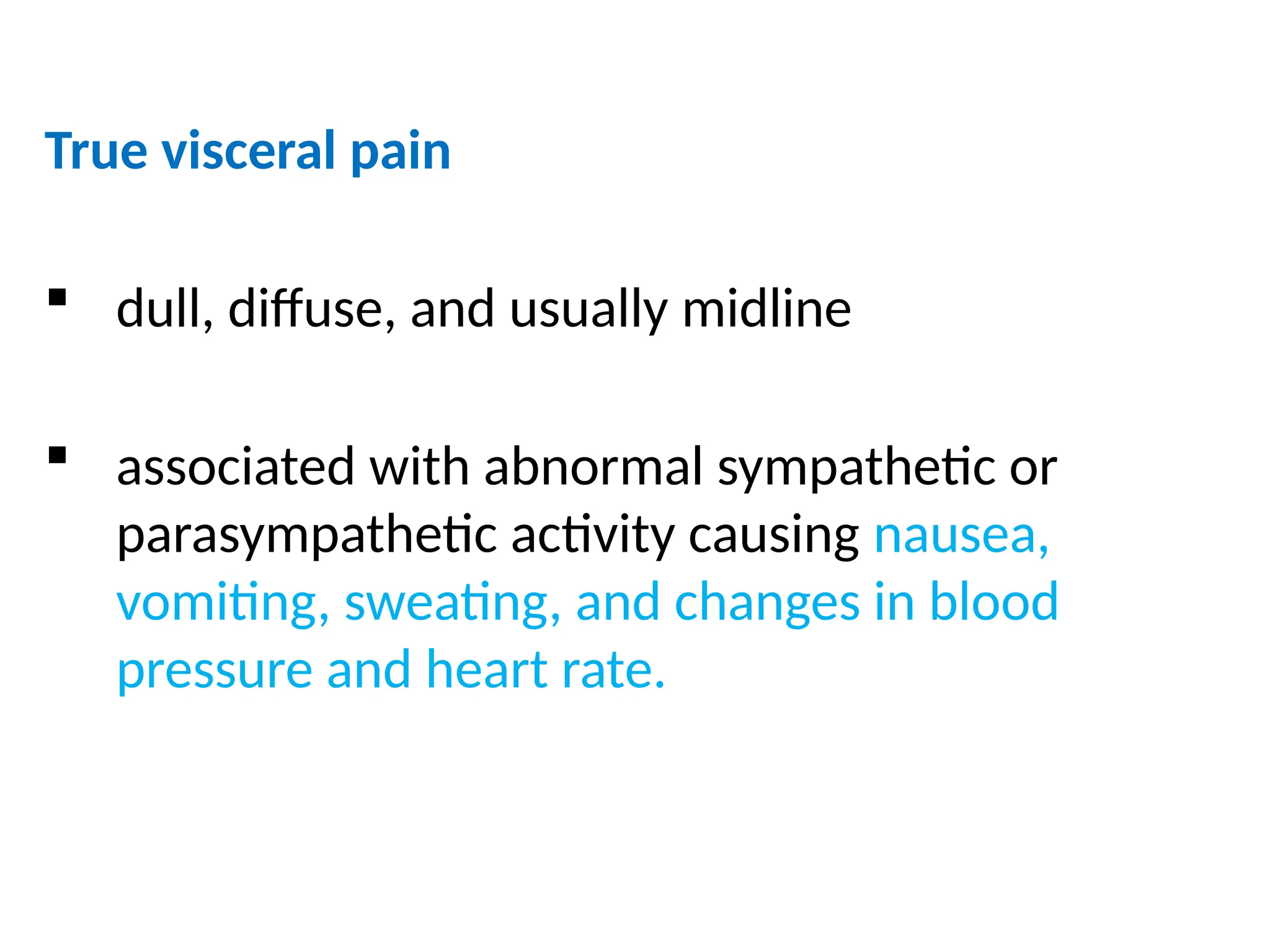
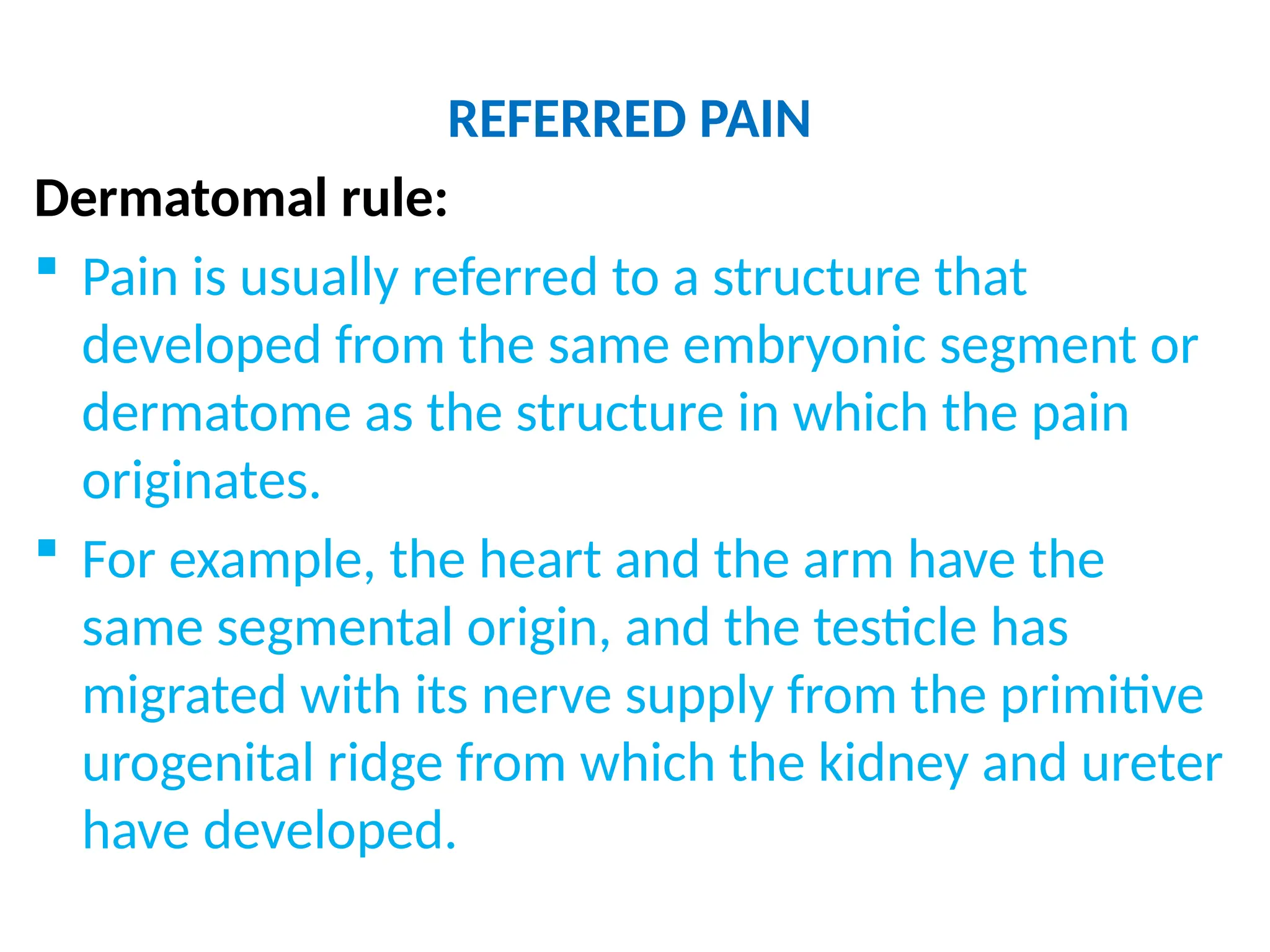
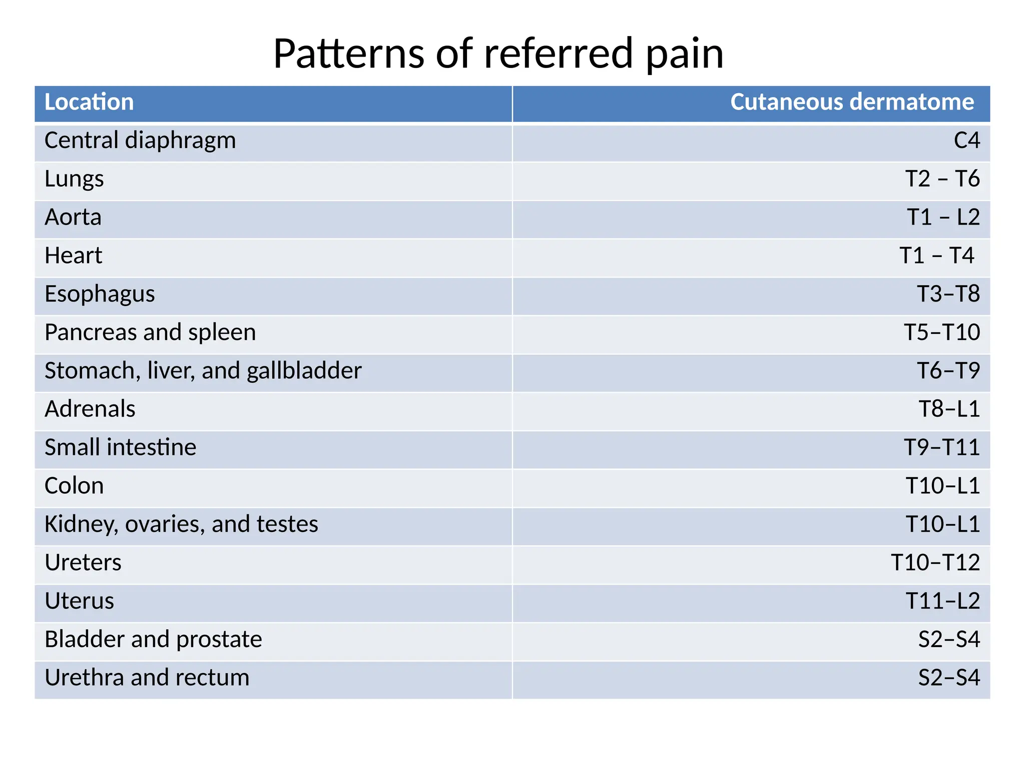
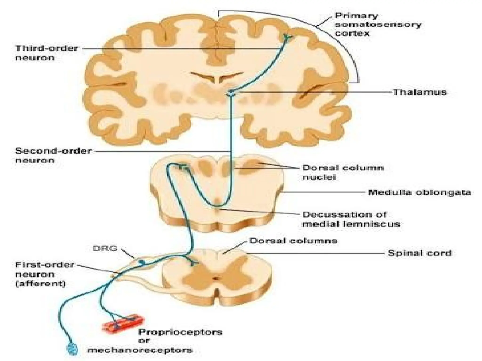
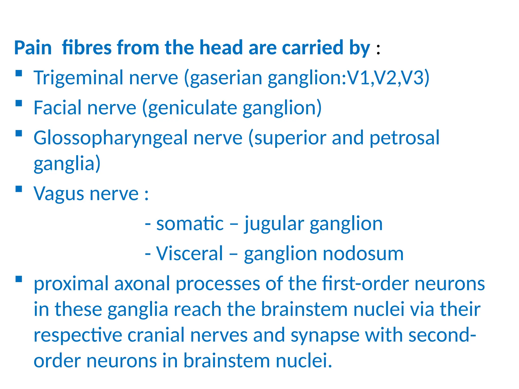
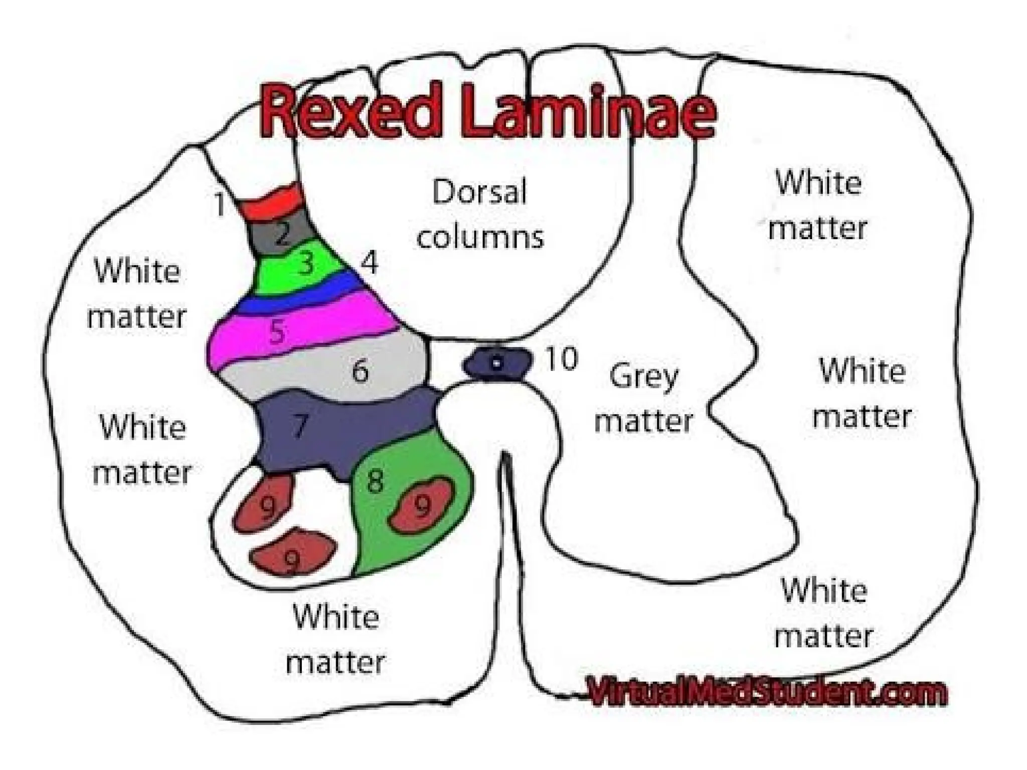
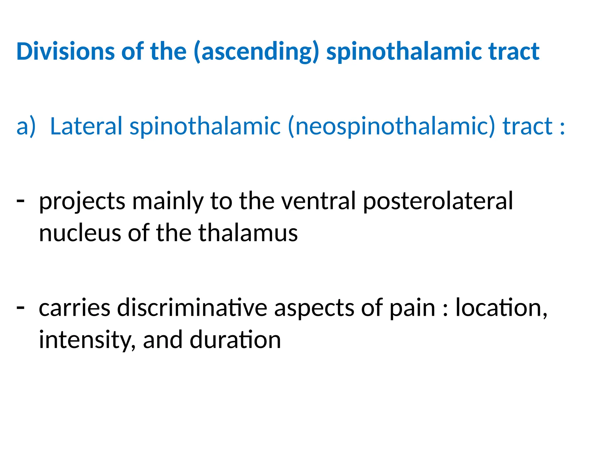
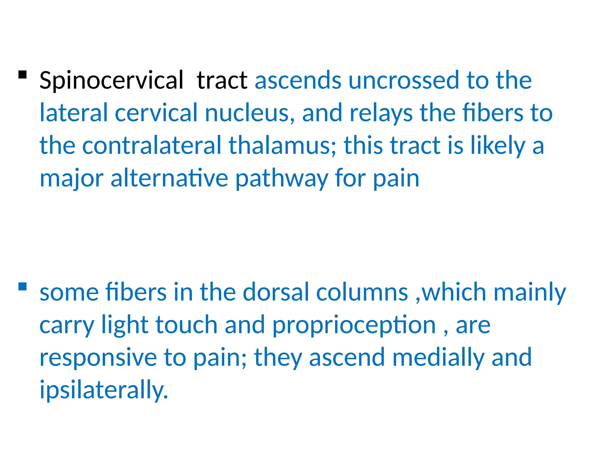
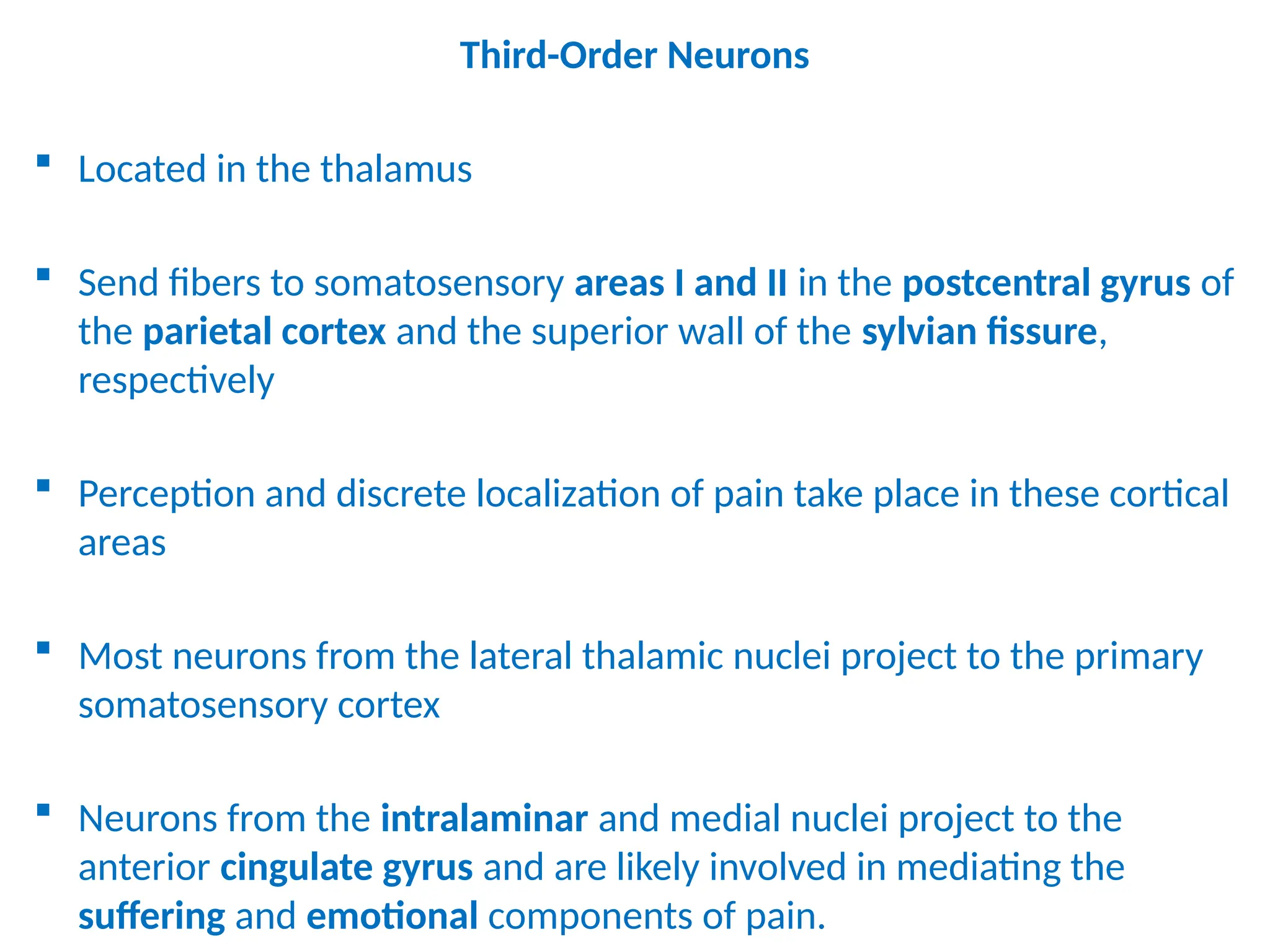
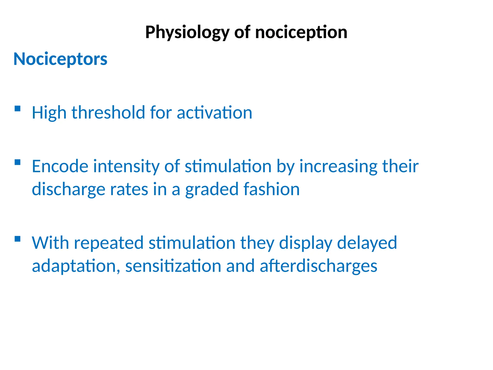
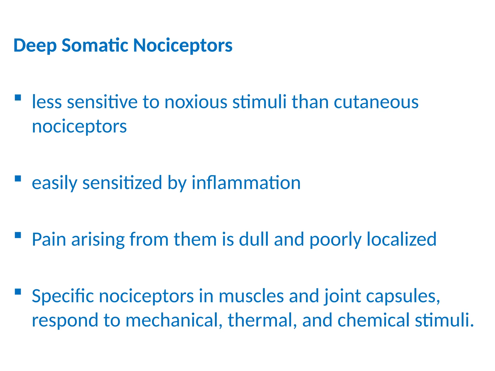
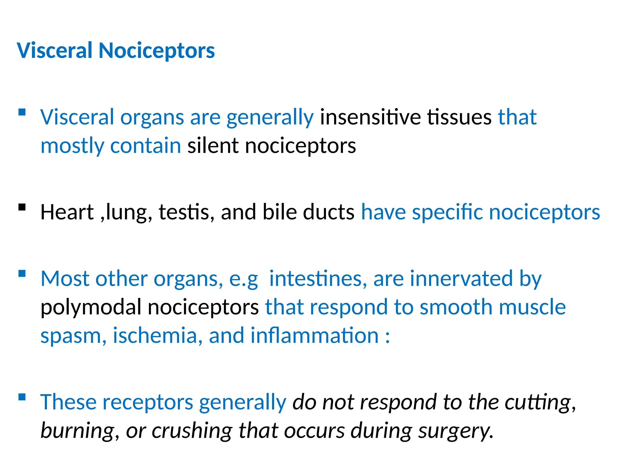
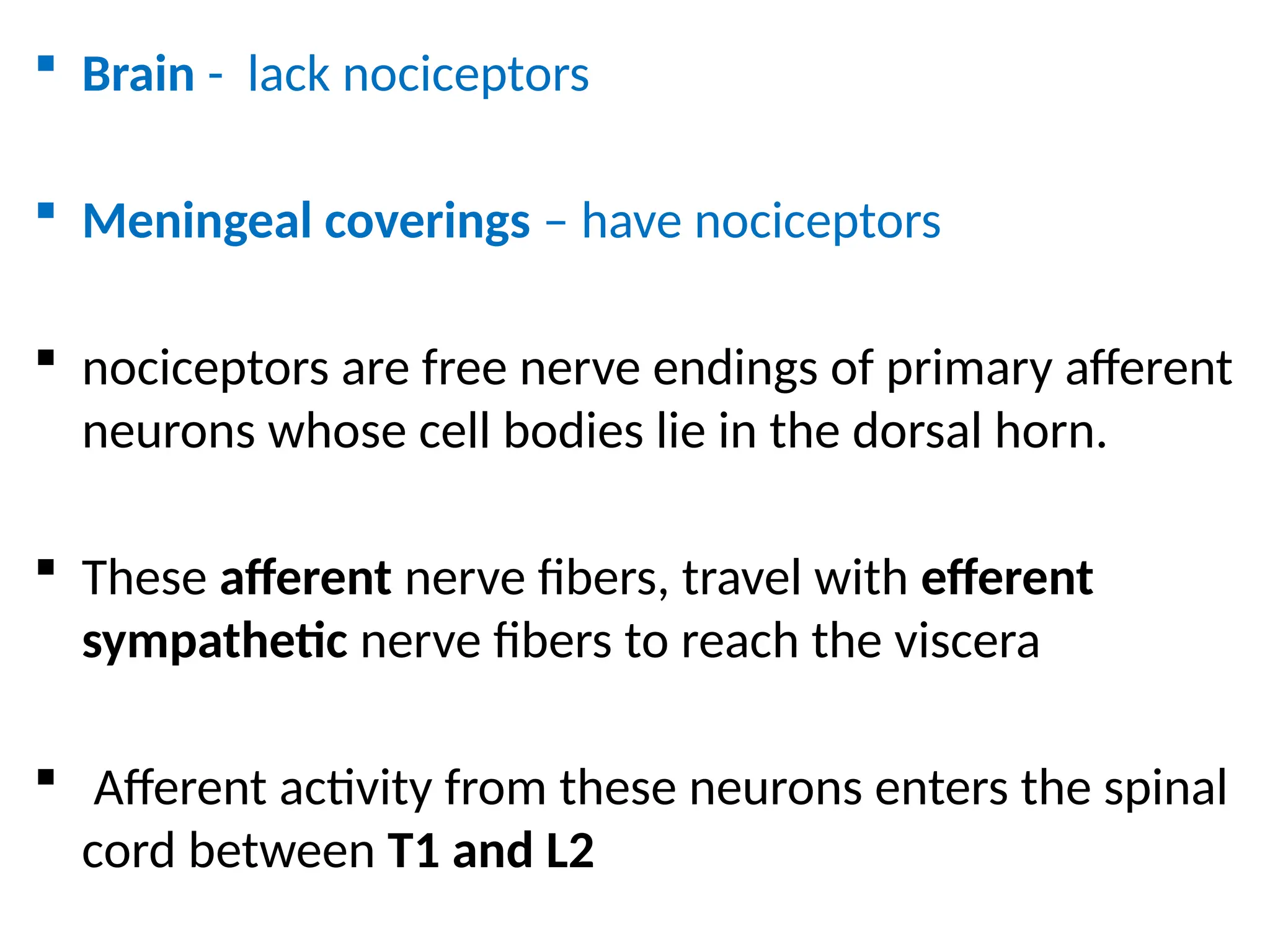
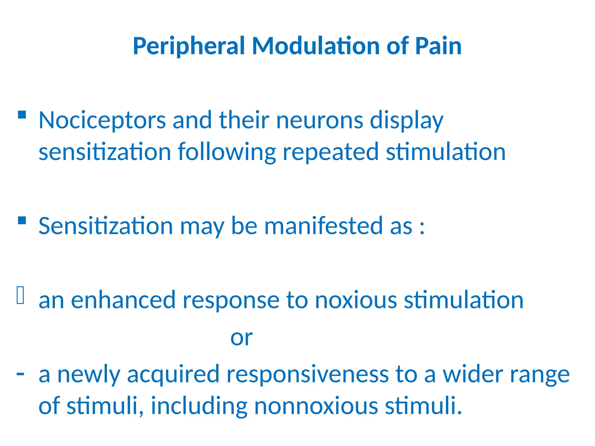
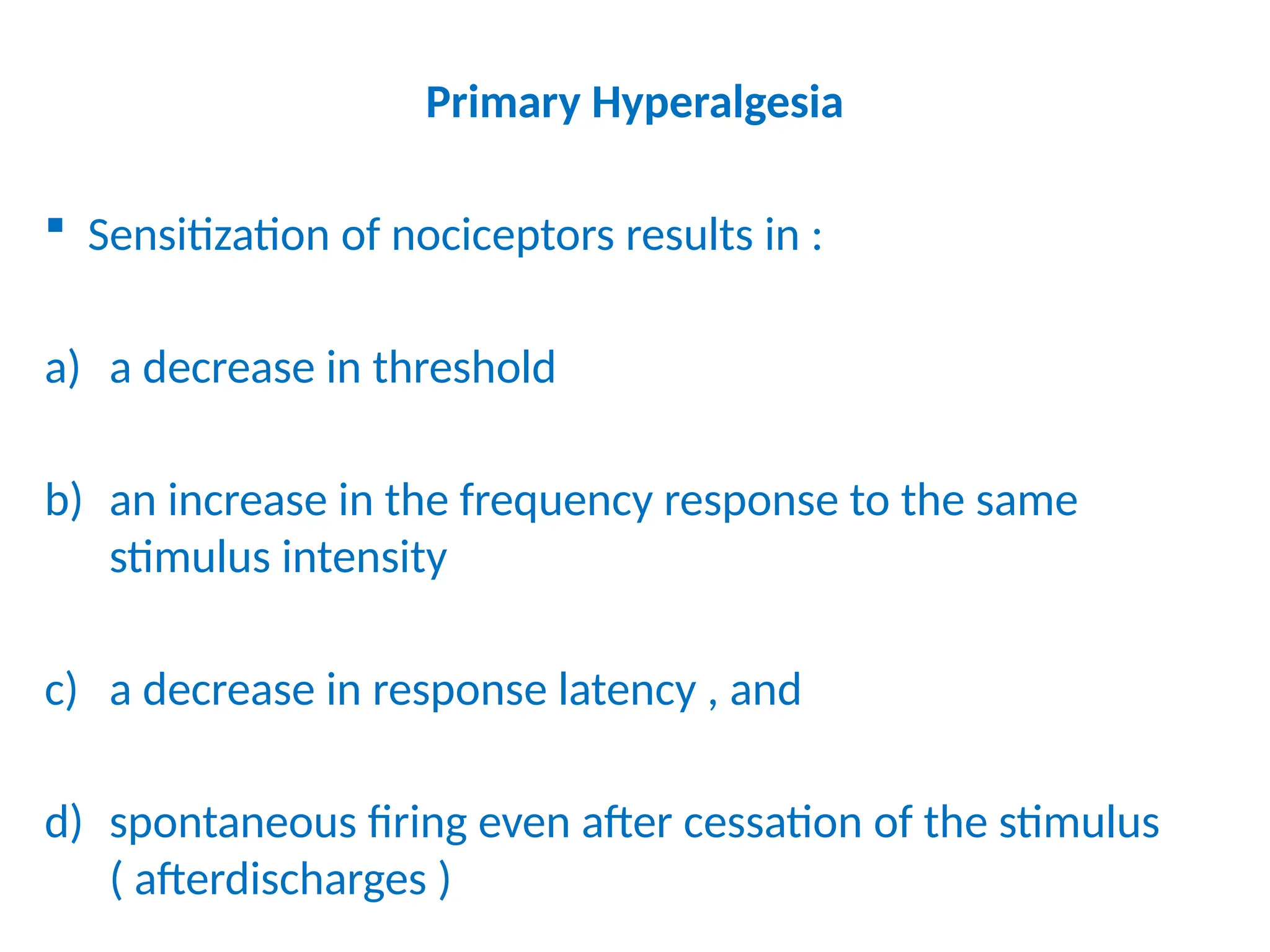
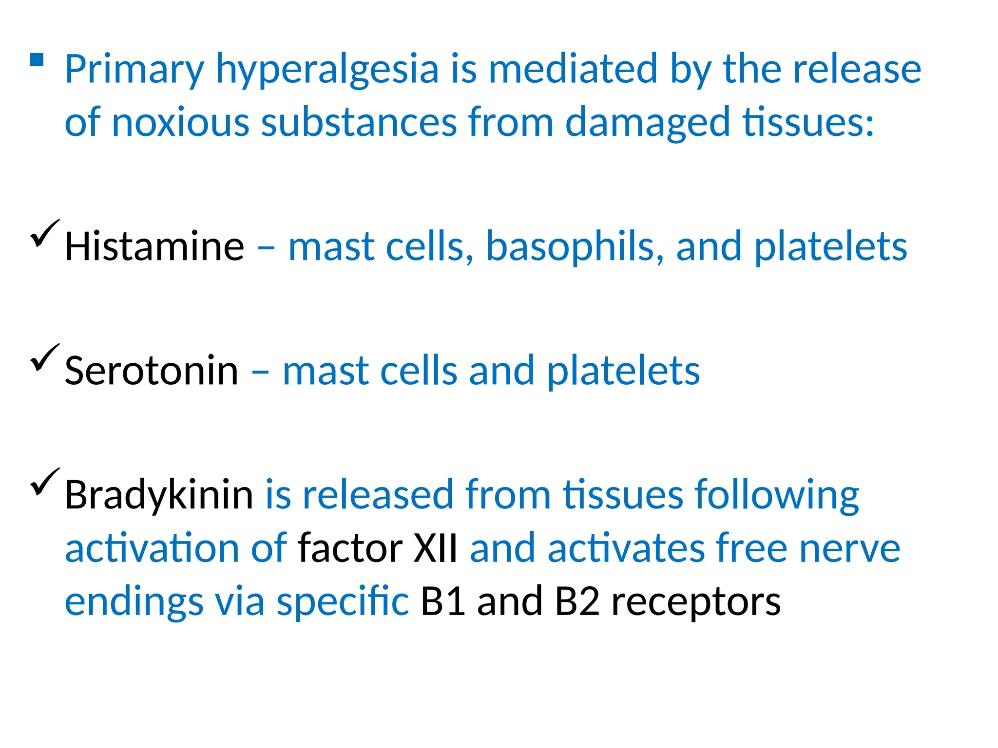
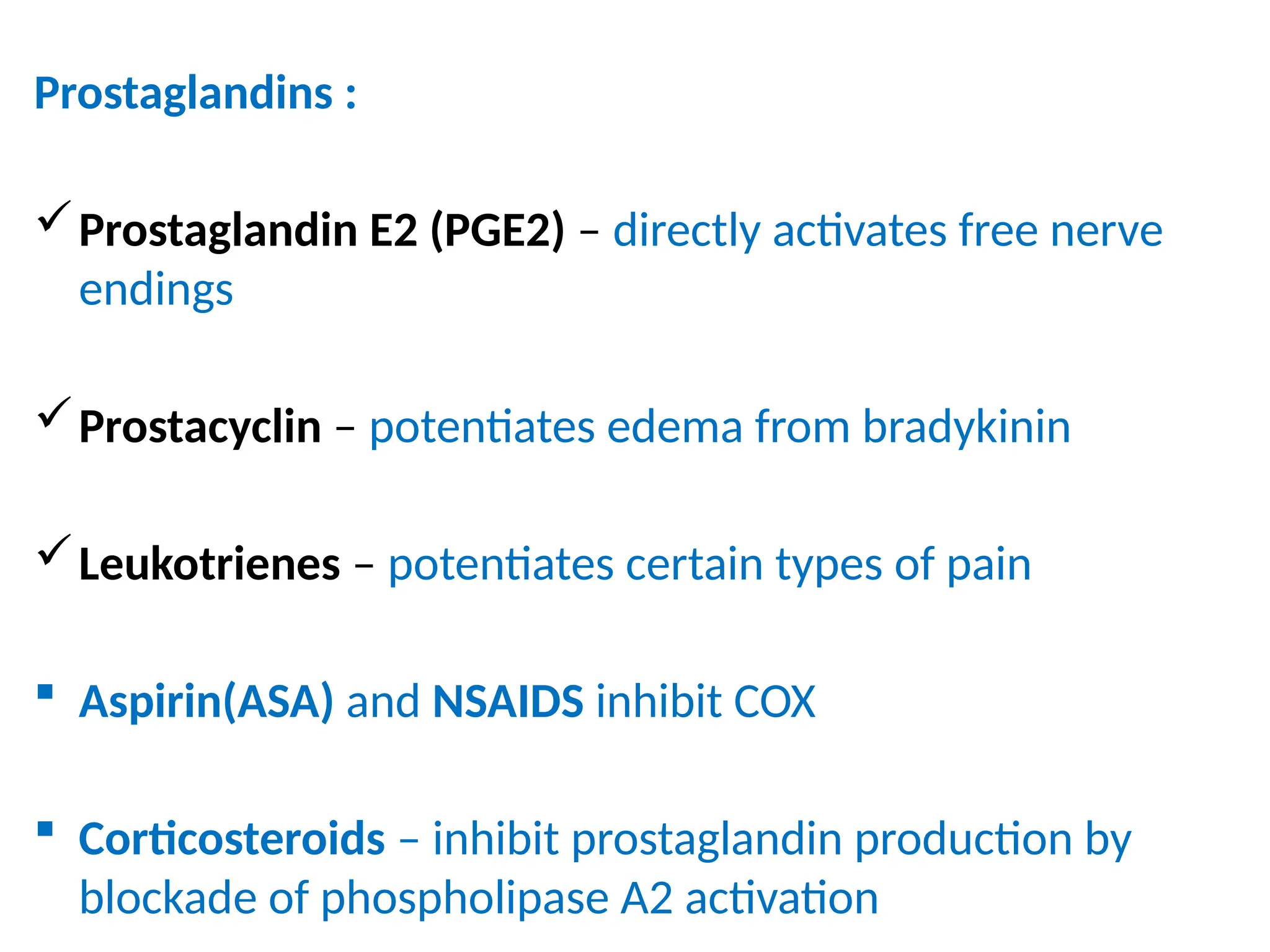
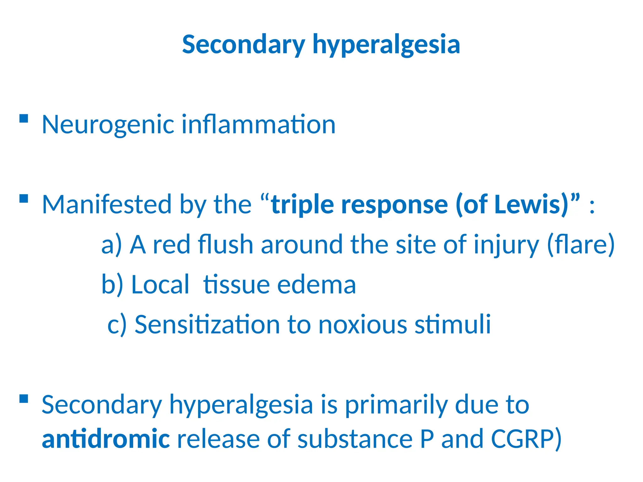
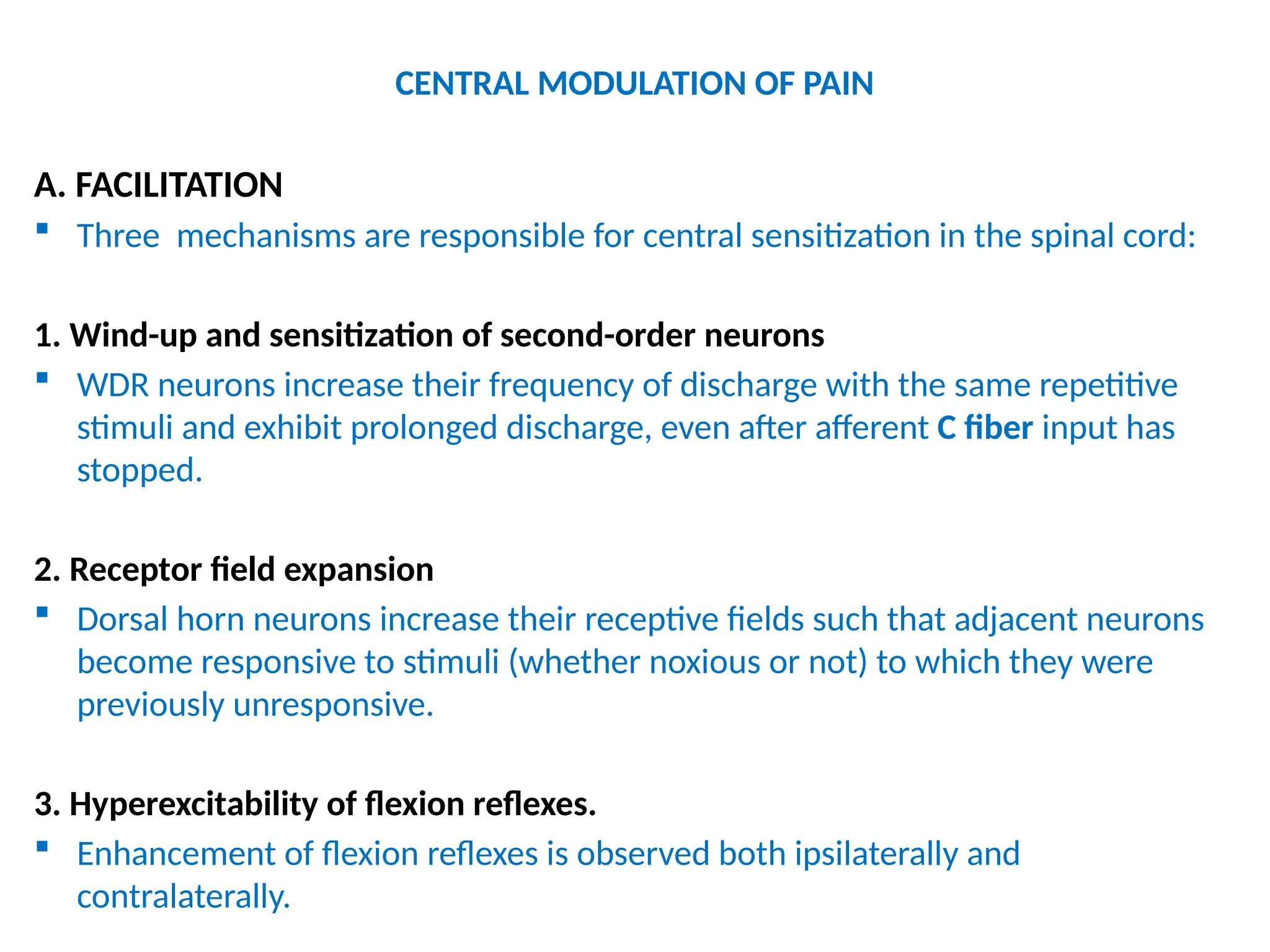
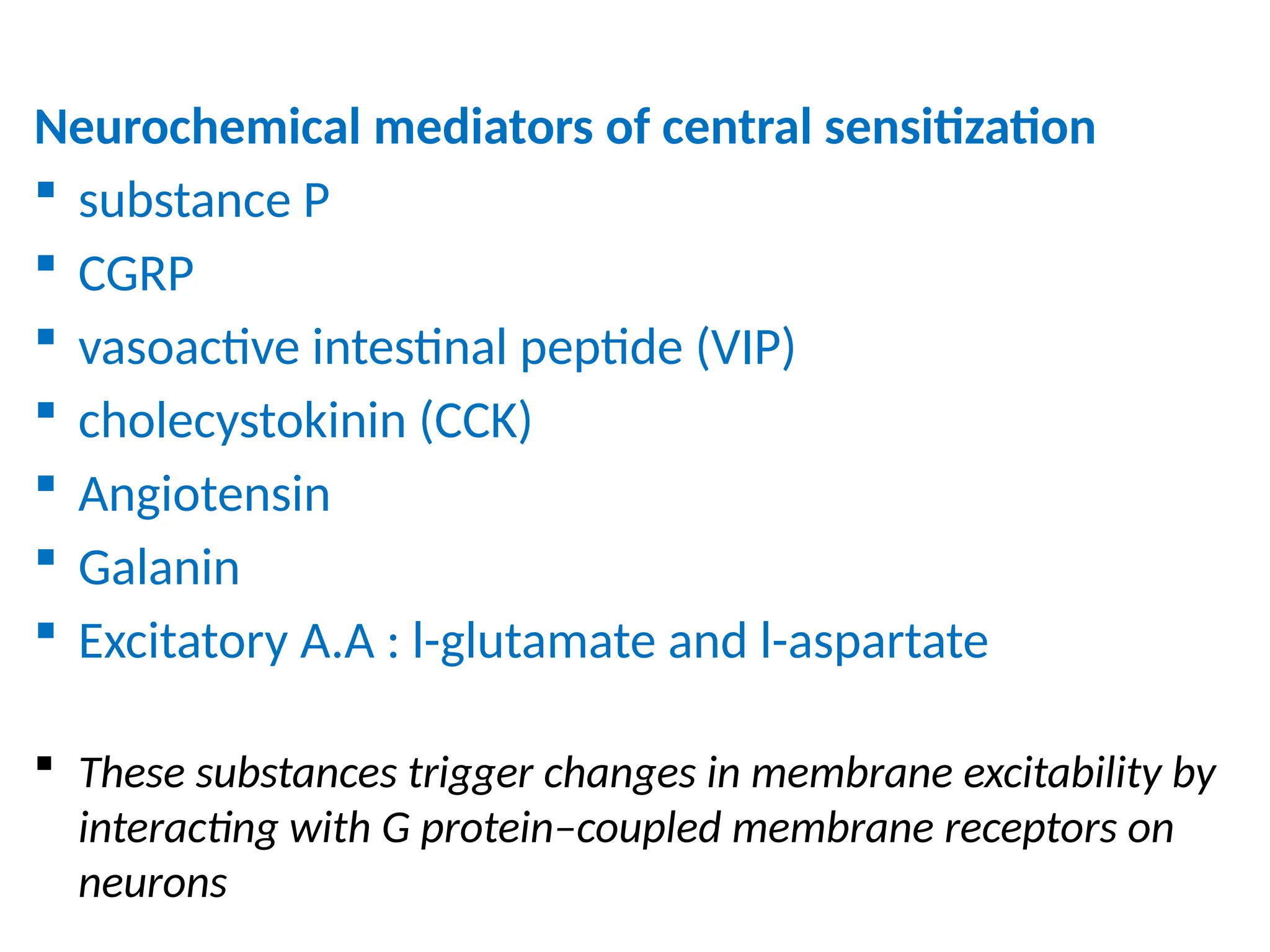
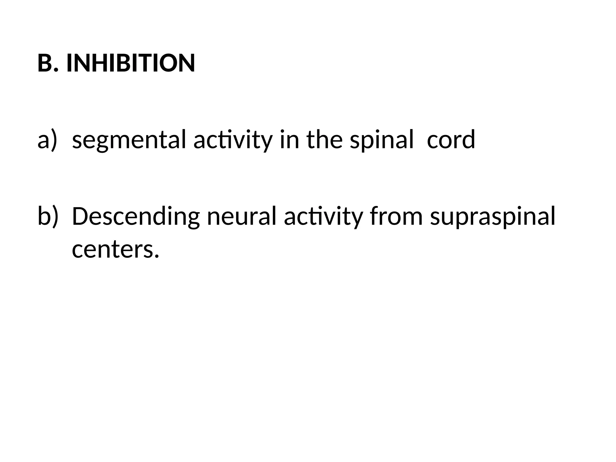
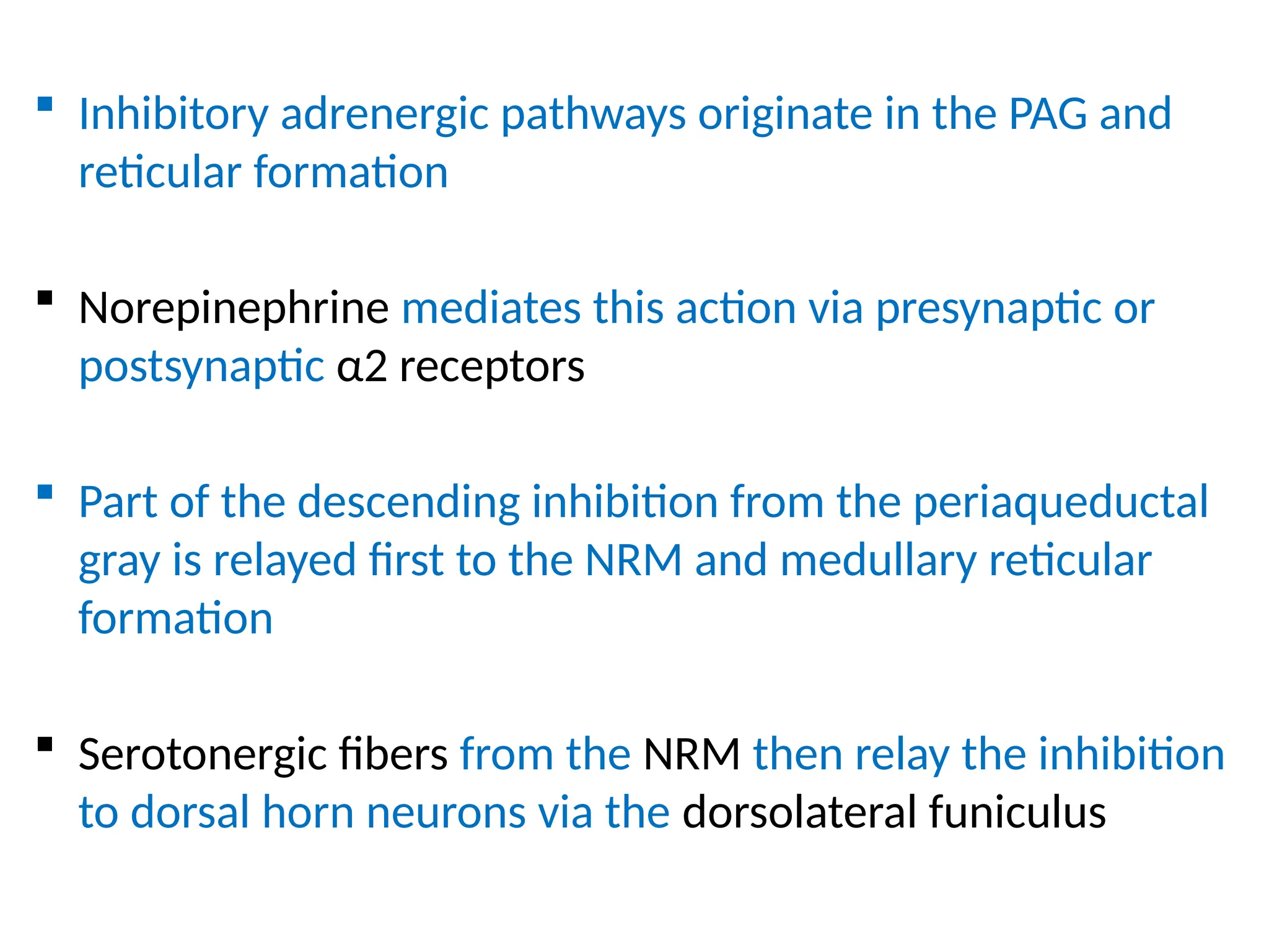
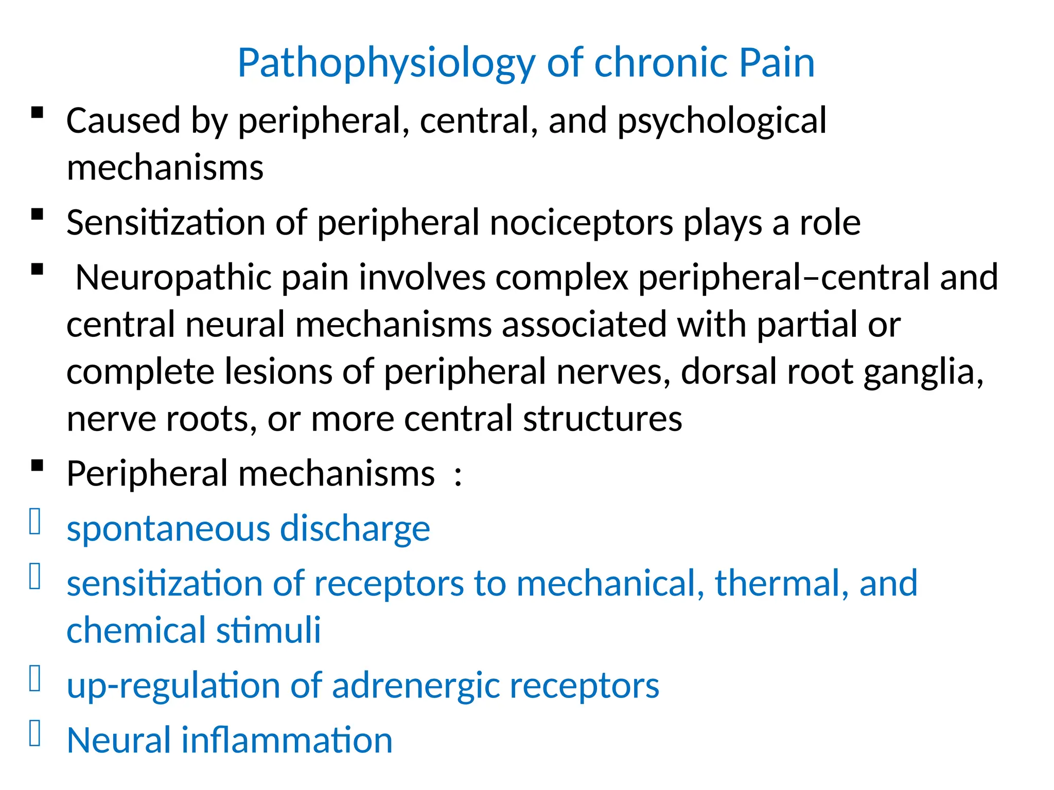
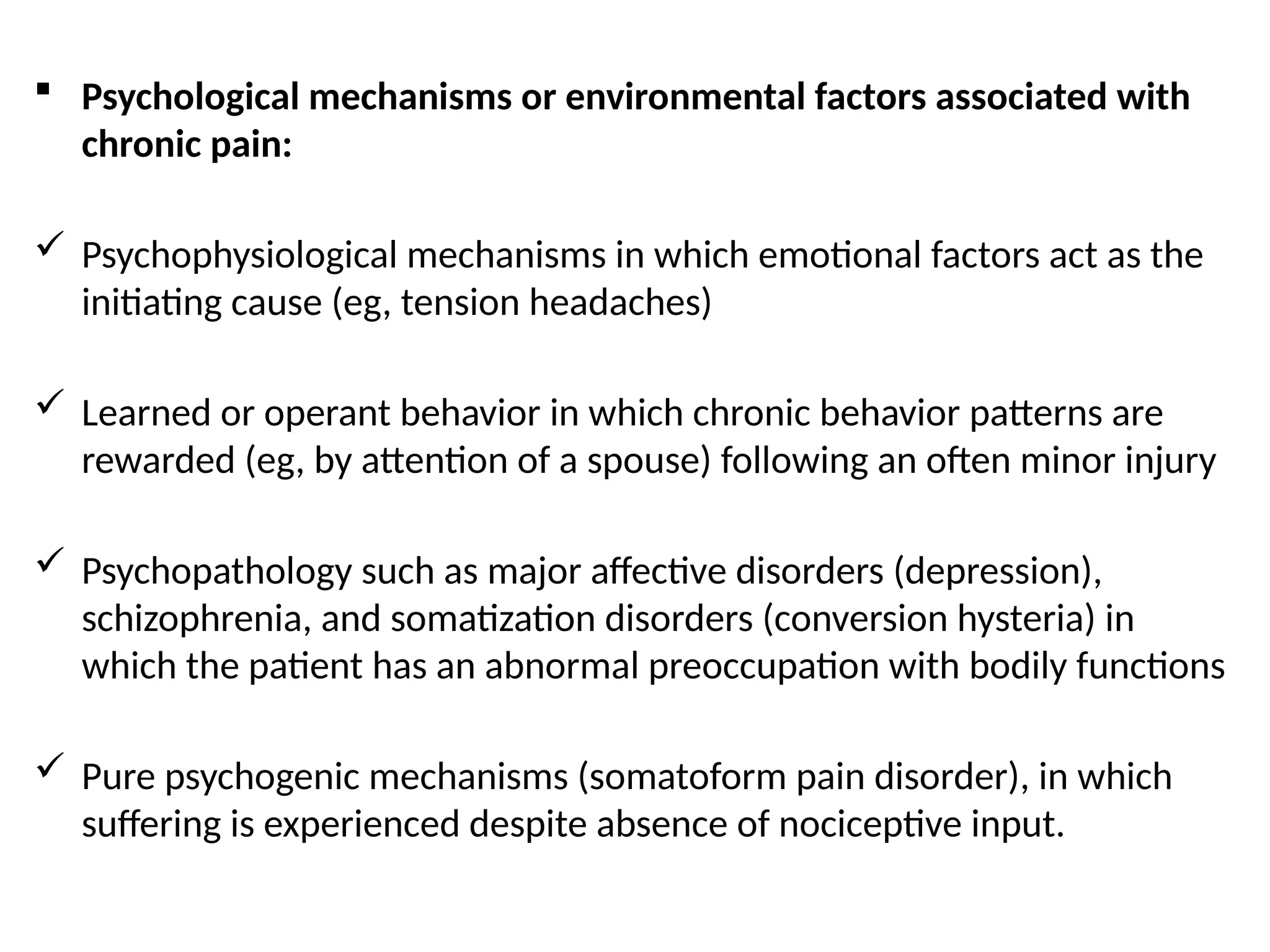
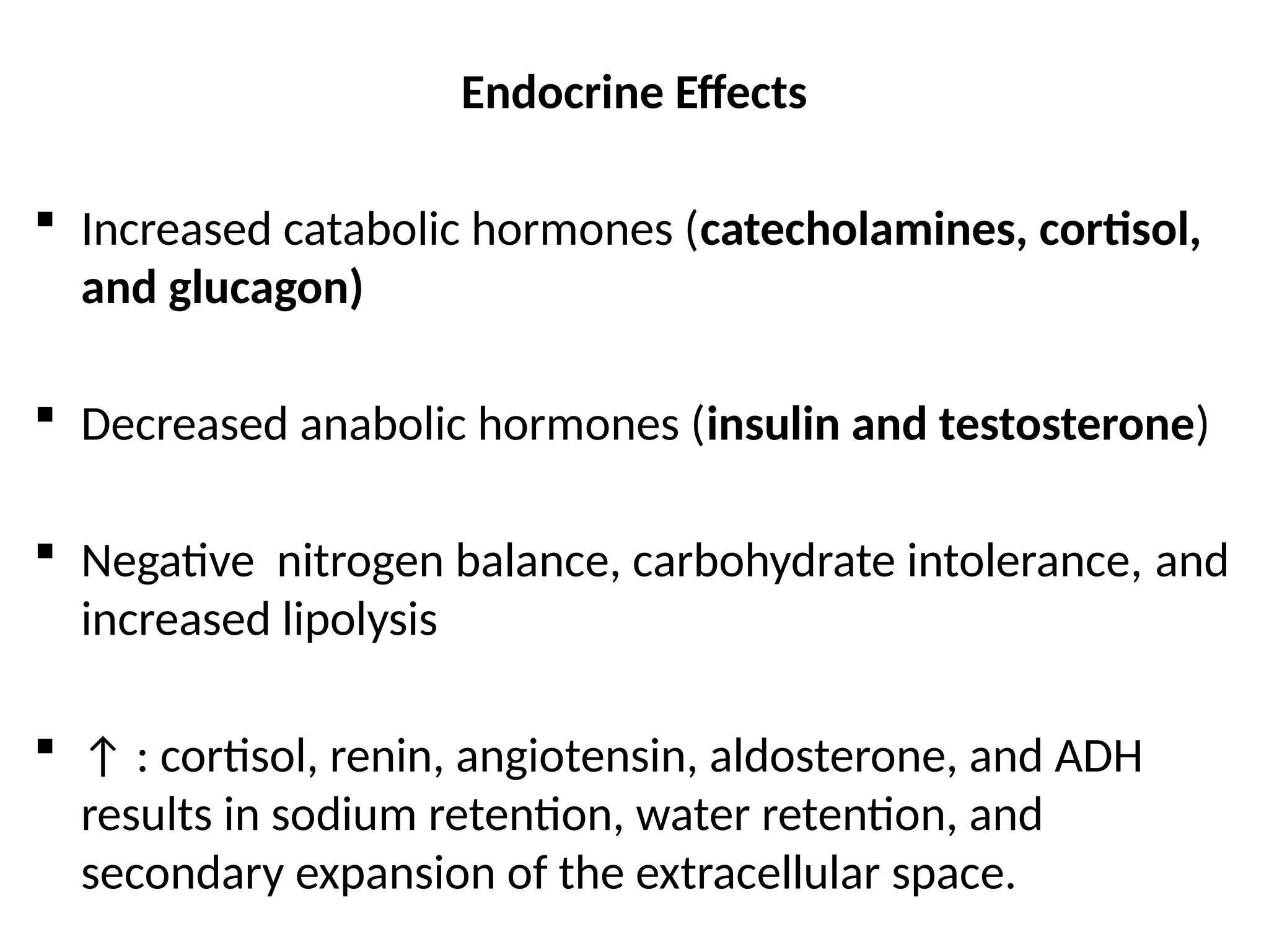
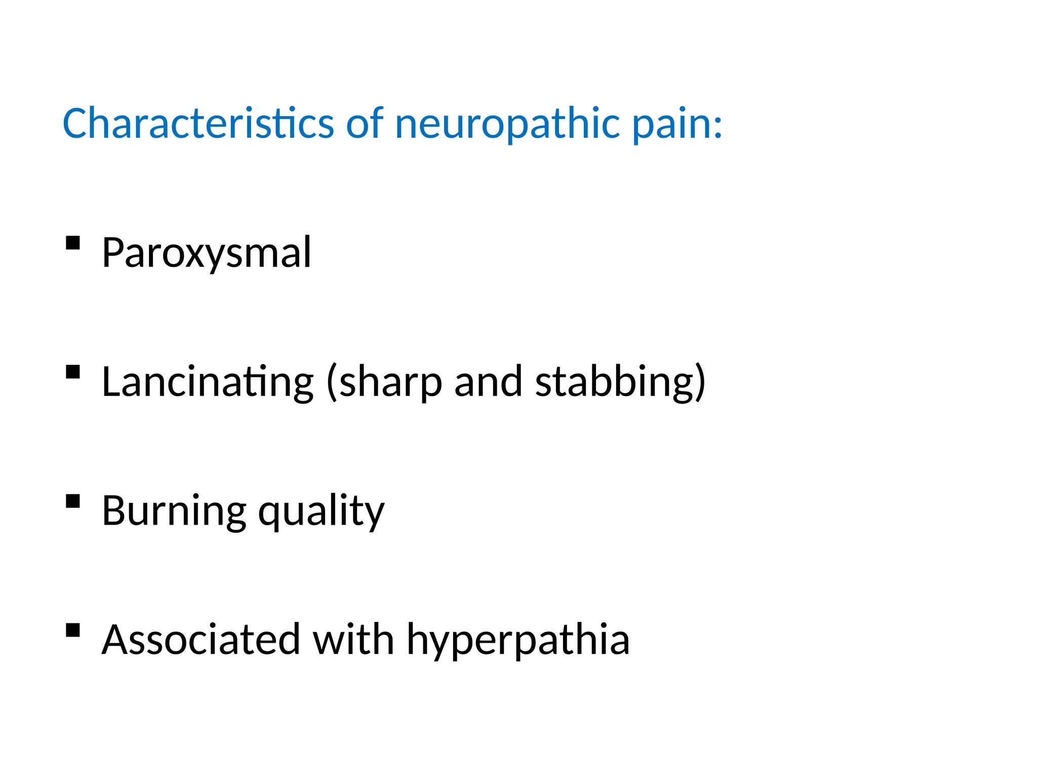
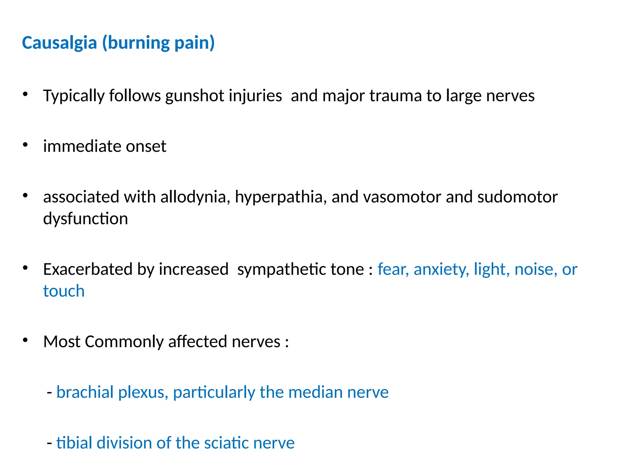
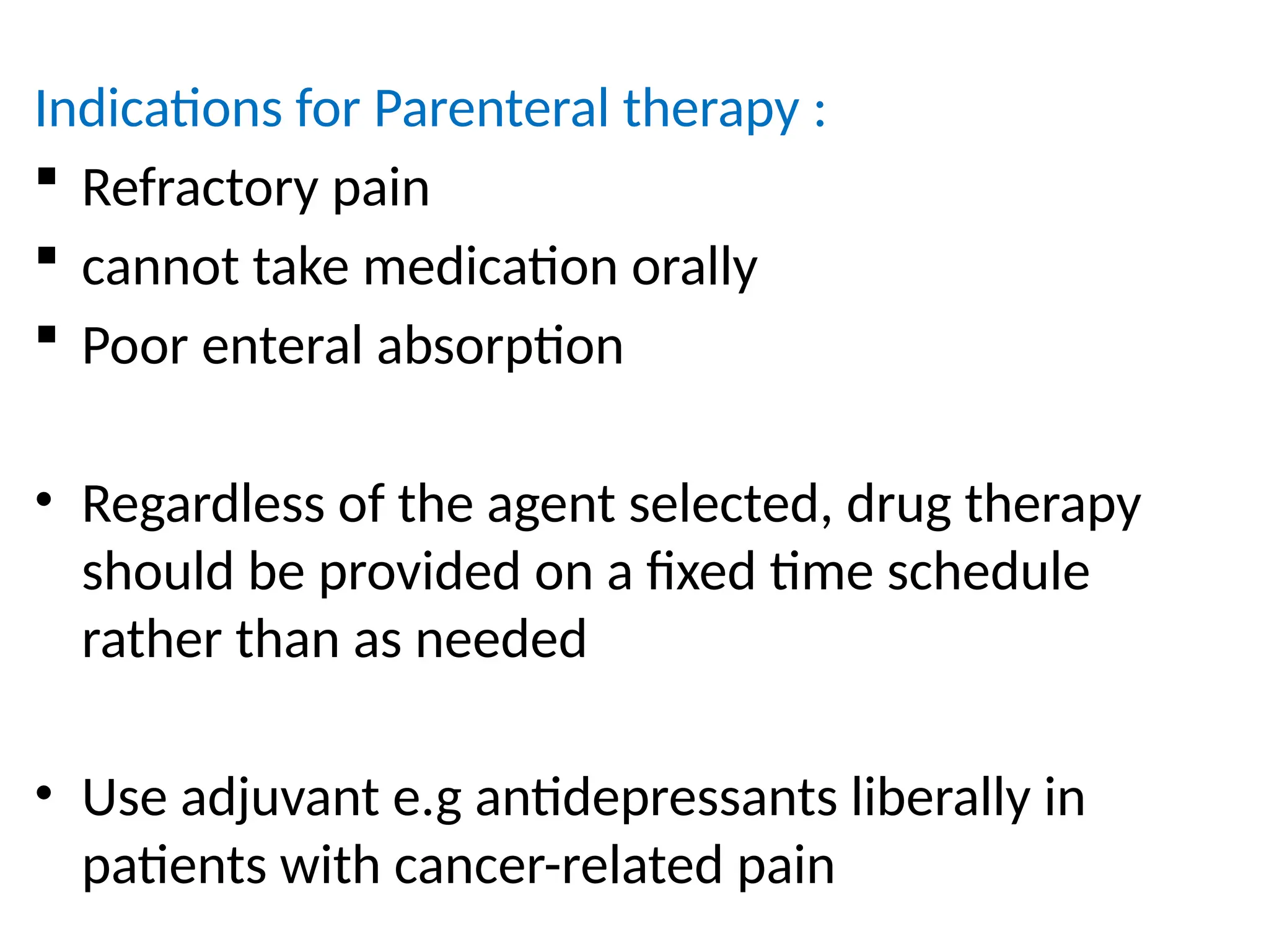
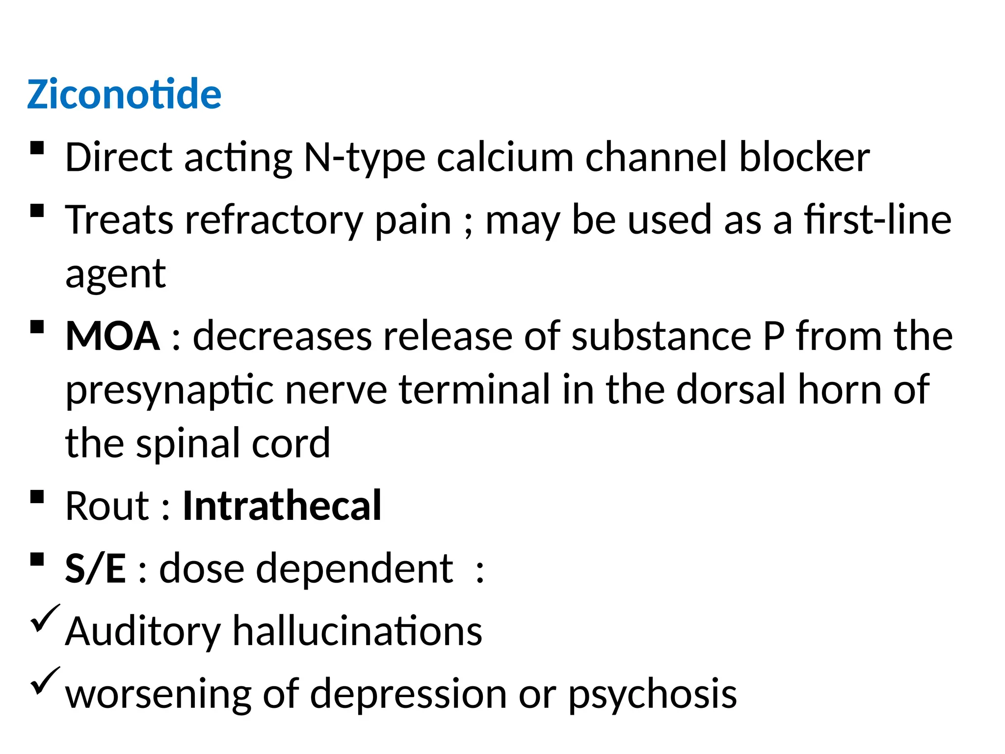
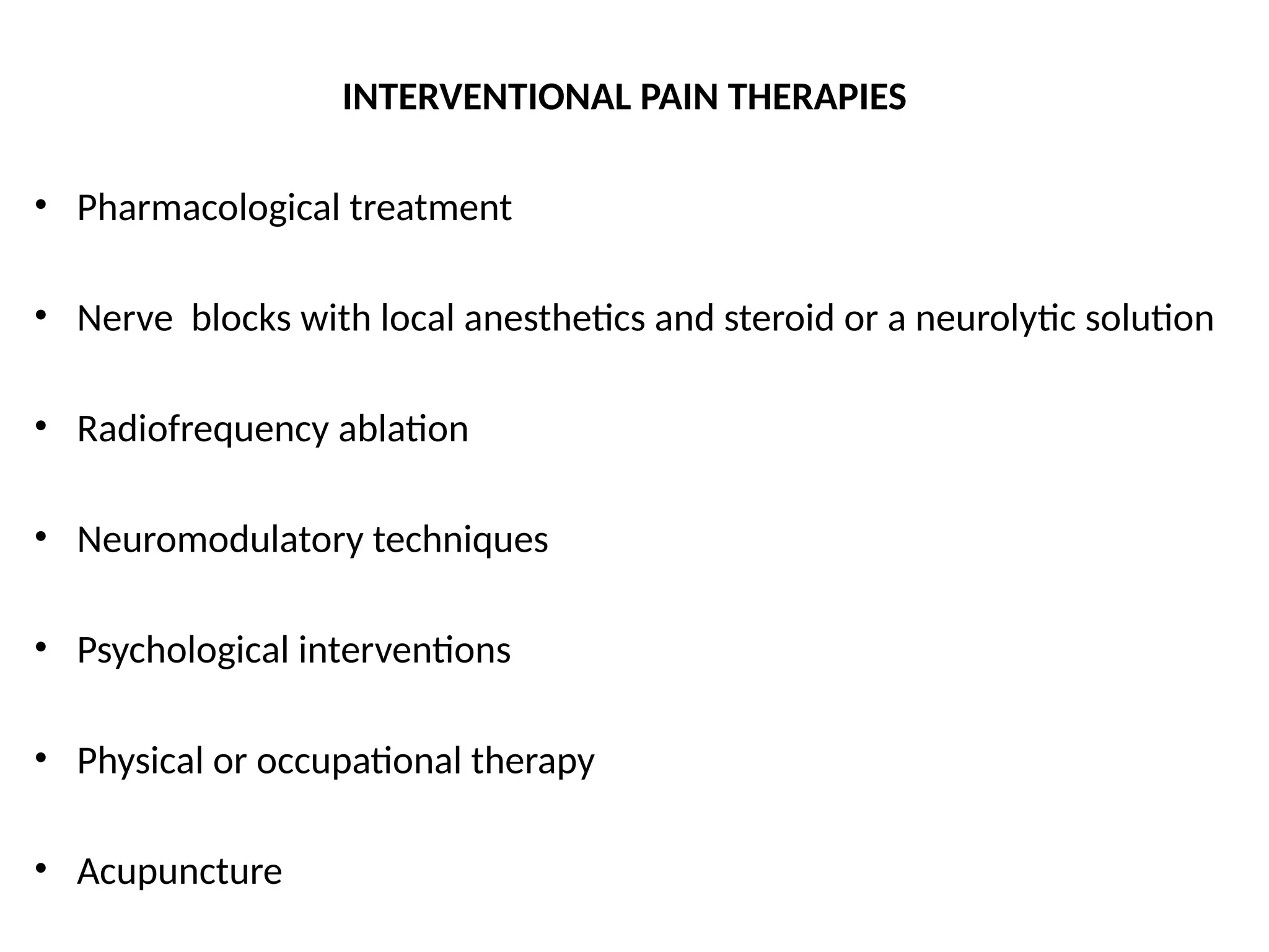
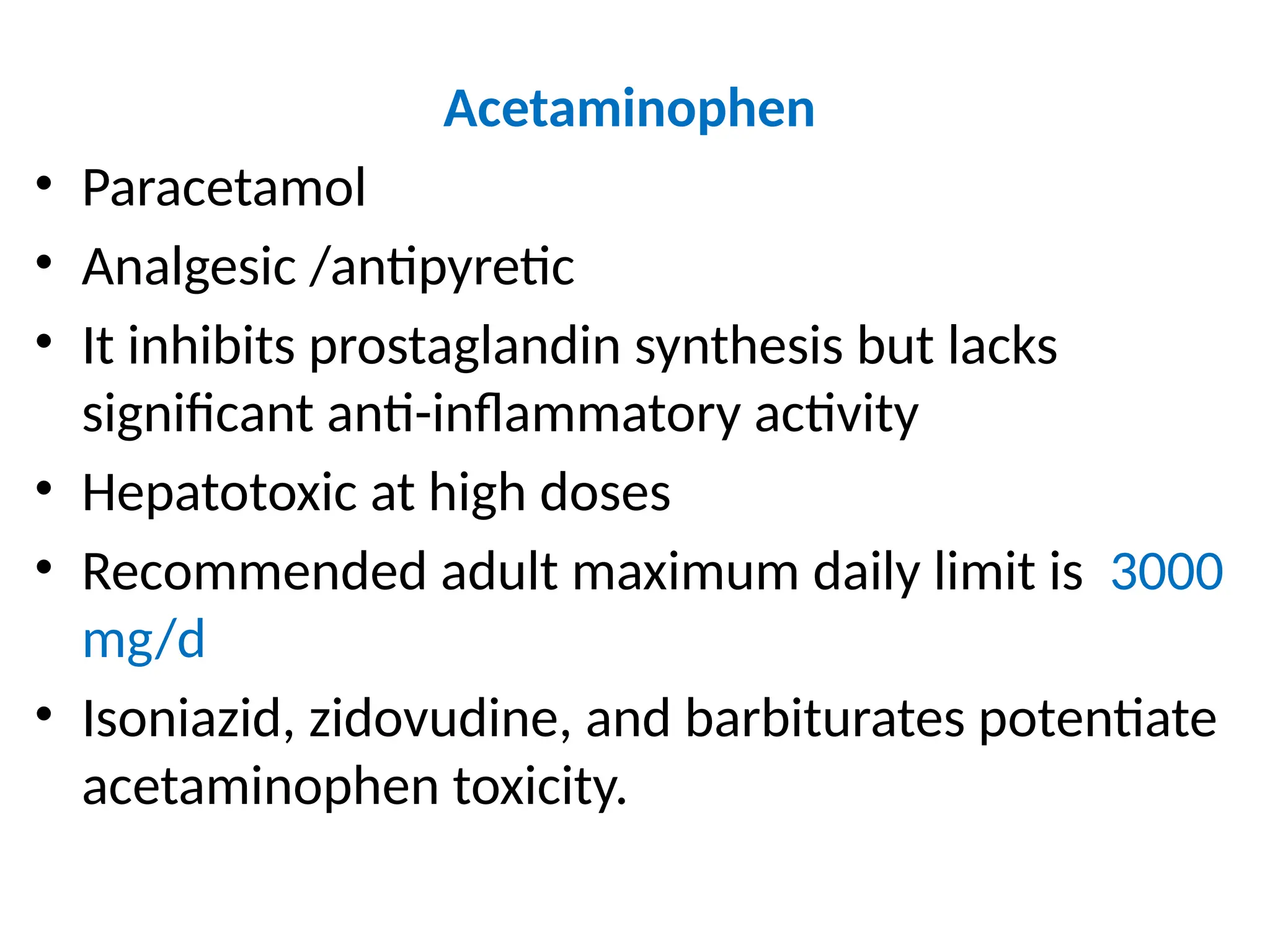
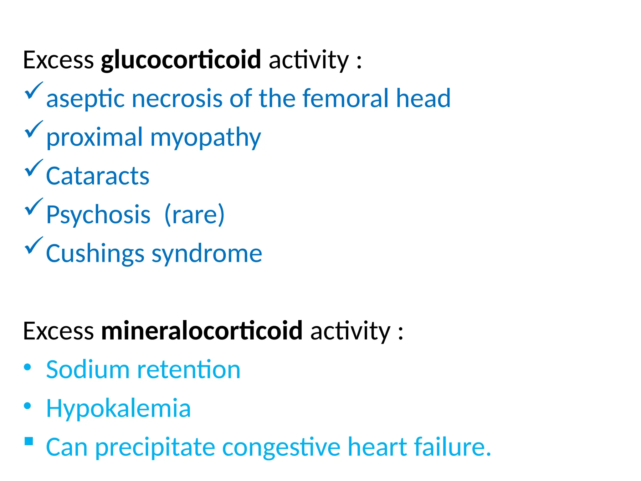
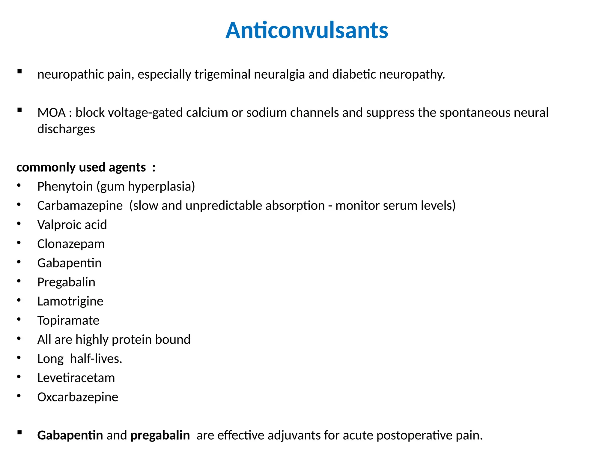
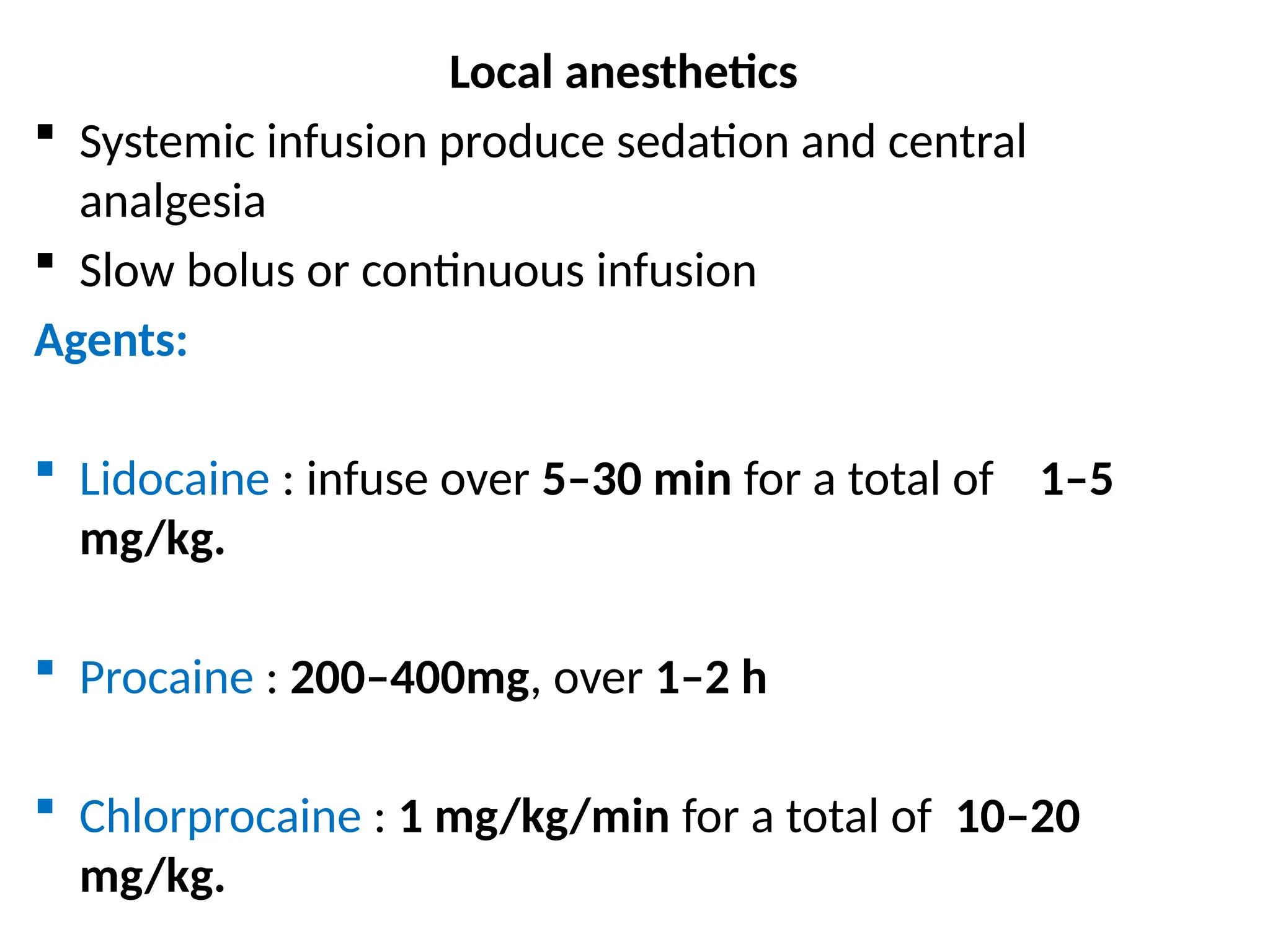
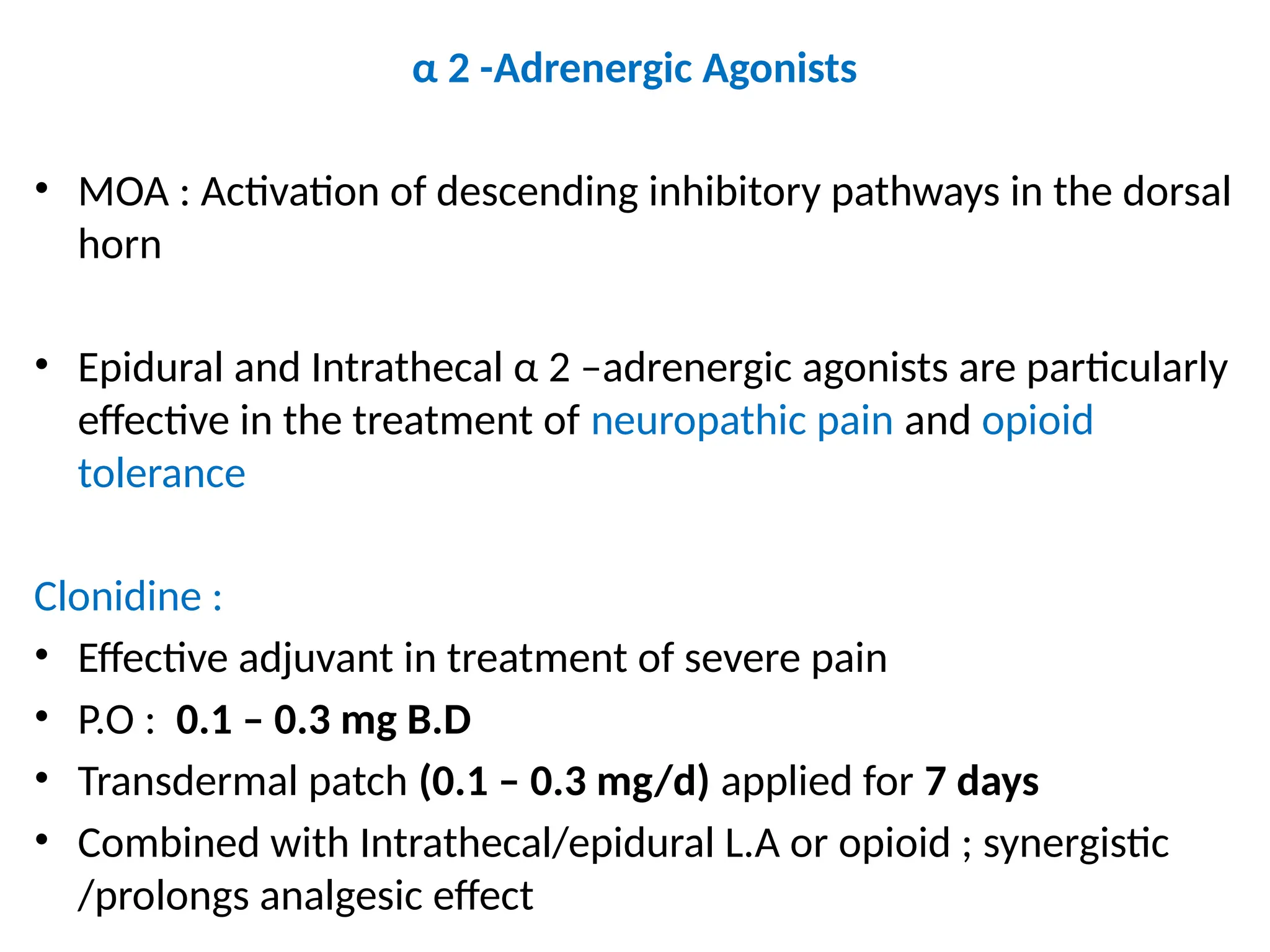
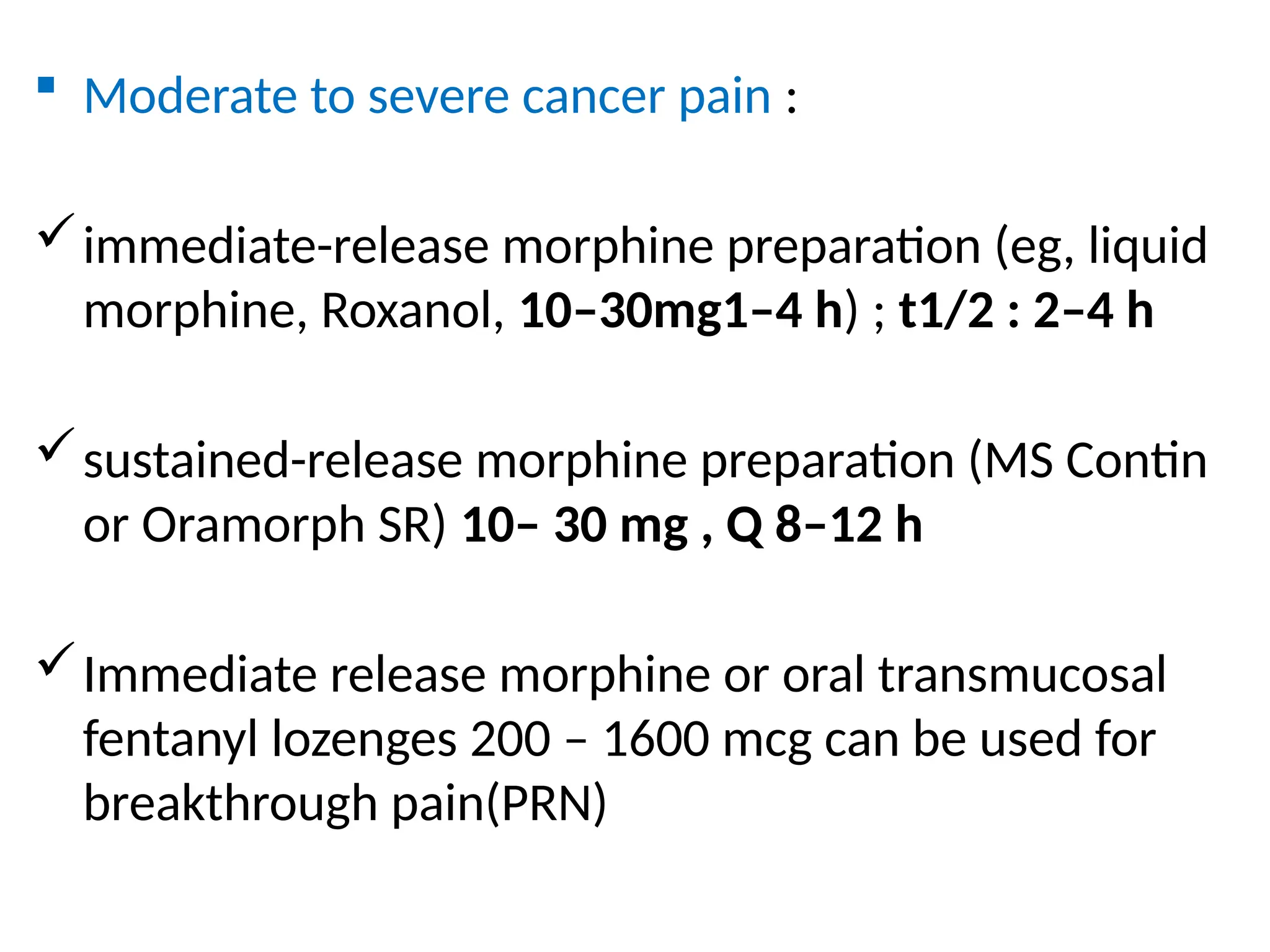
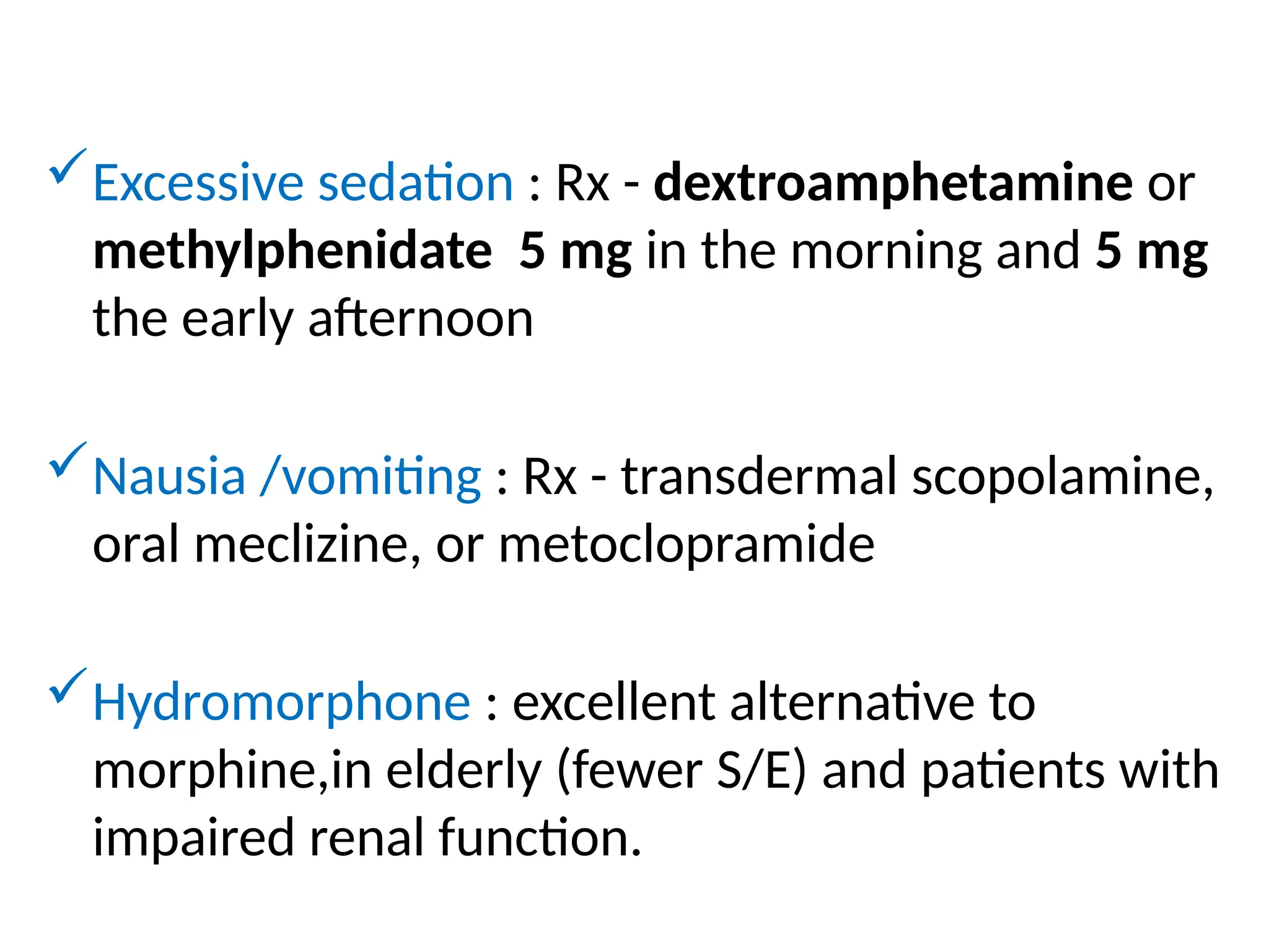
CHRONIC PAIN MANAGEMENT
MONGERA MWAMBA
Sensation :
 Protopathic (noxious e.g pain)
 Epicritic (non-noxious e.g temperature )
Epicritic sensations
 Characterized by low threshold receptors
 Conducted by large myelinated nerve fibres
 Examples :
- light touch
- pressure
- proprioception
- temperature discrimination
Protopathic sensation (noxious)
 Detected by high threshold receptors
 Conducted by smaller, lightly myelinated (Aδ)
and unmyelinated (C) nerve fibers.
 Example : pain
Pain
 an unpleasant sensory and emotional
experience associated with actual or potential
tissue damage, or described in terms of such
damage.
Terms used in the management of pain
 Allodynia : Perception of an ordinarily nonnoxious
stimulus as pain (painful sensation from a warm
shower when the skin is damaged by sunburn)
 Analgesia : Absence of pain perception
 Anesthesia Absence of all sensation
 Anesthesia dolorosa : Pain in an area that lacks
sensation
 Dysesthesia : Unpleasant or abnormal sensation
with or without a stimulus
 Hypalgesia (hypoalgesia) : Diminished response
to noxious stimulation (eg, pinprick)
 Hyperalgesia : Increased response to noxious
stimulation
 Hyperesthesia : Increased response to mild
stimulation
 Hyperpathia : Presence of hyperesthesia,
allodynia, and hyperalgesia usually associated
with overreaction, and persistence of the
sensation after the stimulus
 Hypesthesia (hypoesthesia) Reduced cutaneous
sensation (eg, light touch, pressure, or
temperature)
 Neuralgia : Pain in the distribution of a nerve or a
group of nerves
 Paresthesia : Abnormal sensation perceived
without an apparent stimulus
 Radiculopathy : Functional abnormality of one or
more nerve roots
Classification of pain
a) Acute or chronic pain
b) Pathophysiology (e.g nociceptive or neuropathic
pain)
c) Etiology (e.g arthritis or cancer pain)
d) Affected area (e.g headache or low back pain)
Nociceptive pain
 Caused by activation or sensitization of peripheral
nociceptors
 Nociceptors are specialized receptors that
transduce noxious stimuli
Neuropathic pain
 Result from injury or acquired abnormalities of
peripheral or central neural structures.
ACUTE PAIN
 Caused by noxious stimulation due to injury, a
disease process, or abnormal function of muscle
or viscera
 Usually nociceptive
 Nociceptive pain serves to detect, localize, and
limit tissue damage
 Four physiological processes are involved:
transduction, transmission, modulation, and
perception
 Associated with neuroendocrine stress response
proportional to the pain’s intensity.
 Most common forms include post-traumatic,
postoperative, and obstetric pain ,myocardial infarction,
pancreatitis, and renal calculi.
 Most forms are self-limited or resolve with treatment
in a few days or weeks.
Types of acute (nociceptive) pain
1. Somatic pain
 Superficial somatic pain
 Deep somatic pain
2. Visceral pain
 True localized visceral pain
 Localized parietal pain
 Referred visceral pain
 Referred parietal pain
Somatic pain
a) superficial somatic pain :
 due to nociceptive input arising from skin,
subcutaneous tissues, and mucous membranes
 well localized
 described as a sharp, pricking, throbbing, or
burning sensation
b) Deep somatic pain
 arises from muscles, tendons, joints, or bones.
 dull, aching quality
 Poorly localized
 Both the intensity and duration of the stimulus
affect the degree of localization
Visceral pain
 Visceral acute pain is due to a disease process or
abnormal function involving an internal organ or its
covering (eg, parietal pleura, pericardium, or
peritoneum)
 Four subtypes :
a) true localized visceral pain
b) localized parietal pain
c) Referred visceral pain
d) Referred parietal pain.
True visceral pain
 dull, diffuse, and usually midline
 associated with abnormal sympathetic or
parasympathetic activity causing nausea,
vomiting, sweating, and changes in blood
pressure and heart rate.
Parietal pain
 Sharp
 stabbing sensation localized to the area around
the organ or referred to a distant site
REFERRED PAIN
Dermatomal rule:
 Pain is usually referred to a structure that
developed from the same embryonic segment or
dermatome as the structure in which the pain
originates.
 For example, the heart and the arm have the
same segmental origin, and the testicle has
migrated with its nerve supply from the primitive
urogenital ridge from which the kidney and ureter
have developed.
Convergence - projection theory.
 The basis for referred pain may be convergence of somatic and visceral pain fibers
on the same second-order neurons in the dorsal horn that project to the thalamus
and then to the somatosensory cortex.
 Somatic and visceral neurons converge in lamina I–VI of the ipsilateral dorsal horn
 Neurons in lamina VII receive afferents from both sides of the body—a
requirement if convergence is to explain referral to the side opposite that of the
source of pain
 The somatic nociceptive fibers normally do not activate the second-order neurons,
but when the visceral stimulus is prolonged, facilitation of the somatic fiber
endings occurs
 They now stimulate the second order neurons, and of course the brain cannot
determine whether the stimulus came from the viscera or from the area of
referral.
Patterns of referred pain
Location Cutaneous dermatome
Central diaphragm C4
Lungs T2 – T6
Aorta T1 – L2
Heart T1 – T4
Esophagus T3–T8
Pancreas and spleen T5–T10
Stomach, liver, and gallbladder T6–T9
Adrenals T8–L1
Small intestine T9–T11
Colon T10–L1
Kidney, ovaries, and testes T10–L1
Ureters T10–T12
Uterus T11–L2
Bladder and prostate S2–S4
Urethra and rectum S2–S4
CHRONIC PAIN
 Pain that persists beyond the usual course of an
acute disease or after a reasonable time for healing
to occur, this healing period typically can vary from
1 to 6 months.
 Nociceptive, neuropathic, or mixed
 Psychological mechanisms or environmental factors
frequently play a major role
 attenuated or absent neuroendocrine stress
responses
 Prominent sleep and affective (mood) disturbances
 Neuropathic pain :
- paroxysmal and lancinating
- has a burning quality
- associated with hyperpathia
 When it is also associated with loss of sensory input (eg,
amputation) into the central nervous system, it is
termed deafferentation pain
 When the sympathetic system plays a major role, it is
oft en termed sympathetically maintained pain .
common forms of chronic pain
 Pain associated with musculoskeletal disorders
 Chronic visceral disorders
 Lesions of peripheral nerves, nerve roots, or dorsal root ganglia
(including diabetic neuropathy, causalgia, phantom limb pain,
and postherpetic neuralgia)
 Lesions of the central nervous system (stroke, spinal cord injury,
and multiple sclerosis)
 Cancer pain
 Pain of most musculoskeletal disorders (eg, rheumatoid arthritis
and osteoarthritis) is primarily nociceptive
 Pain associated with peripheral or central neural disorders is
primarily neuropathic
 The pain associated with some disorders, eg, cancer and chronic
back pain (particularly after surgery), is oft en mixed.
PAIN PATHWAYS
 First order neurons
 Second order neurons
 Third order neurons
First order neurons
 first-order neurons send the proximal end of their
axons into the spinal cord via the dorsal (sensory)
spinal root at each cervical, thoracic, lumbar, and
sacral level.
 Some unmyelinated afferent (C) fibers enter the
spinal cord via the ventral nerve (motor) root
 In the dorsal horn : synapse with second-order
neurons, interneurons, sympathetic neurons, and
ventral horn motor neurons.
Pain fibres from the head are carried by :
 Trigeminal nerve (gaserian ganglion:V1,V2,V3)
 Facial nerve (geniculate ganglion)
 Glossopharyngeal nerve (superior and petrosal
ganglia)
 Vagus nerve :
- somatic – jugular ganglion
- Visceral – ganglion nodosum
 proximal axonal processes of the first-order neurons
in these ganglia reach the brainstem nuclei via their
respective cranial nerves and synapse with second-
order neurons in brainstem nuclei.
Second order neurons
 In spinal cord afferent segregate according to size, with
large, myelinated fibers becoming medial, and small,
unmyelinated fibers becoming lateral
 Pain fibers descend or ascend 1 – 3 spinal cord segments
in Lissauers tract before synapsing with second order
neurons in gray matter of the ipsilateral dorsal horn
 First order neurons also communicate with interneurons
before synapsing with second order neurons
Rexed spinal cord lamina
Lamina Predominant function Input Name
I Somatic nociception
thermoreception
Aδ, C Marginal layer
II Somatic nociception
thermoreception
C, Aδ Substantia gelatinosa
III Somatic mechanoreception Aβ, Aδ Nucleus proprius
IV Mechanoreception Aβ, Aδ Nucleus proprius
V Visceral and somatic nociception
and mechanoreception
Aβ, Aδ, C Nucleus proprius WDR neurons
VI Mechanoreception Aβ Nucleus proprius
VII Sympathetic Intermediolateral column
VIII Aβ Motor horn
IX Motor Aβ Motor horn
X Aβ, (Aδ) Central canal
Lamina I – VI (dorsal horn ) :
 Receive all afferent neural activity
 Principal site of modulation of pain by ascending and
descending neural pathways
Classification of Second order neurons :
 Nociceptive specific
 Wide dynamic range (WDR)neurons
Nociceptive –specific neurons :
 Serve only noxious stimuli
 Arranged somatotopically on lamina I
 Have discrete , somatic receptive fields
 are normally silent and respond only to high-
threshold noxious stimulation, poorly encoding
stimulus intensity
Wide Dynamic Range (WDR) neurons :
 Most prevalent cell type in the dorsal horn
 WDR neurons are most abundant in lamina V
 WDR neurons receive noxious, and nonnoxious
afferent input from Aβ, Aδ, and C fibers
 Large receptive fields
 With repeated stimulation, WDR neurons
increase their firing rate exponentially in a graded
fashion (“wind-up”), even with the same stimulus
intensity
 Most nociceptive C fibers send collaterals to, or
terminate on, second-order neurons in laminae I
and II, and, to a lesser extent, in lamina V
 Nociceptive Aδ fibers synapse mainly in laminae I
and V, and, to a lesser degree, in lamina X
 Lamina I responds primarily to noxious
(nociceptive) stimuli from cutaneous and deep
somatic tissues
 Lamina II (substantia gelatinosa ) :
- contains many interneurons and play a major role
in processing and modulating nociceptive input
from cutaneous nociceptors
- major site of action for opioids
 Laminae III and IV – receive nonnociceptive
sensory input
 Laminae VIII and IX make up the anterior (motor)
horn.
 Lamina VII is the Intermediolateral column and
contains the cell bodies of preganglionic
sympathetic neurons
 Visceral afferents terminate primarily in lamina V,
and, to a lesser extent, in lamina I.
 These two lamina represent points of central
convergence between somatic and visceral
inputs (˟referred pain)
 Lamina V responds to both noxious and
nonnoxious sensory input and receives both
visceral and somatic pain afferents.
visceral nociceptive fibers :
- are fewer in number
- more widely distributed
- Proportionately activate a larger number of spinal
neurons
- Are not organized somatotopically
Spinothalamic tract
 axons of most second-order neurons cross the
midline close to their dermatomal level of origin to
the contralateral side of the spinal cord before they
form the Spinothalamic tract and send their fibers to
the :
- Thalamus
- Reticular formation
- Nucleus raphe magnus
- Periaqueductal gray
 The Spinothalamic tract, which is classically
considered the major pain pathway, lies
anterolaterally in the white matter of the spinal cord
Divisions of the (ascending) spinothalamic tract
a) Lateral spinothalamic (neospinothalamic) tract :
- projects mainly to the ventral posterolateral
nucleus of the thalamus
- carries discriminative aspects of pain : location,
intensity, and duration
b) Medial spinothalamic (paleospinothalamic) tract
- projects to the medial thalamus
- mediates autonomic and unpleasant emotional
perceptions of pain
 Spinothalamic tracts to the Periaqueductal gray link
ascending and descending pathways
 Spinothalamic tracts to the RAS and hypothalamus
are responsible for the arousal response of pain
Alternate Pain Pathways
 Pain fibers ascend diffusely, ipsilaterally, and
contralaterally ;
 some patients continue to perceive pain following
ablation of the contralateral spinothalamic tract, and
therefore other ascending pain pathways are also
important:
 Spinoreticular tract mediate arousal and autonomic
responses to pain
 Spinomesencephalic tract with its projections to the
periaqueductal gray activate antinociception
 spinohypothalamic and spinotelencephalic tracts activate
the hypothalamus and evoke emotional behavior.
 Spinocervical tract ascends uncrossed to the
lateral cervical nucleus, and relays the fibers to
the contralateral thalamus; this tract is likely a
major alternative pathway for pain
 some fibers in the dorsal columns ,which mainly
carry light touch and proprioception , are
responsive to pain; they ascend medially and
ipsilaterally.
Integration with the Sympathetic and Motor Systems
 Somatic and visceral afferents are fully integrated with the skeletal
motor and sympathetic systems in the spinal cord, brainstem, and
higher centers.
 Afferent dorsal horn neurons synapse both directly and indirectly with
anterior horn motor neurons.
 These synapses are responsible for normal/ abnormal reflex muscle
activity associated with pain
 Synapses between afferent nociceptive neurons and sympathetic
neurons in the intermediolateral column result in reflex sympathetically
mediated vasoconstriction, smooth muscle spasm, and the release of
catecholamines, both locally and from the adrenal medulla.
Third-Order Neurons
 Located in the thalamus
 Send fibers to somatosensory areas I and II in the postcentral gyrus of
the parietal cortex and the superior wall of the sylvian fissure,
respectively
 Perception and discrete localization of pain take place in these cortical
areas
 Most neurons from the lateral thalamic nuclei project to the primary
somatosensory cortex
 Neurons from the intralaminar and medial nuclei project to the
anterior cingulate gyrus and are likely involved in mediating the
suffering and emotional components of pain.
Physiology of nociception
Nociceptors
 High threshold for activation
 Encode intensity of stimulation by increasing their
discharge rates in a graded fashion
 With repeated stimulation they display delayed
adaptation, sensitization and afterdischarges
Components of noxious sensations:
a) First pain
 Fast, sharp and well localized
 Conducted within 0.1 s by Aδ fibers
 Aδ fibers secrete and release glutamate as neurotransmitter
at presynaptic membrane
 Tested by pinprick
b) Second pain :
 slower onset
 Dull, and often poorly localized
 Conducted by C fibers.
 C fibers release substance P at presynaptic membrane
 Most nociceptors are free nerve endings that sense
heat and mechanical and chemical tissue damage.
 Types of nociceptors :
(1) mechanonociceptors – respond to pinch and
pinprick
(2) Silent nociceptors – respond only in the presence of
inflammation
(3) Polymodal mechanoheat nociceptors
Polymodal mechanoheat nociceptors
 Most prevalent
 Respond to :
- excessive pressure
- extremes of temperature (>42°C and <40°C)
- noxious substances (bradykinin, histamine, serotonin (5-
HT), H + , K + , some prostaglandins, capsaicin and
adenosine triphosphate)
 Polymodal nociceptors are slow to adapt to strong pressure
 They display heat sensitization
 TRPV1 and TRPV2 :
 Contain ion channels in nerve endings
 Both respond to high temperatures
 Capsaicin stimulates the TRPV1 receptor
(TRPV1 : Transient Receptor Potential cation channel subfamily V
member 1/ capsaisin receptor/ vanilloid receptor 1)
Cutaneous Nociceptors
 Present in both somatic and visceral tissues
 Primary afferent neurons reach tissues by traveling along
spinal somatic, sympathetic, or parasympathetic nerves
 Somatic nociceptors : skin (cutaneous) and deep tissues
(muscle, tendons, fascia, and bone)
 Visceral nociceptors – internal organs.
 cornea and tooth pulp are exclusively innervated by
nociceptive Aδ and C fibers.
Deep Somatic Nociceptors
 less sensitive to noxious stimuli than cutaneous
nociceptors
 easily sensitized by inflammation
 Pain arising from them is dull and poorly localized
 Specific nociceptors in muscles and joint capsules,
respond to mechanical, thermal, and chemical stimuli.
Visceral Nociceptors
 Visceral organs are generally insensitive tissues that
mostly contain silent nociceptors
 Heart ,lung, testis, and bile ducts have specific nociceptors
 Most other organs, e.g intestines, are innervated by
polymodal nociceptors that respond to smooth muscle
spasm, ischemia, and inflammation :
 These receptors generally do not respond to the cutting,
burning, or crushing that occurs during surgery.
 Brain - lack nociceptors
 Meningeal coverings – have nociceptors
 nociceptors are free nerve endings of primary afferent
neurons whose cell bodies lie in the dorsal horn.
 These afferent nerve fibers, travel with efferent
sympathetic nerve fibers to reach the viscera
 Afferent activity from these neurons enters the spinal
cord between T1 and L2
 Nociceptive C fibers from the esophagus, larynx, and
trachea travel with the vagus nerve to enter the
nucleus solitarius in the brainstem
 Afferent pain fibers from the bladder, prostate,
rectum, cervix and urethra, and genitalia are
transmitted into the spinal cord via parasympathetic
nerves at the level of the S2–S4 nerve roots.
 Fibers from primary visceral afferent neurons enter the
cord and synapse more diffusely with single fibers,
often synapsing with multiple dermatomal levels and
often crossing to the contralateral dorsal horn.
Chemical mediators of pain
 Neuropeptides and excitatory amino acids
 Most important Neuropeptides :
- substance P
- Calcitonin - gene related peptide (CGRP)
 Most important Excitatory amino acids
- Glutamate
Chemical mediators of pain
Neurotransmitter Receptor Effect of nociception
Substance P Neurokinin–1 Excitatory
Calcitonin gene-related peptide Excitatory
Glutamate NMDA, AMPA, kainate,
quisqualate
Excitatory
Aspartate NMDA, AMPA, kainate,
quisqualate
Excitatory
Adenosine triphosphate (ATP) P 1 , P 2 Excitatory
Somatostatin Inhibitory
Acetylcholine Muscarinic Inhibitory
Enkephalins μ, δ, κ Inhibitory
β-Endorphin μ, δ, κ Inhibitory
Norepinephrine α 2 Inhibitory
Adenosine A 1 Inhibitory
Serotonin 5-HT 1 (5-HT 3 ) Inhibitory
γ-Aminobutyric acid (GABA) A, B Inhibitory
Glycine Inhibitory
Substance P
 11 amino acid peptide
 Synthesized and released by first-order neurons
peripherally and in the dorsal horn
 Also found in intestines
 Facilitates transmission in pain pathways via
neurokinin-1 receptor activation
 Peripherally , substance P neurons send collaterals
that are closely associated with blood vessels,
sweat glands, hair follicles, and mast cells in the
dermis.
Physiologic effects of Substance P
 Sensitizes nociceptors
 Degranulates histamine from mast cells
 Degranulates 5-HT from platelets
 Potent vasodilator
 Chemoattractant for leukocytes.
 Substance P–releasing neurons also innervate
the viscera and send collateral fibers to
Paravertebral sympathetic ganglia
 Intense stimulation of viscera, therefore, can
cause direct postganglionic sympathetic
discharge
PAIN MODULATION
 Nociceptors
 Spinal cord
 Supraspinal structures
 Modulation can either inhibit (suppress) or
facilitate (intensify) pain.
Peripheral Modulation of Pain
 Nociceptors and their neurons display
sensitization following repeated stimulation
 Sensitization may be manifested as :
- an enhanced response to noxious stimulation
or
- a newly acquired responsiveness to a wider range
of stimuli, including nonnoxious stimuli.
Primary Hyperalgesia
 Sensitization of nociceptors results in :
a) a decrease in threshold
b) an increase in the frequency response to the same
stimulus intensity
c) a decrease in response latency , and
d) spontaneous firing even after cessation of the stimulus
( afterdischarges )
 Primary hyperalgesia is mediated by the release
of noxious substances from damaged tissues:
Histamine – mast cells, basophils, and platelets
Serotonin – mast cells and platelets
Bradykinin is released from tissues following
activation of factor XII and activates free nerve
endings via specific B1 and B2 receptors
Prostaglandins :
Prostaglandin E2 (PGE2) – directly activates free nerve
endings
Prostacyclin – potentiates edema from bradykinin
Leukotrienes – potentiates certain types of pain
 Aspirin(ASA) and NSAIDS inhibit COX
 Corticosteroids – inhibit prostaglandin production by
blockade of phospholipase A2 activation
Secondary hyperalgesia
 Neurogenic inflammation
 Manifested by the “triple response (of Lewis)” :
a) A red flush around the site of injury (flare)
b) Local tissue edema
c) Sensitization to noxious stimuli
 Secondary hyperalgesia is primarily due to
antidromic release of substance P and CGRP)
 Substance P degranulates histamine and 5-HT,
vasodilates blood vessels, causes tissue edema,
and induces the formation of leukotrienes
 Findings that support neural origin of this
response :
(1) it can be produced by electrical stimulation of a
sensory nerve
(2) it is not observed in denervated skin
(3) it is diminished by injection of a local anesthetic.
CENTRAL MODULATION OF PAIN
A. FACILITATION
 Three mechanisms are responsible for central sensitization in the spinal cord:
1. Wind-up and sensitization of second-order neurons
 WDR neurons increase their frequency of discharge with the same repetitive
stimuli and exhibit prolonged discharge, even after afferent C fiber input has
stopped.
2. Receptor field expansion
 Dorsal horn neurons increase their receptive fields such that adjacent neurons
become responsive to stimuli (whether noxious or not) to which they were
previously unresponsive.
3. Hyperexcitability of flexion reflexes.
 Enhancement of flexion reflexes is observed both ipsilaterally and
contralaterally.
Neurochemical mediators of central sensitization
 substance P
 CGRP
 vasoactive intestinal peptide (VIP)
 cholecystokinin (CCK)
 Angiotensin
 Galanin
 Excitatory A.A : l-glutamate and l-aspartate
 These substances trigger changes in membrane excitability by
interacting with G protein–coupled membrane receptors on
neurons
Aspartate and glutamate
 Plays a role in windup by activating NMDA
receptor ,and ,induction and maintenance of central
sensitization
 Activation of NMDA receptors also induces nitric
oxide synthetase, increasing formation of nitric oxide
 Both prostaglandins and nitric oxide facilitate the
release of excitatory amino acids in the spinal cord.
B. INHIBITION
a) segmental activity in the spinal cord
b) Descending neural activity from supraspinal
centers.
1. Segmental inhibition
 Activation of large afferent fibers subserving
sensation inhibits WDR neuron and spinothalamic
tract activity
 Additionally activation of noxious stimuli in
noncontiguous parts of the body inhibits WDR
neurons at other levels :
 This may explain why pain in one part of the body
inhibits pain in other parts.
Segmental Inhibitory neurotransmitters :
 Glycine – increases Cl − conductance across
neuronal cell membranes
 γ-aminobutyric acid (GABA) – activation of GABA B
receptors
 Adenosine mediates antinociception by acting on A
1 , which inhibits adenyl cyclase :
-Methylxanthines reverse this effect through
phosphodiesterase inhibition.
Gate control theory (Melzack n Wall 1965)
 Proposes a mechanism for how pain is reduced by activating a non-painful
sensation
 For example deep touch activates pacinian corpuscle that transmit a signal via the
Dorsal Colum Medial Leminiscus (DCML)
 DCML sends collaterals to the substantia gelatinosa
 DCML ascends ipsilaterally to the medula where it crosses over in the medial
leminiscus of the brain stem
 When DCML is activated by deep touch, it activates the inhibitory
neurons(interneurons)
 Inhibitory neurons release inhibitory neurotransmitters e.g enkephalins which :
 Bind to opioid receptors in the presynaptic membrane and cause clossure of Ca2+
channels , leading to less release of excitatory neurotransmitters e.g substance P
and glutamate, hence less excitation of the secondary neuron, hyperpolarization
and a decreased action potential frequency decreasing pain signals to the brain
 Bind to opioid receptors on the dendrites of secondary neuron and cause opening
of K+ channels leading to hyperpolarization of the second order neuron ,
decreasing action potential frequency ,hence less pain signal transmission to the
brain
2. Supraspinal inhibition
 Several supraspinal structures send descending
fibres to inhibit pain in the dorsal column :
a) periaqueductal gray
b) reticular formation
c) nucleus raphe magnus (NRM)
Periaqueductal gray (PAG)
 Stimulation of the periaqueductal gray area in the midbrain
produces widespread analgesia in humans
 Axons from these tracts act presynaptically on primary afferent
neurons and postsynaptically on second-order neurons and/or
interneurons.
 These pathways mediate their antinociceptive action via α 2 -
adrenergic, serotonergic, and opiate (μ, δ, and κ) receptor
mechanisms
 MAOi block reuptake of catecholamines and serotonin hence
express analgesic efficacy
 Inhibitory adrenergic pathways originate in the PAG and
reticular formation
 Norepinephrine mediates this action via presynaptic or
postsynaptic α2 receptors
 Part of the descending inhibition from the periaqueductal
gray is relayed first to the NRM and medullary reticular
formation
 Serotonergic fibers from the NRM then relay the inhibition
to dorsal horn neurons via the dorsolateral funiculus
Effects of noradrenergic/serotonergic fibres
 N.E/5-HTbind on presynaptic receptors inhibiting release
of substance P
 Stimulates an opioid interneuron in substantia gelatinosa.
The interneuron release endogenous opioids (enkephalins
/β-endorphins/ dynorphins ) which :
i) inhibit presynaptic release substance P
ii) prevents depolarization of the postsynaptic membrane
 Endogenous opiate system : NRM and reticular
formation acts via methionine enkephalin, leucine
enkephalin, and β-endorphin (antidote – naloxone
 These opioids act presynaptically to hyperpolarize
primary afferent neurons and inhibit the release of
substance P.
 Exogenous opioids preferentially act
postsynaptically on the second-order neurons or
interneurons in the substantia gelatinosa.
Pathophysiology of chronic Pain
 Caused by peripheral, central, and psychological
mechanisms
 Sensitization of peripheral nociceptors plays a role
 Neuropathic pain involves complex peripheral–central and
central neural mechanisms associated with partial or
complete lesions of peripheral nerves, dorsal root ganglia,
nerve roots, or more central structures
 Peripheral mechanisms :
- spontaneous discharge
- sensitization of receptors to mechanical, thermal, and
chemical stimuli
- up-regulation of adrenergic receptors
- Neural inflammation
 Central mechanisms :
Loss of segmental inhibition
wind-up of WDR neurons
spontaneous discharges in deafferentated
neurons
reorganization of neural connections.
Mechanisms of neuropathic pain
 Spontaneous self-sustaining neuronal activity in the primary
afferent neuron (such as a neuroma)
 Marked mechanosensitivity associated with chronic nerve
compression
 Short-circuits between pain fibers and other types of fibers
following demyelination, resulting in activation of nociceptive fibers
by nonnoxious stimuli at the site of injury (ephaptic transmission)
 Functional reorganization of receptive fields in dorsal horn neurons
such that sensory input from surrounding intact nerves emphasizes
or intensifies any input from the area of injury.
 Spontaneous electrical activity in dorsal horn cells or
thalamic nuclei
 Release of segmental inhibition in the spinal cord
 Loss of descending inhibitory influences that are
dependent on normal sensory input
 Lesions of the thalamus or other supraspinal structures.
 Psychological mechanisms or environmental factors associated with
chronic pain:
 Psychophysiological mechanisms in which emotional factors act as the
initiating cause (eg, tension headaches)
 Learned or operant behavior in which chronic behavior patterns are
rewarded (eg, by attention of a spouse) following an often minor injury
 Psychopathology such as major affective disorders (depression),
schizophrenia, and somatization disorders (conversion hysteria) in
which the patient has an abnormal preoccupation with bodily functions
 Pure psychogenic mechanisms (somatoform pain disorder), in which
suffering is experienced despite absence of nociceptive input.
SYSTEMIC RESPONSE TO ACUTE PAIN
 Triggers a neuroendocrine response proportionate to the
pain intensity
 Hormonal response results from increased sympathetic
tone and hypothalamically mediated reflexes
 moderate – severe acute pain adversely affect
perioperative morbidity and mortality
Cardiovascular Effects
 Hypertension
 Tachycardia
 Enhanced myocardial irritability
 Increased systemic vascular resistance
 ↑ C.O in normal patients but may
 ↓ C.O in patients with compromised ventricular
function
 ↑ myocardial oxygen demand can worsen or
precipitate myocardial ischemia
Respiratory Effects
 ↑O2 consumption
 ↑ CO2 production
 ↑ minute ventilation
 ↑ increased work of breathing
 Pain due to abdominal or thoracic incisions reduce
pulmonary function due to guarding (splinting) :
↓TV ↓FRC
promotes atelectasis, intrapulmonary shunting,
hypoxemia, and hypoventilation
 Reductions in vital capacity impair coughing and clearing
of secretions
Gastrointestinal and Urinary Effects
 Enhanced sympathetic tone increases sphincter tone
and decreases intestinal and urinary motility, promoting
ileus and urinary retention, respectively.
 Hypersecretion of gastric acid can promote stress
ulceration and worsen the consequences of pulmonary
aspiration
 Nausea, vomiting, and constipation
Endocrine Effects
 Increased catabolic hormones (catecholamines, cortisol,
and glucagon)
 Decreased anabolic hormones (insulin and testosterone)
 Negative nitrogen balance, carbohydrate intolerance, and
increased lipolysis
 ↑ : cortisol, renin, angiotensin, aldosterone, and ADH
results in sodium retention, water retention, and
secondary expansion of the extracellular space.
Hematological Effects
 Increased platelet adhesiveness
 Reduced fibrinolysis
 Hypercoagulability
Immune Effects
 Leukocytosis
 Depresses reticuloendothelial system and
predisposing patients to infection
 Stress-induced immunodepression enhance
tumor growth and metastasis.
Psychological Effects
 Anxiety
 sleep disturbances
 Depression
 Frustration and anger that may be directed at
family, friends, or the medical staff .
Systemic Responses to Chronic Pain
 Absent or attenuated neuroendocrine response
 Sleep and affective disturbances (depression)
 Changes in appetite (decease/increase)
 Stresses on social relationships
Evaluation of a patient with chronic pain
 Location ,onset and quality of pain
 Alleviating and exacerbating factors
 Pain history including therapies and changes in
symptoms over time
 Psychological factors
 X-rays/CT-scans/MRIs : physiological causes
Measurement of pain
 Reliable quantitation of pain severity helps determine
therapeutic interventions and evaluate the efficacy of
treatments
 Pain is a subjective experience that is influenced by
psychological, cultural, and other variables
 Scales :
• Numerical rating scale
• Wong-Baker FACES rating scale (paeds > 3years)
• Visual Analog scale (VAS)
• McGill Pain Questionnaire (MPQ)
PSYCHOLOGICAL EVALUATION
Indications :
 Medical evaluation can not reveal cause of pain
 Pain intensity, characteristic and duration are
disproportionate to disease /injury
 Depression/psychological issues are apparent
 Emotional and related disorders commonly
associated with chronic pain:
a) Somatization disorder:
Physical symptoms of a medical condition that
cannot be explained, resulting in involuntary
distress and physical impairment.
b) Conversion disorder
Symptoms of voluntary motor or sensory deficits
that suggest a medical condition; symptoms cannot
be medically explained but are associated with
psychological factors and are not intentionally
feigned.
c) Hypochondriasis
Prolonged (>6 months) preoccupation with the fear of
having a serious illness despite adequate medical
evaluation and reassurance.
d) Malingering
Intentional production of physical or psychological
symptoms that is motivated by external incentives (eg,
avoiding work or financial compensation)
e) Substance related disorders
Habitual misuse of prescribed or illicit substances that
often precedes and drives complaints of pain and drug
seeking behavior.
Entrapment syndromes
• Nerve courses through anatomically narrowed passages
• Sensory, motor, or mixed nerves
• Genetic factors and repetitive macrotrauma or
microtrauma and adjucent tenosynovitis
• Sensory nerve : pain and numbness in its distribution distal
to the site of entrapment ;pain may be referred proximal to
site of entrapment
• Motor nerve :weakness in the muscle(s) it innervates.
Common Entrapment neuropathies
Nerve Entrapment Location of pain
Cranial nerves VII, IX,
and X
Styloid process or stylohyoid
ligament
Ipsilateral tonsil, base of
tongue, temporomandibular
joint, and ear (Eagle’s
syndrome)
Brachial plexus Scalenus anticus muscle or a
cervical rib
Ulnar side of arm and
forearm
(scalenus anticus syndrome)
Suprascapular nerve Suprascapular notch Posterior and lateral shoulder
Median nerve Pronator teres muscle Proximal forearm and palmar
surface of the first
three digits (pronator
syndrome)
Interdigital nerve Deep transverse tarsal
ligament
Between toes and foot
(Morton’s neuroma)
Ulnar nerve Cubital fossa (elbow) Fourth and fifth digits of the
hand (cubital
tunnel syndrome)
Ulnar nerve Guyon’s canal (wrist) Fourth and fifth digits of the
hand
Lateral femoral
cutaneous nerve
Anterior iliac spine under the
inguinal ligament
Anterolateral thigh (meralgia
paresthetica)
Obturator nerve Obturator canal Upper medial thigh
Saphenous nerve Subsartorial tunnel (adductor
canal)
Medial calf
Sciatic nerve Sciatic notch Buttock and leg (piriformis
syndrome)
Common peroneal nerve Fibular neck Lateral distal leg and foot
Deep peroneal nerve Anterior tarsal tunnel Big toe or foot
Superficial peroneal nerve Deep fascia above the ankle Anterior ankle and dorsum of
foot
Posterior tibial nerve Posterior tarsal tunnel Undersurface of foot (tarsal
tunnel syndrome)
Myofascial pain
• pain syndromes characterized by aching muscle pain, muscle
spasm, stiffness, weakness, and, occasionally, autonomic
dysfunction(vasoconstriction or piloerection)
• Discrete trigger points of marked tenderness in one or more
muscles or the associated connective tissue
• Palpation of involved muscles reveal tight, ropy bands over trigger
points
• Pain radiates in a fixed pattern that does not follow dermatomes
• Caused by gross trauma or repetitive microtrauma
FIBROMYALGIA
 American College of Rheumatology criteria for
fibromyalgia :
1. Widespread Pain Index (WPI) score of 7 or higher, and
Symptom Severity (SS) scale score of 5 or higher, or WPI
of 3–6 and SS scale score of 9 or higher
2. Symptoms present at a similar level for at least 3 months
3. Absence of another disorder that would otherwise
explain the pain.
Treatment of fibromyalgia :
• Cardiovascular conditioning
• strength training
• Improving sleep hygiene
• cognitive–behavioral therapy
• Patient education
• Pharmacotherapy:
Pregabalin (gabapentinoid)
Duloxetine (serotonin-norepinephrine reuptake
inhihibitor - SNRIs)
Milnacipran (SNRIs)
Low Back Pain and Associated Syndromes
Causes of low back pain:
• Lumbosacral strain
• Degenerative disc disease
• Myofascial syndromes
• Congenital
• Trauma
• Inflammatory process
• Infectious process
• Metabolic disorder
• Psychological disorder
• Neoplastic process
a) Paravertebral Muscle & Lumbosacral Joint Sprain/Strain
 Sprain/strain cause 80 – 90 % of low back pain
 Associated with :
- lifting heavy objects
- falls
- sudden abnormal movements of the spine
 Sprain : generally used when the pain is related to a well-defined
acute injury :
-self limiting benign process
-resolves in 1-2 weeks
-symptomatic treatment : bed rest and oral analgesics
 Strain : used when pain is more chronic and is likely related to
repetitive minor injuries
 Injury to paravertebral muscles and ligaments results in reflex
muscle spasm ,produces dull and aching pain that radiates to
buttocks or hips
 Sacroiliac joint :
 Rotational injuries ; slippage or subluxation of the joint
 Pain along posterior ilium, and radiates to the hip, posterior thigh
and knee
 L.A intra-articular injection : diagnostic and therapeautic
b) Buttock Pain
• Coccydynia (coccygodynia) : trauma to the coccyx or surrounding
ligaments
Treatment :
• Physical therapy
• Coccygeal nerve blocks to the lateral aspects of the coccyx
• Ablative or neuromodulatory techniques
Piriformies syndrome :
 Pain in the buttock
 Numbness and tingling in the distribution of sciatic nerve
c) Degenerative disease
• Intervertebral discs bear 1/3 of the wt of
spinal colum
• Structure of intervertebral disc:
Nucleus pulposus
Annulus fibrosus
nucleus pulposus :
• Central portion of the intervertebral disc
• composed of gelatinous material early in life
• This material degenerates and becomes fibrotic with advancing
age and following trauma.
annulus fibrosus :
• Surrounds nucleus pulposus
• Thinnest posteriorly
• bounded superiorly and inferiorly by cartilaginous plates.
Mechanisms of Disc (discogenic) pain :
(1) protrusion or extrusion of the nucleus pulposus
posteriorly
(2) Loss of disc height, resulting in the reactive
formation of bony spurs (osteophytes) from the
rims of the vertebral bodies above and below the
disc.
• Why lumbar spine is most vulnerable to Degenerative
disc disease :
1. It is subjected to the greatest motion
2. Posterior longitudinal ligament is thinnest at L2–L5
Factors predisposing to lumber spine disc disease :
3. Increased body weight
4. Cigarette smocking
Treatment of discogenic pain:
• Conservative therapy
• Steroid injections into the disc
• Intradiscal biacplasty, involving heating the posterior
annulus of the disc by way of radiofrequency ablation
• Surgical fusion with bone graft or hardware
placement
d) Herniated (Prolapsed) Intervertebral Disc
• Weakness and degeneration of the annulus fibrosus and
posterior longitudinal ligament can cause herniation of the
nucleus pulposus posteriorly into the spinal canal
• 90% of disc herniations occur at L5–S1 or L4–L5
• Symptoms develop following flexion injuries and heavy lifting ,
and is associated with bulging, protrusion, or extrusion of the
disc
• Disc herniations usually occur posterolaterally and often result
in compression of adjacent nerve roots, producing pain that
radiates along that dermatome ( radiculopathy ).
• When disc material is extruded through the annulus fibrosus
and posterior longitudinal ligament, free fragments can become
wedged in the spinal canal or the intervertebral foramina.
• Less commonly a large disc bulges or large fragments extrude
posteriorly, compressing the cauda equina in the dural sac
producing :
 Bilateral pain
 Urinary retention
 fecal incontinence.
 Factors aggravating disc disease pain :
• Bending
• Lifting
• Prolonged sitting
• Increased intraabdominal pressure (sneezing, coughing,
or straining)
 Factors relieving disc disease pain:
• lying down
 Numbness /weakness – indicate radiculopathy
• A centrally herniated disc will usually cause pain
at the lower level
• Laterally protruded disc cause pain at the same
level as the disc
Lumbar disc radiculopathies :
Disk Level
L3–L4 (L4 Nerve) L4–L5 (L5 Nerve) L5–S1 (S1 Nerve)
Pain distribution Anterolateral thigh,
anteromedial calf to
the ankle
Lateral thigh,
anterolateral
calf, medial dorsum
of foot,
especially between
the first
and second toes
Gluteal region,
posterior thigh,
posterolateral calf,
lateral dorsum
and undersurface of
the foot,
particularly between
fourth
and fifth toes
Weakness Quadriceps femoris Dorsiflexion of the
foot
Plantar flexion of foot
Reflex affected Knee None Ankle
 Treatment of acute back pain due to herniated disc :
 Modification of activity
 NSAIDs
 Acetaminophen
 Opioids
 Stop smocking (nicotine compromises blood flow to the relatively
avascular intervertebral disc)
 Percutaneous disc decompression- extraction of a small amount
of nucleus pulposus to decompress the nerve root
 Surgical decompression
 Physical therapy
 NB : back supports should be discouraged because they may
weaken paraspinal muscles
Spinal Stenosis
• Spinal stenosis is a disease of advancing age
• Degeneration of nucleus pulposus reduces disc height
and leads to osteophyte formation ( spondylosis ) at the
endplates of adjoining vertebral bodies
• In conjunction with facet joint hypertrophy and with
ligamentum flavum hypertrophy and calcification, this
process leads to progressive narrowing of the neural
foramina and spinal canal.
• Neural compression may cause radiculopathy that
mimics a herniated disc.
• Extensive osteophyte formation may compress
multiple nerve roots and cause bilateral pain
• Back pain usually radiates into the buttocks,
thighs, and legs
• Back pain worse with exercise ; relieved by rest,
particularly sitting with the spine flexed
(“shopping cart sign”)
Treatment of mild-moderate spinal stenosis
• Epidural steroids
• Minimally invasive lumbar decompression (MILD)
procedure : percutaneously sculpting of lamina and
ligamentum flavum to reduce central canal
compression.
NEUROPATHIC PAIN
• diabetic neuropathy
• Causalgia (burning pain)
• phantom limbs
• postherpetic neuralgia
• Stroke
• spinal cord injury
• multiple sclerosis
Cancer pain
chronic low back pain
Characteristics of neuropathic pain:
 Paroxysmal
 Lancinating (sharp and stabbing)
 Burning quality
 Associated with hyperpathia
Treatment options for neuropathic pain :
• Anticonvulsants :eg, gabapentin, pregabalin
• TCAs : e.g nortriptyline or desipramine, amitryptyline, etc
• SNRIs : e.g duloxetin ,milnacipran
• antiarrhythmics : e.g mexiletine
• α 2 -adrenergic agonists : e.g clonidine
• topical agents : lidocaine or capsaicin
• analgesics : NSAIDs and opioids
• Sympathetic blocks
• Spinal cord stimulation
Sympathetically Maintained & Sympathetically Independent Pain
• Complex regional pain syndrome (CRPS) is a neuropathic pain
disorder with significant autonomic features
• Divided into two :
a) CRPS 1(reflex sympathetic dystrophy- RSD) – no nerve injury
b) CRPS 2, formerly known as causalgia – documented nerve
injury
Signs and symptoms of CRPS:
• Burning neuropathic pain
• Hyperalgesia
• Allodynia
• Autonomic dysfunction :
- alterations in sweating(sudomotor changes)
- changes in color and skin temperature
- trophic changes in skin, hair, or nails.
• Decreased strength and ROM of affected
extremity
Causalgia (burning pain)
• Typically follows gunshot injuries and major trauma to large nerves
• immediate onset
• associated with allodynia, hyperpathia, and vasomotor and sudomotor
dysfunction
• Exacerbated by increased sympathetic tone : fear, anxiety, light, noise, or
touch
• Most Commonly affected nerves :
- brachial plexus, particularly the median nerve
- tibial division of the sciatic nerve
Treatment of CRPS
• Sympathetic blockade :
- non-selective α blocker : phenoxybenzamine
- α 1 Selective Prazosin
• Physiotherapy
• Transcutaneous electrical nerve stimulation (TENS) therapy
• Intravenous ketamine
ACUTE HERPES ZOSTER & POSTHERPETIC NEURALGIA (PHN)
• During an initial childhood infection (chickenpox), the varicella-
zoster virus (VZV) infects dorsal root ganglia, where it remains latent
until reactivation
• Acute herpes zoster, represents VZV reactivation, and manifests as
an erythematous vesicular rash in a dermatomal distribution ,
usually associated with severe pain
• Pain often precedes the rash by 48–72 h
• Rash lasts1–2 weeks
• T3–L3 : most commonly affected dermatomes
• Common in elderly and immunocompromised patients
• Self-limited in young healthy patients <50 years
Treatment of PHN:
• Oral analgesics
• ARVs: oral acyclovir, famciclovir, ganciclovir, or
valacyclovir
NB : Antiviral therapy reduces the duration of the rash and
speeds healing
• Sympathetic blockade in acute episodes provides
excellent analgesia
• Antidepressants, anticonvulsants, opioids, and TENS
may be useful
• Transdermal lidocaine 5% patch decrease peripheral
sensitization of nerve endings and receptors.
• Capsaicin cream or a transdermal capsaicin 8%
patch
Classification of headaches.
Classic headache syndromes
• Migraine
• Tension
• Cluster
Vascular disorders
• Temporal arteritis
• Stroke
• Venous thrombosis
Neuralgias
• Trigeminal
• Glossopharyngeal
• Occipital
Intracranial pathology
• Tumor
• Cerebrospinal fluid leak
• Pseudomotor cerebri
• Meningitis
• Aneurysm
Eye disorders
• Glaucoma
• Optic neuritis
Sinus disease
• Allergic
• Bacterial
Classification of headaches
• Temporomandibular joint
disease
• Dental disorders
• Trauma
• Miscellaneous : Cold stimulus
(swallowing cold liquid)
Drug-induced
• Acute ingestion
• Withdrawal (eg, caffeine and
alcohol)
Systemic disorders
a) Infections
• Viral (eg, influenza)
• Bacterial
• Fungal
b) Metabolic
• Hypoglycemia
• Hypoxemia
• Hypercarbia
Tension Headache
• Tight bandlike pain or discomfort that is often associated with tightness
in the neck muscles
• may be frontal, temporal, or occipital
• more often bilateral than unilateral
• Intensity typically builds
• Gradually, fluctuating intensity lasting hours to days
• Associated with emotional stress or depression.
• Symptomatic treatment : NSAIDs.
MIGRAINE HEADACHES
• Throbbing or pounding
• often associated with photophobia, scotoma, nausea and
vomiting.
• Classic migraines are preceded by an aura (localized transient
neurological dysfunction : sensory, motor, visual, or olfactory)
• Pain is unilateral ; can be bilateral
• Location – frontotemporal
• lasts 4–72 h
• Primarily affect children (both sexes equally) and young adults
(predominantly females)
• Familial tendency
• Precipitants : odors, certain foods (eg, red wine), menses, and sleep
deprivation
• Sleep relieves the headache
• Complex mechanism involving vasomotor, autonomic (serotonergic
brainstem systems), and trigeminal nucleus dysfunction
Abortive treatment for migraines
• Oxygen
• Sumatriptan (6 mg subcutaneously)
• Dihydroergotamine (1 mg intramuscularly or
subcutaneously)
• Intravenous lidocaine (100 mg)
• nasal butarphanol (1–2 mg)
• Sphenopalatine ganglion block.
• Zolmitriptan nasal spray
• Dihydroergotamine nasal spray
• oral serotonin 5-HT 1B/1D –receptor agonist
(almotriptan, frovatriptan, naratriptan, rizatriptan,
eletriptan, or sumatriptan).
Prophylactic treatment for migraines
 β-adrenergic blockers
 Calcium channel blockers
 Valproic acid
 Amitryptyline
 Botulinum toxin A (Botox) injections.
Cluster Headache
• classically unilateral and periorbital
• Occur in clusters of one to three attacks a day over a 4 – 8
week period
• Painful burning or drilling sensation that may awaken the
patient from sleep
• Each episode lasts 30 –120 min
• headaches are typically episodic but can become chronic
without remissions
• Remissions may last a year
• Primarily affect males (90%)
Signs and symptoms
 Red eye
 Tearing
 Nasal stuffiness
 Ptosis
 Horner’s syndrome
Horner's syndrome (oculosympathetic paresis)
Miosis
Hemifacial
anhidrosis
Partial Ptosis
Enophthalmos
Abortive treatments for Cluster headaches
 Oxygen
 sphenopalatine ganglion block
Prophylaxis :
 Lithium
 Steroid
 Verapamil
Temporal Arteritis
 Inflammatory disorder of extracranial arteries
 headache can be bilateral or unilateral
 Location : temporal area in at least 50% of patients.
 Pain develops over a few hours, is dull and lancinating
 Pain is worse at night and in cold weather
 Scalp tenderness
 Often accompanied by polymyalgia rheumatica, fever, and weight
loss
• Commonly affects older patients (>55 years) ; incidence 1 : 10,000
per year
• Female predominance.
• Complications : blindness (ophthalmic artery) ; early steroid
therapy ameliorates ophthalmic artery involvement
Trigeminal Neuralgia
• Also called tic douloureux
• Unilateral
• usually located in the V2 or V3 distribution of the trigeminal nerve
• has an electric shock quality lasting from seconds to minutes at a
time
• often provoked by contact with a discrete trigger
• Facial muscle spasm may be present
• common in middle-aged and elderly with a 2:1, F : M ratio
• Common causes :
- compression of the nerve by the superior cerebellar artery as it
exits the brainstem
- cerebellopontine angle tumor
- Multiple sclerosis.
Treatment :
a) Carbamazepine (DOC) ; S/E agranulocytosis.
b) Phenytoin
c) Baclofen
d) Glycerol injection
e) Radiofrequency ablation
f) Balloon compression of the gasserian ganglion
g) Microvascular decompression of the trigeminal
nerve.
Cancer Related Pain
Causes :
• Cancerous lesion itself
• Metastatic disease
• Complications such as neural compression or infection
• Treatment such as chemotherapy or radiation therapy
 Patient may have acute/chronic pain that is unrelated
to cancer
WHO Analgesic Ladder
STRONG
OPIOIDS
(morphine
hydromorph
one)
WEAK ORAL OPIOIDS
(codeine,oxycodone)
NONOPIOID
(Acetaminophen,NSAIDs)
Indications for Parenteral therapy :
 Refractory pain
 cannot take medication orally
 Poor enteral absorption
• Regardless of the agent selected, drug therapy
should be provided on a fixed time schedule
rather than as needed
• Use adjuvant e.g antidepressants liberally in
patients with cancer-related pain
Ziconotide
 Direct acting N-type calcium channel blocker
 Treats refractory pain ; may be used as a first-line
agent
 MOA : decreases release of substance P from the
presynaptic nerve terminal in the dorsal horn of
the spinal cord
 Rout : Intrathecal
 S/E : dose dependent :
Auditory hallucinations
worsening of depression or psychosis
INTERVENTIONAL PAIN THERAPIES
• Pharmacological treatment
• Nerve blocks with local anesthetics and steroid or a neurolytic solution
• Radiofrequency ablation
• Neuromodulatory techniques
• Psychological interventions
• Physical or occupational therapy
• Acupuncture
PHARMACOLOGICAL INTERVENTIONS
• Acetaminophen
• Cyclooxygenase (COX) inhibitors
• Opioids
• Antidepressants
• Neuroleptic agents
• Anticonvulsants
• Corticosteroids
• Systemic administration of local anesthetics.
Acetaminophen
• Paracetamol
• Analgesic /antipyretic
• It inhibits prostaglandin synthesis but lacks
significant anti-inflammatory activity
• Hepatotoxic at high doses
• Recommended adult maximum daily limit is 3000
mg/d
• Isoniazid, zidovudine, and barbiturates potentiate
acetaminophen toxicity.
Children
 <12yrs 10-15mg/kg/dose q 4hrs : do not to
exceed 2.6gm/24hrs
 Weight ≤10 kg: 7.5 mg kg−1
 Weight >10 kg: 15 mg kg−1
Adult
• 325-650mg q 4hrs : do not to exceed 3gm/24hrs.
• Onset 30 min
• Duration is 3 – 4 hr
Nonsteroidal Anti-inflammatory Drugs (NSAIDs)
• inhibit prostaglandin synthesis
• Prostaglandins sensitize and amplify nociceptive
input
• analgesic, antipyretic, and anti-inflammatory
effects
• COX 1 and COX 2
• COX-1 is constitutive and widespread throughout
the body.
• COX-2 is expressed primarily with inflammation.
• COX-2 inhibitors do not interfere with platelet
aggregation
• Well absorbed enterally
• Food delays absorption but has no effect on
bioavailability
• Highly protein binding >80% ; displaces Wafarin
• Hepatic metabolism ; renal excretion
Side effects of NSAIDs
• Stomach upset
• Heartburn
• Nausea
• Dyspepsia
• dizziness,
• Headache
• Drowsiness
• Gastric ulceration due to inhibition of
prostaglandin-mediated mucus and bicarbonate
secretion.
• Except selective COX-2 inhibitors, all other COX inhibitors induce
platelet dysfunction
• Aspirin irreversibly acetylates platelets, inhibiting platelet
adhesiveness for 1–2 weeks
• Antiplatelet effect of other NSAIDs is reversible and lasts
approximately five elimination half-lives (24–96 h)
• NSAIDs can exacerbate bronchospasm in patients with the triad of
nasal polyps, rhinitis, and asthma
• ASA can precipitate Reye’s syndrome in children with varicella or
influenza.
• NSAIDs can cause acute renal insufficiency and renal papillary necrosis
Antidepressants
• Lower doses than for antidepressant activity
• Useful for neuropathic pain
• MOA : blockade of presynaptic reuptake of serotonin,
norepinephrine, or both : SSRIs and SNRIs
• Antidepressants potentiate the action of opioids
• Normalize sleep patterns
• All undergo first pass metabolism
• Highly protein bound
• Large volume of distribution
• Elimination half-lives vary between 1 – 4 days
• Many have active metabolites
Side effects of antidepressants
 antimuscarinic effects:
 dry mouth
 impaired visual accommodation
 urinary retention
 constipation
 antihistaminic effects :
 Sedation
 increased gastric pH
 α-adrenergic blockade:
 orthostatic hypotension
 quinidine-like effect
 atrioventricular block
 QT prolongation
 Torsades de pointes
Serotonin & Norepinephrine Reuptake Inhibitors (SNRIs)
a) Milnacipran :
 Indications : fibromyalgia
 elimination half-life : 8 h
 minimally metabolized by the liver
 Primarily excreted unchanged in urine
b) Duloxetine :
 Indications : neuropathic pain, depression, and
 half-life : 12 h
 metabolized by the liver
 metabolites are excreted in urine.
Absolute and relative contraindications to SNRIs
• Hypersensitivity
• Usage of other drugs that act on the central nervous
system e.g MAOIs
• Hepatic and renal impairment
• Uncontrolled narrow-angle glaucoma
• Suicidal ideation.
Side effects
 Nausea
 Headache
 Dizziness
 Constipation
 Insomnia
 Hyperhydrosis
 Hot flashes
 Vomiting
 Palpitations
 Dry mouth
 Hypertension
Neuroleptics
• Indications : refractory neuropathic pain especially
with marked agitation or psychotic symptoms.
• Commonly used agents
- Fluphenazine
- Haloperidol
- Chlorpromazine
- perphenazine.
 MOA : blockade of dopaminergic receptors in
mesolimbic sites.
 Blockade of dopiaminergic neurons in nigrostriatal pathways
produce undesirable extrapyramidal side effects :
• Masklike facies
• Festinating gait
• Cogwheel rigidity
• Bradykinesia
• Acute dystonic reactions e.g oculogyric crisis and torticollis
• Akathisia (extreme restlessness)
• Tardive dyskinesia (involuntary choreoathetoid movements
of the tongue, lip smacking, and truncal instability)
 Antihistaminic , antimuscarinic, and α-adrenergic–blocking
effects
Antispasmodics & Muscle Relaxants
Indications :
 musculoskeletal sprain
 Pain associated with spasm or contractures
a) Tizanidine
• centrally acting α 2 -adrenergic agonist.
• Indications :muscle spasm in multiple sclerosis, low back pain, and
spastic diplegia.
b) Cyclobenzaprine :
 MOA : unknown
 Indications :muscle spasm in multiple sclerosis, low back pain,
and spastic diplegia.
c) Baclofen :
• GABA B agonist
• Indications : muscle spasm associated with multiple sclerosis or
spinal cord injury
Effects of Abrupt discontinuation of Baclofen
• Fever
• Altered mental status
• Pronounced muscle spasticity or rigidity
• Rhabdomyolysis
• Death
Corticosteroids
• Anti-inflammatory and possibly analgesic actions
• Agents differ in potency, relative glucocorticoid and
mineralocorticoid activities, and duration or action
Excess glucocorticoid activity :
Hypertension
Hyperglycemia
increased susceptibility to infection
peptic ulcers
Osteoporosis
Excess glucocorticoid activity :
aseptic necrosis of the femoral head
proximal myopathy
Cataracts
Psychosis (rare)
Cushings syndrome
Excess mineralocorticoid activity :
• Sodium retention
• Hypokalemia
 Can precipitate congestive heart failure.
Anticonvulsants
 neuropathic pain, especially trigeminal neuralgia and diabetic neuropathy.
 MOA : block voltage-gated calcium or sodium channels and suppress the spontaneous neural
discharges
commonly used agents :
• Phenytoin (gum hyperplasia)
• Carbamazepine (slow and unpredictable absorption - monitor serum levels)
• Valproic acid
• Clonazepam
• Gabapentin
• Pregabalin
• Lamotrigine
• Topiramate
• All are highly protein bound
• Long half-lives.
• Levetiracetam
• Oxcarbazepine
 Gabapentin and pregabalin are effective adjuvants for acute postoperative pain.
Local anesthetics
 Systemic infusion produce sedation and central
analgesia
 Slow bolus or continuous infusion
Agents:
 Lidocaine : infuse over 5–30 min for a total of 1–5
mg/kg.
 Procaine : 200–400mg, over 1–2 h
 Chlorprocaine : 1 mg/kg/min for a total of 10–20
mg/kg.
Monitoring :
• electrocardiographic data
• blood pressure
• Respiration
• pulse oximetry
• mental status
 full resuscitation equipment should be immediately
available
 Signs of toxicity: tinnitus, slurring of speech,
excessive sedation, or nystagmus.
 5% lidocaine transdermal patch (Lidoderm)
containing 700 mg of lidocaine has been
approved treats PHN :
 1- 3 patches applied alternating 12h on and 12h
off
 Patients who are unresponsive to anticonvulsants
but respond to L.A may benefit from chronic P.O
antiarrythmic agents e.g mexiletine 150 – 300mg
Q6 – 8 h
α 2 -Adrenergic Agonists
• MOA : Activation of descending inhibitory pathways in the dorsal
horn
• Epidural and Intrathecal α 2 –adrenergic agonists are particularly
effective in the treatment of neuropathic pain and opioid
tolerance
Clonidine :
• Effective adjuvant in treatment of severe pain
• P.O : 0.1 – 0.3 mg B.D
• Transdermal patch (0.1 – 0.3 mg/d) applied for 7 days
• Combined with Intrathecal/epidural L.A or opioid ; synergistic
/prolongs analgesic effect
Opioids
a) Oral opioids :
 Agents : codeine, oxycodone, and hydrocodone
 Readily absorbed
 hepatic first-pass metabolism limits systemic
delivery
 Hepatic biotransformation and conjugation ; renal
elimination
 Codeine is transformed by liver into morphine.
 When prescribed on a fixed schedule, stool
softeners or laxatives are oft en indicated.
 Tramadol
- synthetic oral opioid that also blocks neuronal
reuptake of norepinephrine and serotonin.
- same efficacy as the combination of codeine and
acetaminophen
- significantly less respiratory depression
- little effect on gastric emptying.
 Moderate to severe cancer pain :
immediate-release morphine preparation (eg, liquid
morphine, Roxanol, 10–30mg1–4 h) ; t1/2 : 2–4 h
sustained-release morphine preparation (MS Contin
or Oramorph SR) 10– 30 mg , Q 8–12 h
Immediate release morphine or oral transmucosal
fentanyl lozenges 200 – 1600 mcg can be used for
breakthrough pain(PRN)
Excessive sedation : Rx - dextroamphetamine or
methylphenidate 5 mg in the morning and 5 mg
the early afternoon
Nausia /vomiting : Rx - transdermal scopolamine,
oral meclizine, or metoclopramide
Hydromorphone : excellent alternative to
morphine,in elderly (fewer S/E) and patients with
impaired renal function.
 Methadone : half-life 15 – 30h; clinical duration 6 – 8h
 Opioid tolerance: escalate doses of opioid to maintain the same
analgesic effect
 Physical dependence :
- manifests in opioid withdrawal when the opioid medication is
either abruptly discontinued or the dose is abruptly and
significantly decreased
- occurs in all patients receiving large doses of opioids for extended
periods
- Results in decreased opioid-related sedation, nausea, and
respiratory depression
 Psychological dependence :
- characterized by behavioral changes focusing on
drug craving
- rare in cancer patients
 Tapentadol:
- μ-opioid receptor agonist
- Has norepinephrine reuptake inhibition properties
- less nausea and vomiting ; less constipation
- Indications: acute and chronic pain
- It should not be used concomitantly MAOIs
(↑norepinephrine)
Opioid antagonists :
 Naloxone
 Methylnatrexone
 Alvimopan
Botulinum Toxin (Botox)
• Treatment of conditions associated with involuntary
muscle contraction eg, focal dystonia and spasticity
• Prophylactic treatment of chronic migraine headache
• MOA : blocks acetylcholine released at the synapse in
motor nerve endings but not sensory nerve fibers
• Mechanisms of analgesia : improved local blood flow,
relief of muscle spasms, and release of muscular
compression of nerve fibers

CHRONIC PAIN MANAGEMENT IN ANESTHESIA PPT

  • 1.
  • 2.
    Sensation :  Protopathic(noxious e.g pain)  Epicritic (non-noxious e.g temperature ) Epicritic sensations  Characterized by low threshold receptors  Conducted by large myelinated nerve fibres  Examples : - light touch - pressure - proprioception - temperature discrimination
  • 3.
    Protopathic sensation (noxious) Detected by high threshold receptors  Conducted by smaller, lightly myelinated (Aδ) and unmyelinated (C) nerve fibers.  Example : pain
  • 4.
    Pain  an unpleasantsensory and emotional experience associated with actual or potential tissue damage, or described in terms of such damage.
  • 5.
    Terms used inthe management of pain  Allodynia : Perception of an ordinarily nonnoxious stimulus as pain (painful sensation from a warm shower when the skin is damaged by sunburn)  Analgesia : Absence of pain perception  Anesthesia Absence of all sensation
  • 6.
     Anesthesia dolorosa: Pain in an area that lacks sensation  Dysesthesia : Unpleasant or abnormal sensation with or without a stimulus  Hypalgesia (hypoalgesia) : Diminished response to noxious stimulation (eg, pinprick)
  • 7.
     Hyperalgesia :Increased response to noxious stimulation  Hyperesthesia : Increased response to mild stimulation  Hyperpathia : Presence of hyperesthesia, allodynia, and hyperalgesia usually associated with overreaction, and persistence of the sensation after the stimulus
  • 8.
     Hypesthesia (hypoesthesia)Reduced cutaneous sensation (eg, light touch, pressure, or temperature)  Neuralgia : Pain in the distribution of a nerve or a group of nerves  Paresthesia : Abnormal sensation perceived without an apparent stimulus  Radiculopathy : Functional abnormality of one or more nerve roots
  • 9.
    Classification of pain a)Acute or chronic pain b) Pathophysiology (e.g nociceptive or neuropathic pain) c) Etiology (e.g arthritis or cancer pain) d) Affected area (e.g headache or low back pain)
  • 10.
    Nociceptive pain  Causedby activation or sensitization of peripheral nociceptors  Nociceptors are specialized receptors that transduce noxious stimuli Neuropathic pain  Result from injury or acquired abnormalities of peripheral or central neural structures.
  • 11.
    ACUTE PAIN  Causedby noxious stimulation due to injury, a disease process, or abnormal function of muscle or viscera  Usually nociceptive  Nociceptive pain serves to detect, localize, and limit tissue damage
  • 12.
     Four physiologicalprocesses are involved: transduction, transmission, modulation, and perception  Associated with neuroendocrine stress response proportional to the pain’s intensity.  Most common forms include post-traumatic, postoperative, and obstetric pain ,myocardial infarction, pancreatitis, and renal calculi.  Most forms are self-limited or resolve with treatment in a few days or weeks.
  • 13.
    Types of acute(nociceptive) pain 1. Somatic pain  Superficial somatic pain  Deep somatic pain 2. Visceral pain  True localized visceral pain  Localized parietal pain  Referred visceral pain  Referred parietal pain
  • 14.
    Somatic pain a) superficialsomatic pain :  due to nociceptive input arising from skin, subcutaneous tissues, and mucous membranes  well localized  described as a sharp, pricking, throbbing, or burning sensation
  • 15.
    b) Deep somaticpain  arises from muscles, tendons, joints, or bones.  dull, aching quality  Poorly localized  Both the intensity and duration of the stimulus affect the degree of localization
  • 16.
    Visceral pain  Visceralacute pain is due to a disease process or abnormal function involving an internal organ or its covering (eg, parietal pleura, pericardium, or peritoneum)  Four subtypes : a) true localized visceral pain b) localized parietal pain c) Referred visceral pain d) Referred parietal pain.
  • 17.
    True visceral pain dull, diffuse, and usually midline  associated with abnormal sympathetic or parasympathetic activity causing nausea, vomiting, sweating, and changes in blood pressure and heart rate.
  • 18.
    Parietal pain  Sharp stabbing sensation localized to the area around the organ or referred to a distant site
  • 19.
    REFERRED PAIN Dermatomal rule: Pain is usually referred to a structure that developed from the same embryonic segment or dermatome as the structure in which the pain originates.  For example, the heart and the arm have the same segmental origin, and the testicle has migrated with its nerve supply from the primitive urogenital ridge from which the kidney and ureter have developed.
  • 20.
    Convergence - projectiontheory.  The basis for referred pain may be convergence of somatic and visceral pain fibers on the same second-order neurons in the dorsal horn that project to the thalamus and then to the somatosensory cortex.  Somatic and visceral neurons converge in lamina I–VI of the ipsilateral dorsal horn  Neurons in lamina VII receive afferents from both sides of the body—a requirement if convergence is to explain referral to the side opposite that of the source of pain  The somatic nociceptive fibers normally do not activate the second-order neurons, but when the visceral stimulus is prolonged, facilitation of the somatic fiber endings occurs  They now stimulate the second order neurons, and of course the brain cannot determine whether the stimulus came from the viscera or from the area of referral.
  • 21.
    Patterns of referredpain Location Cutaneous dermatome Central diaphragm C4 Lungs T2 – T6 Aorta T1 – L2 Heart T1 – T4 Esophagus T3–T8 Pancreas and spleen T5–T10 Stomach, liver, and gallbladder T6–T9 Adrenals T8–L1 Small intestine T9–T11 Colon T10–L1 Kidney, ovaries, and testes T10–L1 Ureters T10–T12 Uterus T11–L2 Bladder and prostate S2–S4 Urethra and rectum S2–S4
  • 22.
    CHRONIC PAIN  Painthat persists beyond the usual course of an acute disease or after a reasonable time for healing to occur, this healing period typically can vary from 1 to 6 months.  Nociceptive, neuropathic, or mixed  Psychological mechanisms or environmental factors frequently play a major role  attenuated or absent neuroendocrine stress responses  Prominent sleep and affective (mood) disturbances
  • 23.
     Neuropathic pain: - paroxysmal and lancinating - has a burning quality - associated with hyperpathia  When it is also associated with loss of sensory input (eg, amputation) into the central nervous system, it is termed deafferentation pain  When the sympathetic system plays a major role, it is oft en termed sympathetically maintained pain .
  • 24.
    common forms ofchronic pain  Pain associated with musculoskeletal disorders  Chronic visceral disorders  Lesions of peripheral nerves, nerve roots, or dorsal root ganglia (including diabetic neuropathy, causalgia, phantom limb pain, and postherpetic neuralgia)  Lesions of the central nervous system (stroke, spinal cord injury, and multiple sclerosis)  Cancer pain  Pain of most musculoskeletal disorders (eg, rheumatoid arthritis and osteoarthritis) is primarily nociceptive  Pain associated with peripheral or central neural disorders is primarily neuropathic  The pain associated with some disorders, eg, cancer and chronic back pain (particularly after surgery), is oft en mixed.
  • 25.
    PAIN PATHWAYS  Firstorder neurons  Second order neurons  Third order neurons
  • 27.
    First order neurons first-order neurons send the proximal end of their axons into the spinal cord via the dorsal (sensory) spinal root at each cervical, thoracic, lumbar, and sacral level.  Some unmyelinated afferent (C) fibers enter the spinal cord via the ventral nerve (motor) root  In the dorsal horn : synapse with second-order neurons, interneurons, sympathetic neurons, and ventral horn motor neurons.
  • 28.
    Pain fibres fromthe head are carried by :  Trigeminal nerve (gaserian ganglion:V1,V2,V3)  Facial nerve (geniculate ganglion)  Glossopharyngeal nerve (superior and petrosal ganglia)  Vagus nerve : - somatic – jugular ganglion - Visceral – ganglion nodosum  proximal axonal processes of the first-order neurons in these ganglia reach the brainstem nuclei via their respective cranial nerves and synapse with second- order neurons in brainstem nuclei.
  • 29.
    Second order neurons In spinal cord afferent segregate according to size, with large, myelinated fibers becoming medial, and small, unmyelinated fibers becoming lateral  Pain fibers descend or ascend 1 – 3 spinal cord segments in Lissauers tract before synapsing with second order neurons in gray matter of the ipsilateral dorsal horn  First order neurons also communicate with interneurons before synapsing with second order neurons
  • 31.
    Rexed spinal cordlamina Lamina Predominant function Input Name I Somatic nociception thermoreception Aδ, C Marginal layer II Somatic nociception thermoreception C, Aδ Substantia gelatinosa III Somatic mechanoreception Aβ, Aδ Nucleus proprius IV Mechanoreception Aβ, Aδ Nucleus proprius V Visceral and somatic nociception and mechanoreception Aβ, Aδ, C Nucleus proprius WDR neurons VI Mechanoreception Aβ Nucleus proprius VII Sympathetic Intermediolateral column VIII Aβ Motor horn IX Motor Aβ Motor horn X Aβ, (Aδ) Central canal
  • 32.
    Lamina I –VI (dorsal horn ) :  Receive all afferent neural activity  Principal site of modulation of pain by ascending and descending neural pathways Classification of Second order neurons :  Nociceptive specific  Wide dynamic range (WDR)neurons
  • 33.
    Nociceptive –specific neurons:  Serve only noxious stimuli  Arranged somatotopically on lamina I  Have discrete , somatic receptive fields  are normally silent and respond only to high- threshold noxious stimulation, poorly encoding stimulus intensity
  • 36.
    Wide Dynamic Range(WDR) neurons :  Most prevalent cell type in the dorsal horn  WDR neurons are most abundant in lamina V  WDR neurons receive noxious, and nonnoxious afferent input from Aβ, Aδ, and C fibers  Large receptive fields  With repeated stimulation, WDR neurons increase their firing rate exponentially in a graded fashion (“wind-up”), even with the same stimulus intensity
  • 38.
     Most nociceptiveC fibers send collaterals to, or terminate on, second-order neurons in laminae I and II, and, to a lesser extent, in lamina V  Nociceptive Aδ fibers synapse mainly in laminae I and V, and, to a lesser degree, in lamina X  Lamina I responds primarily to noxious (nociceptive) stimuli from cutaneous and deep somatic tissues
  • 39.
     Lamina II(substantia gelatinosa ) : - contains many interneurons and play a major role in processing and modulating nociceptive input from cutaneous nociceptors - major site of action for opioids  Laminae III and IV – receive nonnociceptive sensory input  Laminae VIII and IX make up the anterior (motor) horn.  Lamina VII is the Intermediolateral column and contains the cell bodies of preganglionic sympathetic neurons
  • 40.
     Visceral afferentsterminate primarily in lamina V, and, to a lesser extent, in lamina I.  These two lamina represent points of central convergence between somatic and visceral inputs (˟referred pain)  Lamina V responds to both noxious and nonnoxious sensory input and receives both visceral and somatic pain afferents.
  • 41.
    visceral nociceptive fibers: - are fewer in number - more widely distributed - Proportionately activate a larger number of spinal neurons - Are not organized somatotopically
  • 42.
    Spinothalamic tract  axonsof most second-order neurons cross the midline close to their dermatomal level of origin to the contralateral side of the spinal cord before they form the Spinothalamic tract and send their fibers to the : - Thalamus - Reticular formation - Nucleus raphe magnus - Periaqueductal gray  The Spinothalamic tract, which is classically considered the major pain pathway, lies anterolaterally in the white matter of the spinal cord
  • 45.
    Divisions of the(ascending) spinothalamic tract a) Lateral spinothalamic (neospinothalamic) tract : - projects mainly to the ventral posterolateral nucleus of the thalamus - carries discriminative aspects of pain : location, intensity, and duration
  • 46.
    b) Medial spinothalamic(paleospinothalamic) tract - projects to the medial thalamus - mediates autonomic and unpleasant emotional perceptions of pain  Spinothalamic tracts to the Periaqueductal gray link ascending and descending pathways  Spinothalamic tracts to the RAS and hypothalamus are responsible for the arousal response of pain
  • 47.
    Alternate Pain Pathways Pain fibers ascend diffusely, ipsilaterally, and contralaterally ;  some patients continue to perceive pain following ablation of the contralateral spinothalamic tract, and therefore other ascending pain pathways are also important:  Spinoreticular tract mediate arousal and autonomic responses to pain  Spinomesencephalic tract with its projections to the periaqueductal gray activate antinociception  spinohypothalamic and spinotelencephalic tracts activate the hypothalamus and evoke emotional behavior.
  • 48.
     Spinocervical tractascends uncrossed to the lateral cervical nucleus, and relays the fibers to the contralateral thalamus; this tract is likely a major alternative pathway for pain  some fibers in the dorsal columns ,which mainly carry light touch and proprioception , are responsive to pain; they ascend medially and ipsilaterally.
  • 49.
    Integration with theSympathetic and Motor Systems  Somatic and visceral afferents are fully integrated with the skeletal motor and sympathetic systems in the spinal cord, brainstem, and higher centers.  Afferent dorsal horn neurons synapse both directly and indirectly with anterior horn motor neurons.  These synapses are responsible for normal/ abnormal reflex muscle activity associated with pain  Synapses between afferent nociceptive neurons and sympathetic neurons in the intermediolateral column result in reflex sympathetically mediated vasoconstriction, smooth muscle spasm, and the release of catecholamines, both locally and from the adrenal medulla.
  • 50.
    Third-Order Neurons  Locatedin the thalamus  Send fibers to somatosensory areas I and II in the postcentral gyrus of the parietal cortex and the superior wall of the sylvian fissure, respectively  Perception and discrete localization of pain take place in these cortical areas  Most neurons from the lateral thalamic nuclei project to the primary somatosensory cortex  Neurons from the intralaminar and medial nuclei project to the anterior cingulate gyrus and are likely involved in mediating the suffering and emotional components of pain.
  • 51.
    Physiology of nociception Nociceptors High threshold for activation  Encode intensity of stimulation by increasing their discharge rates in a graded fashion  With repeated stimulation they display delayed adaptation, sensitization and afterdischarges
  • 52.
    Components of noxioussensations: a) First pain  Fast, sharp and well localized  Conducted within 0.1 s by Aδ fibers  Aδ fibers secrete and release glutamate as neurotransmitter at presynaptic membrane  Tested by pinprick b) Second pain :  slower onset  Dull, and often poorly localized  Conducted by C fibers.  C fibers release substance P at presynaptic membrane
  • 53.
     Most nociceptorsare free nerve endings that sense heat and mechanical and chemical tissue damage.  Types of nociceptors : (1) mechanonociceptors – respond to pinch and pinprick (2) Silent nociceptors – respond only in the presence of inflammation (3) Polymodal mechanoheat nociceptors
  • 54.
    Polymodal mechanoheat nociceptors Most prevalent  Respond to : - excessive pressure - extremes of temperature (>42°C and <40°C) - noxious substances (bradykinin, histamine, serotonin (5- HT), H + , K + , some prostaglandins, capsaicin and adenosine triphosphate)
  • 55.
     Polymodal nociceptorsare slow to adapt to strong pressure  They display heat sensitization  TRPV1 and TRPV2 :  Contain ion channels in nerve endings  Both respond to high temperatures  Capsaicin stimulates the TRPV1 receptor (TRPV1 : Transient Receptor Potential cation channel subfamily V member 1/ capsaisin receptor/ vanilloid receptor 1)
  • 56.
    Cutaneous Nociceptors  Presentin both somatic and visceral tissues  Primary afferent neurons reach tissues by traveling along spinal somatic, sympathetic, or parasympathetic nerves  Somatic nociceptors : skin (cutaneous) and deep tissues (muscle, tendons, fascia, and bone)  Visceral nociceptors – internal organs.  cornea and tooth pulp are exclusively innervated by nociceptive Aδ and C fibers.
  • 57.
    Deep Somatic Nociceptors less sensitive to noxious stimuli than cutaneous nociceptors  easily sensitized by inflammation  Pain arising from them is dull and poorly localized  Specific nociceptors in muscles and joint capsules, respond to mechanical, thermal, and chemical stimuli.
  • 58.
    Visceral Nociceptors  Visceralorgans are generally insensitive tissues that mostly contain silent nociceptors  Heart ,lung, testis, and bile ducts have specific nociceptors  Most other organs, e.g intestines, are innervated by polymodal nociceptors that respond to smooth muscle spasm, ischemia, and inflammation :  These receptors generally do not respond to the cutting, burning, or crushing that occurs during surgery.
  • 59.
     Brain -lack nociceptors  Meningeal coverings – have nociceptors  nociceptors are free nerve endings of primary afferent neurons whose cell bodies lie in the dorsal horn.  These afferent nerve fibers, travel with efferent sympathetic nerve fibers to reach the viscera  Afferent activity from these neurons enters the spinal cord between T1 and L2
  • 60.
     Nociceptive Cfibers from the esophagus, larynx, and trachea travel with the vagus nerve to enter the nucleus solitarius in the brainstem  Afferent pain fibers from the bladder, prostate, rectum, cervix and urethra, and genitalia are transmitted into the spinal cord via parasympathetic nerves at the level of the S2–S4 nerve roots.  Fibers from primary visceral afferent neurons enter the cord and synapse more diffusely with single fibers, often synapsing with multiple dermatomal levels and often crossing to the contralateral dorsal horn.
  • 61.
    Chemical mediators ofpain  Neuropeptides and excitatory amino acids  Most important Neuropeptides : - substance P - Calcitonin - gene related peptide (CGRP)  Most important Excitatory amino acids - Glutamate
  • 62.
    Chemical mediators ofpain Neurotransmitter Receptor Effect of nociception Substance P Neurokinin–1 Excitatory Calcitonin gene-related peptide Excitatory Glutamate NMDA, AMPA, kainate, quisqualate Excitatory Aspartate NMDA, AMPA, kainate, quisqualate Excitatory Adenosine triphosphate (ATP) P 1 , P 2 Excitatory Somatostatin Inhibitory Acetylcholine Muscarinic Inhibitory Enkephalins μ, δ, κ Inhibitory β-Endorphin μ, δ, κ Inhibitory Norepinephrine α 2 Inhibitory Adenosine A 1 Inhibitory Serotonin 5-HT 1 (5-HT 3 ) Inhibitory γ-Aminobutyric acid (GABA) A, B Inhibitory Glycine Inhibitory
  • 63.
    Substance P  11amino acid peptide  Synthesized and released by first-order neurons peripherally and in the dorsal horn  Also found in intestines  Facilitates transmission in pain pathways via neurokinin-1 receptor activation
  • 64.
     Peripherally ,substance P neurons send collaterals that are closely associated with blood vessels, sweat glands, hair follicles, and mast cells in the dermis. Physiologic effects of Substance P  Sensitizes nociceptors  Degranulates histamine from mast cells  Degranulates 5-HT from platelets  Potent vasodilator  Chemoattractant for leukocytes.
  • 65.
     Substance P–releasingneurons also innervate the viscera and send collateral fibers to Paravertebral sympathetic ganglia  Intense stimulation of viscera, therefore, can cause direct postganglionic sympathetic discharge
  • 66.
    PAIN MODULATION  Nociceptors Spinal cord  Supraspinal structures  Modulation can either inhibit (suppress) or facilitate (intensify) pain.
  • 67.
    Peripheral Modulation ofPain  Nociceptors and their neurons display sensitization following repeated stimulation  Sensitization may be manifested as : - an enhanced response to noxious stimulation or - a newly acquired responsiveness to a wider range of stimuli, including nonnoxious stimuli.
  • 68.
    Primary Hyperalgesia  Sensitizationof nociceptors results in : a) a decrease in threshold b) an increase in the frequency response to the same stimulus intensity c) a decrease in response latency , and d) spontaneous firing even after cessation of the stimulus ( afterdischarges )
  • 69.
     Primary hyperalgesiais mediated by the release of noxious substances from damaged tissues: Histamine – mast cells, basophils, and platelets Serotonin – mast cells and platelets Bradykinin is released from tissues following activation of factor XII and activates free nerve endings via specific B1 and B2 receptors
  • 70.
    Prostaglandins : Prostaglandin E2(PGE2) – directly activates free nerve endings Prostacyclin – potentiates edema from bradykinin Leukotrienes – potentiates certain types of pain  Aspirin(ASA) and NSAIDS inhibit COX  Corticosteroids – inhibit prostaglandin production by blockade of phospholipase A2 activation
  • 71.
    Secondary hyperalgesia  Neurogenicinflammation  Manifested by the “triple response (of Lewis)” : a) A red flush around the site of injury (flare) b) Local tissue edema c) Sensitization to noxious stimuli  Secondary hyperalgesia is primarily due to antidromic release of substance P and CGRP)
  • 72.
     Substance Pdegranulates histamine and 5-HT, vasodilates blood vessels, causes tissue edema, and induces the formation of leukotrienes  Findings that support neural origin of this response : (1) it can be produced by electrical stimulation of a sensory nerve (2) it is not observed in denervated skin (3) it is diminished by injection of a local anesthetic.
  • 73.
    CENTRAL MODULATION OFPAIN A. FACILITATION  Three mechanisms are responsible for central sensitization in the spinal cord: 1. Wind-up and sensitization of second-order neurons  WDR neurons increase their frequency of discharge with the same repetitive stimuli and exhibit prolonged discharge, even after afferent C fiber input has stopped. 2. Receptor field expansion  Dorsal horn neurons increase their receptive fields such that adjacent neurons become responsive to stimuli (whether noxious or not) to which they were previously unresponsive. 3. Hyperexcitability of flexion reflexes.  Enhancement of flexion reflexes is observed both ipsilaterally and contralaterally.
  • 74.
    Neurochemical mediators ofcentral sensitization  substance P  CGRP  vasoactive intestinal peptide (VIP)  cholecystokinin (CCK)  Angiotensin  Galanin  Excitatory A.A : l-glutamate and l-aspartate  These substances trigger changes in membrane excitability by interacting with G protein–coupled membrane receptors on neurons
  • 75.
    Aspartate and glutamate Plays a role in windup by activating NMDA receptor ,and ,induction and maintenance of central sensitization  Activation of NMDA receptors also induces nitric oxide synthetase, increasing formation of nitric oxide  Both prostaglandins and nitric oxide facilitate the release of excitatory amino acids in the spinal cord.
  • 76.
    B. INHIBITION a) segmentalactivity in the spinal cord b) Descending neural activity from supraspinal centers.
  • 77.
    1. Segmental inhibition Activation of large afferent fibers subserving sensation inhibits WDR neuron and spinothalamic tract activity  Additionally activation of noxious stimuli in noncontiguous parts of the body inhibits WDR neurons at other levels :  This may explain why pain in one part of the body inhibits pain in other parts.
  • 78.
    Segmental Inhibitory neurotransmitters:  Glycine – increases Cl − conductance across neuronal cell membranes  γ-aminobutyric acid (GABA) – activation of GABA B receptors  Adenosine mediates antinociception by acting on A 1 , which inhibits adenyl cyclase : -Methylxanthines reverse this effect through phosphodiesterase inhibition.
  • 79.
    Gate control theory(Melzack n Wall 1965)  Proposes a mechanism for how pain is reduced by activating a non-painful sensation  For example deep touch activates pacinian corpuscle that transmit a signal via the Dorsal Colum Medial Leminiscus (DCML)  DCML sends collaterals to the substantia gelatinosa  DCML ascends ipsilaterally to the medula where it crosses over in the medial leminiscus of the brain stem  When DCML is activated by deep touch, it activates the inhibitory neurons(interneurons)  Inhibitory neurons release inhibitory neurotransmitters e.g enkephalins which :  Bind to opioid receptors in the presynaptic membrane and cause clossure of Ca2+ channels , leading to less release of excitatory neurotransmitters e.g substance P and glutamate, hence less excitation of the secondary neuron, hyperpolarization and a decreased action potential frequency decreasing pain signals to the brain  Bind to opioid receptors on the dendrites of secondary neuron and cause opening of K+ channels leading to hyperpolarization of the second order neuron , decreasing action potential frequency ,hence less pain signal transmission to the brain
  • 82.
    2. Supraspinal inhibition Several supraspinal structures send descending fibres to inhibit pain in the dorsal column : a) periaqueductal gray b) reticular formation c) nucleus raphe magnus (NRM)
  • 83.
    Periaqueductal gray (PAG) Stimulation of the periaqueductal gray area in the midbrain produces widespread analgesia in humans  Axons from these tracts act presynaptically on primary afferent neurons and postsynaptically on second-order neurons and/or interneurons.  These pathways mediate their antinociceptive action via α 2 - adrenergic, serotonergic, and opiate (μ, δ, and κ) receptor mechanisms  MAOi block reuptake of catecholamines and serotonin hence express analgesic efficacy
  • 84.
     Inhibitory adrenergicpathways originate in the PAG and reticular formation  Norepinephrine mediates this action via presynaptic or postsynaptic α2 receptors  Part of the descending inhibition from the periaqueductal gray is relayed first to the NRM and medullary reticular formation  Serotonergic fibers from the NRM then relay the inhibition to dorsal horn neurons via the dorsolateral funiculus
  • 85.
    Effects of noradrenergic/serotonergicfibres  N.E/5-HTbind on presynaptic receptors inhibiting release of substance P  Stimulates an opioid interneuron in substantia gelatinosa. The interneuron release endogenous opioids (enkephalins /β-endorphins/ dynorphins ) which : i) inhibit presynaptic release substance P ii) prevents depolarization of the postsynaptic membrane
  • 86.
     Endogenous opiatesystem : NRM and reticular formation acts via methionine enkephalin, leucine enkephalin, and β-endorphin (antidote – naloxone  These opioids act presynaptically to hyperpolarize primary afferent neurons and inhibit the release of substance P.  Exogenous opioids preferentially act postsynaptically on the second-order neurons or interneurons in the substantia gelatinosa.
  • 87.
    Pathophysiology of chronicPain  Caused by peripheral, central, and psychological mechanisms  Sensitization of peripheral nociceptors plays a role  Neuropathic pain involves complex peripheral–central and central neural mechanisms associated with partial or complete lesions of peripheral nerves, dorsal root ganglia, nerve roots, or more central structures  Peripheral mechanisms : - spontaneous discharge - sensitization of receptors to mechanical, thermal, and chemical stimuli - up-regulation of adrenergic receptors - Neural inflammation
  • 88.
     Central mechanisms: Loss of segmental inhibition wind-up of WDR neurons spontaneous discharges in deafferentated neurons reorganization of neural connections.
  • 89.
    Mechanisms of neuropathicpain  Spontaneous self-sustaining neuronal activity in the primary afferent neuron (such as a neuroma)  Marked mechanosensitivity associated with chronic nerve compression  Short-circuits between pain fibers and other types of fibers following demyelination, resulting in activation of nociceptive fibers by nonnoxious stimuli at the site of injury (ephaptic transmission)  Functional reorganization of receptive fields in dorsal horn neurons such that sensory input from surrounding intact nerves emphasizes or intensifies any input from the area of injury.
  • 90.
     Spontaneous electricalactivity in dorsal horn cells or thalamic nuclei  Release of segmental inhibition in the spinal cord  Loss of descending inhibitory influences that are dependent on normal sensory input  Lesions of the thalamus or other supraspinal structures.
  • 91.
     Psychological mechanismsor environmental factors associated with chronic pain:  Psychophysiological mechanisms in which emotional factors act as the initiating cause (eg, tension headaches)  Learned or operant behavior in which chronic behavior patterns are rewarded (eg, by attention of a spouse) following an often minor injury  Psychopathology such as major affective disorders (depression), schizophrenia, and somatization disorders (conversion hysteria) in which the patient has an abnormal preoccupation with bodily functions  Pure psychogenic mechanisms (somatoform pain disorder), in which suffering is experienced despite absence of nociceptive input.
  • 92.
    SYSTEMIC RESPONSE TOACUTE PAIN  Triggers a neuroendocrine response proportionate to the pain intensity  Hormonal response results from increased sympathetic tone and hypothalamically mediated reflexes  moderate – severe acute pain adversely affect perioperative morbidity and mortality
  • 93.
    Cardiovascular Effects  Hypertension Tachycardia  Enhanced myocardial irritability  Increased systemic vascular resistance  ↑ C.O in normal patients but may  ↓ C.O in patients with compromised ventricular function  ↑ myocardial oxygen demand can worsen or precipitate myocardial ischemia
  • 94.
    Respiratory Effects  ↑O2consumption  ↑ CO2 production  ↑ minute ventilation  ↑ increased work of breathing  Pain due to abdominal or thoracic incisions reduce pulmonary function due to guarding (splinting) : ↓TV ↓FRC promotes atelectasis, intrapulmonary shunting, hypoxemia, and hypoventilation  Reductions in vital capacity impair coughing and clearing of secretions
  • 95.
    Gastrointestinal and UrinaryEffects  Enhanced sympathetic tone increases sphincter tone and decreases intestinal and urinary motility, promoting ileus and urinary retention, respectively.  Hypersecretion of gastric acid can promote stress ulceration and worsen the consequences of pulmonary aspiration  Nausea, vomiting, and constipation
  • 96.
    Endocrine Effects  Increasedcatabolic hormones (catecholamines, cortisol, and glucagon)  Decreased anabolic hormones (insulin and testosterone)  Negative nitrogen balance, carbohydrate intolerance, and increased lipolysis  ↑ : cortisol, renin, angiotensin, aldosterone, and ADH results in sodium retention, water retention, and secondary expansion of the extracellular space.
  • 97.
    Hematological Effects  Increasedplatelet adhesiveness  Reduced fibrinolysis  Hypercoagulability
  • 98.
    Immune Effects  Leukocytosis Depresses reticuloendothelial system and predisposing patients to infection  Stress-induced immunodepression enhance tumor growth and metastasis.
  • 99.
    Psychological Effects  Anxiety sleep disturbances  Depression  Frustration and anger that may be directed at family, friends, or the medical staff .
  • 100.
    Systemic Responses toChronic Pain  Absent or attenuated neuroendocrine response  Sleep and affective disturbances (depression)  Changes in appetite (decease/increase)  Stresses on social relationships
  • 101.
    Evaluation of apatient with chronic pain  Location ,onset and quality of pain  Alleviating and exacerbating factors  Pain history including therapies and changes in symptoms over time  Psychological factors  X-rays/CT-scans/MRIs : physiological causes
  • 102.
    Measurement of pain Reliable quantitation of pain severity helps determine therapeutic interventions and evaluate the efficacy of treatments  Pain is a subjective experience that is influenced by psychological, cultural, and other variables  Scales : • Numerical rating scale • Wong-Baker FACES rating scale (paeds > 3years) • Visual Analog scale (VAS) • McGill Pain Questionnaire (MPQ)
  • 103.
    PSYCHOLOGICAL EVALUATION Indications : Medical evaluation can not reveal cause of pain  Pain intensity, characteristic and duration are disproportionate to disease /injury  Depression/psychological issues are apparent
  • 104.
     Emotional andrelated disorders commonly associated with chronic pain: a) Somatization disorder: Physical symptoms of a medical condition that cannot be explained, resulting in involuntary distress and physical impairment. b) Conversion disorder Symptoms of voluntary motor or sensory deficits that suggest a medical condition; symptoms cannot be medically explained but are associated with psychological factors and are not intentionally feigned.
  • 105.
    c) Hypochondriasis Prolonged (>6months) preoccupation with the fear of having a serious illness despite adequate medical evaluation and reassurance. d) Malingering Intentional production of physical or psychological symptoms that is motivated by external incentives (eg, avoiding work or financial compensation) e) Substance related disorders Habitual misuse of prescribed or illicit substances that often precedes and drives complaints of pain and drug seeking behavior.
  • 106.
    Entrapment syndromes • Nervecourses through anatomically narrowed passages • Sensory, motor, or mixed nerves • Genetic factors and repetitive macrotrauma or microtrauma and adjucent tenosynovitis • Sensory nerve : pain and numbness in its distribution distal to the site of entrapment ;pain may be referred proximal to site of entrapment • Motor nerve :weakness in the muscle(s) it innervates.
  • 107.
    Common Entrapment neuropathies NerveEntrapment Location of pain Cranial nerves VII, IX, and X Styloid process or stylohyoid ligament Ipsilateral tonsil, base of tongue, temporomandibular joint, and ear (Eagle’s syndrome) Brachial plexus Scalenus anticus muscle or a cervical rib Ulnar side of arm and forearm (scalenus anticus syndrome) Suprascapular nerve Suprascapular notch Posterior and lateral shoulder Median nerve Pronator teres muscle Proximal forearm and palmar surface of the first three digits (pronator syndrome) Interdigital nerve Deep transverse tarsal ligament Between toes and foot (Morton’s neuroma)
  • 108.
    Ulnar nerve Cubitalfossa (elbow) Fourth and fifth digits of the hand (cubital tunnel syndrome) Ulnar nerve Guyon’s canal (wrist) Fourth and fifth digits of the hand Lateral femoral cutaneous nerve Anterior iliac spine under the inguinal ligament Anterolateral thigh (meralgia paresthetica) Obturator nerve Obturator canal Upper medial thigh Saphenous nerve Subsartorial tunnel (adductor canal) Medial calf Sciatic nerve Sciatic notch Buttock and leg (piriformis syndrome) Common peroneal nerve Fibular neck Lateral distal leg and foot Deep peroneal nerve Anterior tarsal tunnel Big toe or foot Superficial peroneal nerve Deep fascia above the ankle Anterior ankle and dorsum of foot Posterior tibial nerve Posterior tarsal tunnel Undersurface of foot (tarsal tunnel syndrome)
  • 109.
    Myofascial pain • painsyndromes characterized by aching muscle pain, muscle spasm, stiffness, weakness, and, occasionally, autonomic dysfunction(vasoconstriction or piloerection) • Discrete trigger points of marked tenderness in one or more muscles or the associated connective tissue • Palpation of involved muscles reveal tight, ropy bands over trigger points • Pain radiates in a fixed pattern that does not follow dermatomes • Caused by gross trauma or repetitive microtrauma
  • 110.
    FIBROMYALGIA  American Collegeof Rheumatology criteria for fibromyalgia : 1. Widespread Pain Index (WPI) score of 7 or higher, and Symptom Severity (SS) scale score of 5 or higher, or WPI of 3–6 and SS scale score of 9 or higher 2. Symptoms present at a similar level for at least 3 months 3. Absence of another disorder that would otherwise explain the pain.
  • 111.
    Treatment of fibromyalgia: • Cardiovascular conditioning • strength training • Improving sleep hygiene • cognitive–behavioral therapy • Patient education • Pharmacotherapy: Pregabalin (gabapentinoid) Duloxetine (serotonin-norepinephrine reuptake inhihibitor - SNRIs) Milnacipran (SNRIs)
  • 112.
    Low Back Painand Associated Syndromes Causes of low back pain: • Lumbosacral strain • Degenerative disc disease • Myofascial syndromes • Congenital • Trauma • Inflammatory process • Infectious process • Metabolic disorder • Psychological disorder • Neoplastic process
  • 113.
    a) Paravertebral Muscle& Lumbosacral Joint Sprain/Strain  Sprain/strain cause 80 – 90 % of low back pain  Associated with : - lifting heavy objects - falls - sudden abnormal movements of the spine  Sprain : generally used when the pain is related to a well-defined acute injury : -self limiting benign process -resolves in 1-2 weeks -symptomatic treatment : bed rest and oral analgesics
  • 114.
     Strain :used when pain is more chronic and is likely related to repetitive minor injuries  Injury to paravertebral muscles and ligaments results in reflex muscle spasm ,produces dull and aching pain that radiates to buttocks or hips  Sacroiliac joint :  Rotational injuries ; slippage or subluxation of the joint  Pain along posterior ilium, and radiates to the hip, posterior thigh and knee  L.A intra-articular injection : diagnostic and therapeautic
  • 115.
    b) Buttock Pain •Coccydynia (coccygodynia) : trauma to the coccyx or surrounding ligaments Treatment : • Physical therapy • Coccygeal nerve blocks to the lateral aspects of the coccyx • Ablative or neuromodulatory techniques Piriformies syndrome :  Pain in the buttock  Numbness and tingling in the distribution of sciatic nerve
  • 116.
    c) Degenerative disease •Intervertebral discs bear 1/3 of the wt of spinal colum • Structure of intervertebral disc: Nucleus pulposus Annulus fibrosus
  • 117.
    nucleus pulposus : •Central portion of the intervertebral disc • composed of gelatinous material early in life • This material degenerates and becomes fibrotic with advancing age and following trauma. annulus fibrosus : • Surrounds nucleus pulposus • Thinnest posteriorly • bounded superiorly and inferiorly by cartilaginous plates.
  • 121.
    Mechanisms of Disc(discogenic) pain : (1) protrusion or extrusion of the nucleus pulposus posteriorly (2) Loss of disc height, resulting in the reactive formation of bony spurs (osteophytes) from the rims of the vertebral bodies above and below the disc.
  • 122.
    • Why lumbarspine is most vulnerable to Degenerative disc disease : 1. It is subjected to the greatest motion 2. Posterior longitudinal ligament is thinnest at L2–L5 Factors predisposing to lumber spine disc disease : 3. Increased body weight 4. Cigarette smocking
  • 123.
    Treatment of discogenicpain: • Conservative therapy • Steroid injections into the disc • Intradiscal biacplasty, involving heating the posterior annulus of the disc by way of radiofrequency ablation • Surgical fusion with bone graft or hardware placement
  • 124.
    d) Herniated (Prolapsed)Intervertebral Disc • Weakness and degeneration of the annulus fibrosus and posterior longitudinal ligament can cause herniation of the nucleus pulposus posteriorly into the spinal canal • 90% of disc herniations occur at L5–S1 or L4–L5 • Symptoms develop following flexion injuries and heavy lifting , and is associated with bulging, protrusion, or extrusion of the disc • Disc herniations usually occur posterolaterally and often result in compression of adjacent nerve roots, producing pain that radiates along that dermatome ( radiculopathy ).
  • 125.
    • When discmaterial is extruded through the annulus fibrosus and posterior longitudinal ligament, free fragments can become wedged in the spinal canal or the intervertebral foramina. • Less commonly a large disc bulges or large fragments extrude posteriorly, compressing the cauda equina in the dural sac producing :  Bilateral pain  Urinary retention  fecal incontinence.
  • 126.
     Factors aggravatingdisc disease pain : • Bending • Lifting • Prolonged sitting • Increased intraabdominal pressure (sneezing, coughing, or straining)  Factors relieving disc disease pain: • lying down  Numbness /weakness – indicate radiculopathy
  • 127.
    • A centrallyherniated disc will usually cause pain at the lower level • Laterally protruded disc cause pain at the same level as the disc
  • 128.
    Lumbar disc radiculopathies: Disk Level L3–L4 (L4 Nerve) L4–L5 (L5 Nerve) L5–S1 (S1 Nerve) Pain distribution Anterolateral thigh, anteromedial calf to the ankle Lateral thigh, anterolateral calf, medial dorsum of foot, especially between the first and second toes Gluteal region, posterior thigh, posterolateral calf, lateral dorsum and undersurface of the foot, particularly between fourth and fifth toes Weakness Quadriceps femoris Dorsiflexion of the foot Plantar flexion of foot Reflex affected Knee None Ankle
  • 129.
     Treatment ofacute back pain due to herniated disc :  Modification of activity  NSAIDs  Acetaminophen  Opioids  Stop smocking (nicotine compromises blood flow to the relatively avascular intervertebral disc)  Percutaneous disc decompression- extraction of a small amount of nucleus pulposus to decompress the nerve root  Surgical decompression  Physical therapy  NB : back supports should be discouraged because they may weaken paraspinal muscles
  • 130.
    Spinal Stenosis • Spinalstenosis is a disease of advancing age • Degeneration of nucleus pulposus reduces disc height and leads to osteophyte formation ( spondylosis ) at the endplates of adjoining vertebral bodies • In conjunction with facet joint hypertrophy and with ligamentum flavum hypertrophy and calcification, this process leads to progressive narrowing of the neural foramina and spinal canal.
  • 131.
    • Neural compressionmay cause radiculopathy that mimics a herniated disc. • Extensive osteophyte formation may compress multiple nerve roots and cause bilateral pain • Back pain usually radiates into the buttocks, thighs, and legs • Back pain worse with exercise ; relieved by rest, particularly sitting with the spine flexed (“shopping cart sign”)
  • 132.
    Treatment of mild-moderatespinal stenosis • Epidural steroids • Minimally invasive lumbar decompression (MILD) procedure : percutaneously sculpting of lamina and ligamentum flavum to reduce central canal compression.
  • 133.
    NEUROPATHIC PAIN • diabeticneuropathy • Causalgia (burning pain) • phantom limbs • postherpetic neuralgia • Stroke • spinal cord injury • multiple sclerosis Cancer pain chronic low back pain
  • 134.
    Characteristics of neuropathicpain:  Paroxysmal  Lancinating (sharp and stabbing)  Burning quality  Associated with hyperpathia
  • 135.
    Treatment options forneuropathic pain : • Anticonvulsants :eg, gabapentin, pregabalin • TCAs : e.g nortriptyline or desipramine, amitryptyline, etc • SNRIs : e.g duloxetin ,milnacipran • antiarrhythmics : e.g mexiletine • α 2 -adrenergic agonists : e.g clonidine • topical agents : lidocaine or capsaicin • analgesics : NSAIDs and opioids • Sympathetic blocks • Spinal cord stimulation
  • 136.
    Sympathetically Maintained &Sympathetically Independent Pain • Complex regional pain syndrome (CRPS) is a neuropathic pain disorder with significant autonomic features • Divided into two : a) CRPS 1(reflex sympathetic dystrophy- RSD) – no nerve injury b) CRPS 2, formerly known as causalgia – documented nerve injury
  • 137.
    Signs and symptomsof CRPS: • Burning neuropathic pain • Hyperalgesia • Allodynia • Autonomic dysfunction : - alterations in sweating(sudomotor changes) - changes in color and skin temperature - trophic changes in skin, hair, or nails. • Decreased strength and ROM of affected extremity
  • 138.
    Causalgia (burning pain) •Typically follows gunshot injuries and major trauma to large nerves • immediate onset • associated with allodynia, hyperpathia, and vasomotor and sudomotor dysfunction • Exacerbated by increased sympathetic tone : fear, anxiety, light, noise, or touch • Most Commonly affected nerves : - brachial plexus, particularly the median nerve - tibial division of the sciatic nerve
  • 139.
    Treatment of CRPS •Sympathetic blockade : - non-selective α blocker : phenoxybenzamine - α 1 Selective Prazosin • Physiotherapy • Transcutaneous electrical nerve stimulation (TENS) therapy • Intravenous ketamine
  • 140.
    ACUTE HERPES ZOSTER& POSTHERPETIC NEURALGIA (PHN) • During an initial childhood infection (chickenpox), the varicella- zoster virus (VZV) infects dorsal root ganglia, where it remains latent until reactivation • Acute herpes zoster, represents VZV reactivation, and manifests as an erythematous vesicular rash in a dermatomal distribution , usually associated with severe pain • Pain often precedes the rash by 48–72 h • Rash lasts1–2 weeks • T3–L3 : most commonly affected dermatomes
  • 141.
    • Common inelderly and immunocompromised patients • Self-limited in young healthy patients <50 years Treatment of PHN: • Oral analgesics • ARVs: oral acyclovir, famciclovir, ganciclovir, or valacyclovir NB : Antiviral therapy reduces the duration of the rash and speeds healing
  • 142.
    • Sympathetic blockadein acute episodes provides excellent analgesia • Antidepressants, anticonvulsants, opioids, and TENS may be useful • Transdermal lidocaine 5% patch decrease peripheral sensitization of nerve endings and receptors. • Capsaicin cream or a transdermal capsaicin 8% patch
  • 143.
    Classification of headaches. Classicheadache syndromes • Migraine • Tension • Cluster Vascular disorders • Temporal arteritis • Stroke • Venous thrombosis Neuralgias • Trigeminal • Glossopharyngeal • Occipital Intracranial pathology • Tumor • Cerebrospinal fluid leak • Pseudomotor cerebri • Meningitis • Aneurysm Eye disorders • Glaucoma • Optic neuritis Sinus disease • Allergic • Bacterial
  • 144.
    Classification of headaches •Temporomandibular joint disease • Dental disorders • Trauma • Miscellaneous : Cold stimulus (swallowing cold liquid) Drug-induced • Acute ingestion • Withdrawal (eg, caffeine and alcohol) Systemic disorders a) Infections • Viral (eg, influenza) • Bacterial • Fungal b) Metabolic • Hypoglycemia • Hypoxemia • Hypercarbia
  • 145.
    Tension Headache • Tightbandlike pain or discomfort that is often associated with tightness in the neck muscles • may be frontal, temporal, or occipital • more often bilateral than unilateral • Intensity typically builds • Gradually, fluctuating intensity lasting hours to days • Associated with emotional stress or depression. • Symptomatic treatment : NSAIDs.
  • 146.
    MIGRAINE HEADACHES • Throbbingor pounding • often associated with photophobia, scotoma, nausea and vomiting. • Classic migraines are preceded by an aura (localized transient neurological dysfunction : sensory, motor, visual, or olfactory) • Pain is unilateral ; can be bilateral • Location – frontotemporal
  • 147.
    • lasts 4–72h • Primarily affect children (both sexes equally) and young adults (predominantly females) • Familial tendency • Precipitants : odors, certain foods (eg, red wine), menses, and sleep deprivation • Sleep relieves the headache • Complex mechanism involving vasomotor, autonomic (serotonergic brainstem systems), and trigeminal nucleus dysfunction
  • 148.
    Abortive treatment formigraines • Oxygen • Sumatriptan (6 mg subcutaneously) • Dihydroergotamine (1 mg intramuscularly or subcutaneously) • Intravenous lidocaine (100 mg) • nasal butarphanol (1–2 mg) • Sphenopalatine ganglion block. • Zolmitriptan nasal spray • Dihydroergotamine nasal spray • oral serotonin 5-HT 1B/1D –receptor agonist (almotriptan, frovatriptan, naratriptan, rizatriptan, eletriptan, or sumatriptan).
  • 149.
    Prophylactic treatment formigraines  β-adrenergic blockers  Calcium channel blockers  Valproic acid  Amitryptyline  Botulinum toxin A (Botox) injections.
  • 150.
    Cluster Headache • classicallyunilateral and periorbital • Occur in clusters of one to three attacks a day over a 4 – 8 week period • Painful burning or drilling sensation that may awaken the patient from sleep • Each episode lasts 30 –120 min • headaches are typically episodic but can become chronic without remissions
  • 151.
    • Remissions maylast a year • Primarily affect males (90%) Signs and symptoms  Red eye  Tearing  Nasal stuffiness  Ptosis  Horner’s syndrome
  • 152.
    Horner's syndrome (oculosympatheticparesis) Miosis Hemifacial anhidrosis Partial Ptosis Enophthalmos
  • 153.
    Abortive treatments forCluster headaches  Oxygen  sphenopalatine ganglion block Prophylaxis :  Lithium  Steroid  Verapamil
  • 154.
    Temporal Arteritis  Inflammatorydisorder of extracranial arteries  headache can be bilateral or unilateral  Location : temporal area in at least 50% of patients.  Pain develops over a few hours, is dull and lancinating  Pain is worse at night and in cold weather  Scalp tenderness  Often accompanied by polymyalgia rheumatica, fever, and weight loss • Commonly affects older patients (>55 years) ; incidence 1 : 10,000 per year • Female predominance. • Complications : blindness (ophthalmic artery) ; early steroid therapy ameliorates ophthalmic artery involvement
  • 155.
    Trigeminal Neuralgia • Alsocalled tic douloureux • Unilateral • usually located in the V2 or V3 distribution of the trigeminal nerve • has an electric shock quality lasting from seconds to minutes at a time • often provoked by contact with a discrete trigger • Facial muscle spasm may be present • common in middle-aged and elderly with a 2:1, F : M ratio • Common causes : - compression of the nerve by the superior cerebellar artery as it exits the brainstem - cerebellopontine angle tumor - Multiple sclerosis.
  • 156.
    Treatment : a) Carbamazepine(DOC) ; S/E agranulocytosis. b) Phenytoin c) Baclofen d) Glycerol injection e) Radiofrequency ablation f) Balloon compression of the gasserian ganglion g) Microvascular decompression of the trigeminal nerve.
  • 157.
    Cancer Related Pain Causes: • Cancerous lesion itself • Metastatic disease • Complications such as neural compression or infection • Treatment such as chemotherapy or radiation therapy  Patient may have acute/chronic pain that is unrelated to cancer
  • 158.
    WHO Analgesic Ladder STRONG OPIOIDS (morphine hydromorph one) WEAKORAL OPIOIDS (codeine,oxycodone) NONOPIOID (Acetaminophen,NSAIDs)
  • 159.
    Indications for Parenteraltherapy :  Refractory pain  cannot take medication orally  Poor enteral absorption • Regardless of the agent selected, drug therapy should be provided on a fixed time schedule rather than as needed • Use adjuvant e.g antidepressants liberally in patients with cancer-related pain
  • 160.
    Ziconotide  Direct actingN-type calcium channel blocker  Treats refractory pain ; may be used as a first-line agent  MOA : decreases release of substance P from the presynaptic nerve terminal in the dorsal horn of the spinal cord  Rout : Intrathecal  S/E : dose dependent : Auditory hallucinations worsening of depression or psychosis
  • 161.
    INTERVENTIONAL PAIN THERAPIES •Pharmacological treatment • Nerve blocks with local anesthetics and steroid or a neurolytic solution • Radiofrequency ablation • Neuromodulatory techniques • Psychological interventions • Physical or occupational therapy • Acupuncture
  • 162.
    PHARMACOLOGICAL INTERVENTIONS • Acetaminophen •Cyclooxygenase (COX) inhibitors • Opioids • Antidepressants • Neuroleptic agents • Anticonvulsants • Corticosteroids • Systemic administration of local anesthetics.
  • 163.
    Acetaminophen • Paracetamol • Analgesic/antipyretic • It inhibits prostaglandin synthesis but lacks significant anti-inflammatory activity • Hepatotoxic at high doses • Recommended adult maximum daily limit is 3000 mg/d • Isoniazid, zidovudine, and barbiturates potentiate acetaminophen toxicity.
  • 164.
    Children  <12yrs 10-15mg/kg/doseq 4hrs : do not to exceed 2.6gm/24hrs  Weight ≤10 kg: 7.5 mg kg−1  Weight >10 kg: 15 mg kg−1 Adult • 325-650mg q 4hrs : do not to exceed 3gm/24hrs. • Onset 30 min • Duration is 3 – 4 hr
  • 165.
    Nonsteroidal Anti-inflammatory Drugs(NSAIDs) • inhibit prostaglandin synthesis • Prostaglandins sensitize and amplify nociceptive input • analgesic, antipyretic, and anti-inflammatory effects • COX 1 and COX 2 • COX-1 is constitutive and widespread throughout the body. • COX-2 is expressed primarily with inflammation.
  • 166.
    • COX-2 inhibitorsdo not interfere with platelet aggregation • Well absorbed enterally • Food delays absorption but has no effect on bioavailability • Highly protein binding >80% ; displaces Wafarin • Hepatic metabolism ; renal excretion
  • 167.
    Side effects ofNSAIDs • Stomach upset • Heartburn • Nausea • Dyspepsia • dizziness, • Headache • Drowsiness • Gastric ulceration due to inhibition of prostaglandin-mediated mucus and bicarbonate secretion.
  • 168.
    • Except selectiveCOX-2 inhibitors, all other COX inhibitors induce platelet dysfunction • Aspirin irreversibly acetylates platelets, inhibiting platelet adhesiveness for 1–2 weeks • Antiplatelet effect of other NSAIDs is reversible and lasts approximately five elimination half-lives (24–96 h) • NSAIDs can exacerbate bronchospasm in patients with the triad of nasal polyps, rhinitis, and asthma • ASA can precipitate Reye’s syndrome in children with varicella or influenza. • NSAIDs can cause acute renal insufficiency and renal papillary necrosis
  • 169.
    Antidepressants • Lower dosesthan for antidepressant activity • Useful for neuropathic pain • MOA : blockade of presynaptic reuptake of serotonin, norepinephrine, or both : SSRIs and SNRIs • Antidepressants potentiate the action of opioids • Normalize sleep patterns • All undergo first pass metabolism • Highly protein bound • Large volume of distribution • Elimination half-lives vary between 1 – 4 days • Many have active metabolites
  • 170.
    Side effects ofantidepressants  antimuscarinic effects:  dry mouth  impaired visual accommodation  urinary retention  constipation  antihistaminic effects :  Sedation  increased gastric pH  α-adrenergic blockade:  orthostatic hypotension  quinidine-like effect  atrioventricular block  QT prolongation  Torsades de pointes
  • 171.
    Serotonin & NorepinephrineReuptake Inhibitors (SNRIs) a) Milnacipran :  Indications : fibromyalgia  elimination half-life : 8 h  minimally metabolized by the liver  Primarily excreted unchanged in urine b) Duloxetine :  Indications : neuropathic pain, depression, and  half-life : 12 h  metabolized by the liver  metabolites are excreted in urine.
  • 172.
    Absolute and relativecontraindications to SNRIs • Hypersensitivity • Usage of other drugs that act on the central nervous system e.g MAOIs • Hepatic and renal impairment • Uncontrolled narrow-angle glaucoma • Suicidal ideation.
  • 173.
    Side effects  Nausea Headache  Dizziness  Constipation  Insomnia  Hyperhydrosis  Hot flashes  Vomiting  Palpitations  Dry mouth  Hypertension
  • 174.
    Neuroleptics • Indications :refractory neuropathic pain especially with marked agitation or psychotic symptoms. • Commonly used agents - Fluphenazine - Haloperidol - Chlorpromazine - perphenazine.  MOA : blockade of dopaminergic receptors in mesolimbic sites.
  • 175.
     Blockade ofdopiaminergic neurons in nigrostriatal pathways produce undesirable extrapyramidal side effects : • Masklike facies • Festinating gait • Cogwheel rigidity • Bradykinesia • Acute dystonic reactions e.g oculogyric crisis and torticollis • Akathisia (extreme restlessness) • Tardive dyskinesia (involuntary choreoathetoid movements of the tongue, lip smacking, and truncal instability)  Antihistaminic , antimuscarinic, and α-adrenergic–blocking effects
  • 176.
    Antispasmodics & MuscleRelaxants Indications :  musculoskeletal sprain  Pain associated with spasm or contractures a) Tizanidine • centrally acting α 2 -adrenergic agonist. • Indications :muscle spasm in multiple sclerosis, low back pain, and spastic diplegia.
  • 177.
    b) Cyclobenzaprine : MOA : unknown  Indications :muscle spasm in multiple sclerosis, low back pain, and spastic diplegia. c) Baclofen : • GABA B agonist • Indications : muscle spasm associated with multiple sclerosis or spinal cord injury
  • 178.
    Effects of Abruptdiscontinuation of Baclofen • Fever • Altered mental status • Pronounced muscle spasticity or rigidity • Rhabdomyolysis • Death
  • 179.
    Corticosteroids • Anti-inflammatory andpossibly analgesic actions • Agents differ in potency, relative glucocorticoid and mineralocorticoid activities, and duration or action Excess glucocorticoid activity : Hypertension Hyperglycemia increased susceptibility to infection peptic ulcers Osteoporosis
  • 180.
    Excess glucocorticoid activity: aseptic necrosis of the femoral head proximal myopathy Cataracts Psychosis (rare) Cushings syndrome Excess mineralocorticoid activity : • Sodium retention • Hypokalemia  Can precipitate congestive heart failure.
  • 181.
    Anticonvulsants  neuropathic pain,especially trigeminal neuralgia and diabetic neuropathy.  MOA : block voltage-gated calcium or sodium channels and suppress the spontaneous neural discharges commonly used agents : • Phenytoin (gum hyperplasia) • Carbamazepine (slow and unpredictable absorption - monitor serum levels) • Valproic acid • Clonazepam • Gabapentin • Pregabalin • Lamotrigine • Topiramate • All are highly protein bound • Long half-lives. • Levetiracetam • Oxcarbazepine  Gabapentin and pregabalin are effective adjuvants for acute postoperative pain.
  • 182.
    Local anesthetics  Systemicinfusion produce sedation and central analgesia  Slow bolus or continuous infusion Agents:  Lidocaine : infuse over 5–30 min for a total of 1–5 mg/kg.  Procaine : 200–400mg, over 1–2 h  Chlorprocaine : 1 mg/kg/min for a total of 10–20 mg/kg.
  • 183.
    Monitoring : • electrocardiographicdata • blood pressure • Respiration • pulse oximetry • mental status  full resuscitation equipment should be immediately available  Signs of toxicity: tinnitus, slurring of speech, excessive sedation, or nystagmus.
  • 184.
     5% lidocainetransdermal patch (Lidoderm) containing 700 mg of lidocaine has been approved treats PHN :  1- 3 patches applied alternating 12h on and 12h off  Patients who are unresponsive to anticonvulsants but respond to L.A may benefit from chronic P.O antiarrythmic agents e.g mexiletine 150 – 300mg Q6 – 8 h
  • 185.
    α 2 -AdrenergicAgonists • MOA : Activation of descending inhibitory pathways in the dorsal horn • Epidural and Intrathecal α 2 –adrenergic agonists are particularly effective in the treatment of neuropathic pain and opioid tolerance Clonidine : • Effective adjuvant in treatment of severe pain • P.O : 0.1 – 0.3 mg B.D • Transdermal patch (0.1 – 0.3 mg/d) applied for 7 days • Combined with Intrathecal/epidural L.A or opioid ; synergistic /prolongs analgesic effect
  • 186.
    Opioids a) Oral opioids:  Agents : codeine, oxycodone, and hydrocodone  Readily absorbed  hepatic first-pass metabolism limits systemic delivery  Hepatic biotransformation and conjugation ; renal elimination  Codeine is transformed by liver into morphine.  When prescribed on a fixed schedule, stool softeners or laxatives are oft en indicated.
  • 187.
     Tramadol - syntheticoral opioid that also blocks neuronal reuptake of norepinephrine and serotonin. - same efficacy as the combination of codeine and acetaminophen - significantly less respiratory depression - little effect on gastric emptying.
  • 188.
     Moderate tosevere cancer pain : immediate-release morphine preparation (eg, liquid morphine, Roxanol, 10–30mg1–4 h) ; t1/2 : 2–4 h sustained-release morphine preparation (MS Contin or Oramorph SR) 10– 30 mg , Q 8–12 h Immediate release morphine or oral transmucosal fentanyl lozenges 200 – 1600 mcg can be used for breakthrough pain(PRN)
  • 189.
    Excessive sedation :Rx - dextroamphetamine or methylphenidate 5 mg in the morning and 5 mg the early afternoon Nausia /vomiting : Rx - transdermal scopolamine, oral meclizine, or metoclopramide Hydromorphone : excellent alternative to morphine,in elderly (fewer S/E) and patients with impaired renal function.
  • 190.
     Methadone :half-life 15 – 30h; clinical duration 6 – 8h  Opioid tolerance: escalate doses of opioid to maintain the same analgesic effect  Physical dependence : - manifests in opioid withdrawal when the opioid medication is either abruptly discontinued or the dose is abruptly and significantly decreased - occurs in all patients receiving large doses of opioids for extended periods - Results in decreased opioid-related sedation, nausea, and respiratory depression
  • 191.
     Psychological dependence: - characterized by behavioral changes focusing on drug craving - rare in cancer patients  Tapentadol: - μ-opioid receptor agonist - Has norepinephrine reuptake inhibition properties - less nausea and vomiting ; less constipation - Indications: acute and chronic pain - It should not be used concomitantly MAOIs (↑norepinephrine)
  • 192.
    Opioid antagonists : Naloxone  Methylnatrexone  Alvimopan
  • 193.
    Botulinum Toxin (Botox) •Treatment of conditions associated with involuntary muscle contraction eg, focal dystonia and spasticity • Prophylactic treatment of chronic migraine headache • MOA : blocks acetylcholine released at the synapse in motor nerve endings but not sensory nerve fibers • Mechanisms of analgesia : improved local blood flow, relief of muscle spasms, and release of muscular compression of nerve fibers