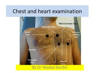
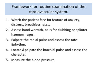
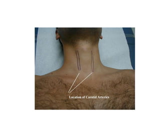
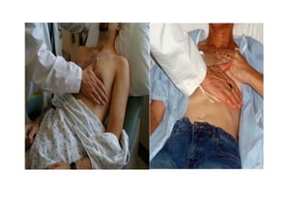
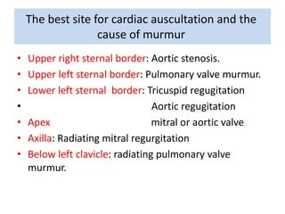
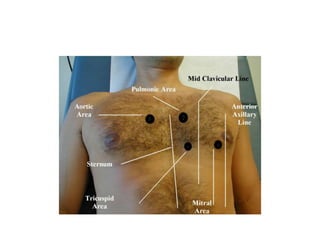
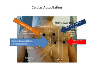
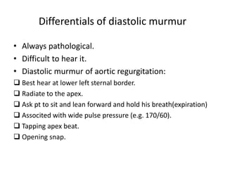
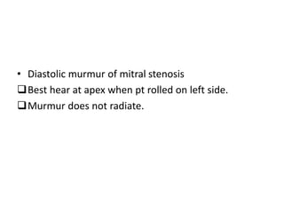
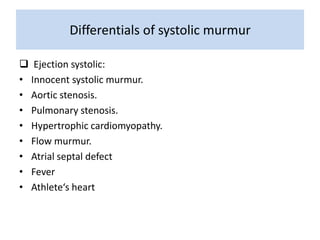
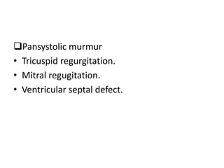
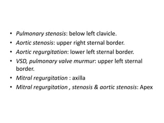
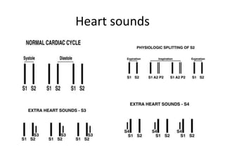
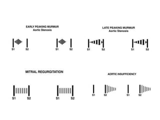
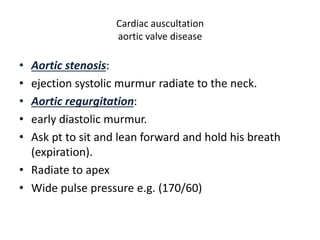
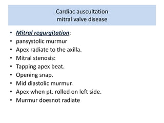
Chest and heart examination 
By Dr Wedad Bardisi
Framework for routine examination of the 
cardiovascular system. 
1. Watch the patient face for feature of anxiety, 
distress, breathlessness… 
2. Assess hand wormth, nails for clubbing or splinter 
haemorrhages. 
3. Palpate the radial pulse and assess the rate 
&rhythm. 
4. Locate &palpate the brachial pulse and assess the 
character. 
5. Measure the blood pressure.
Frame work for routine examination of the 
cardiovascular system 
6. With the patient lying supine at 45°, assess the 
jugular venous pressure and the jugular venous 
pulse form. 
7. Palpate the carotid pulse and assess its character. 
8. With the patient‘s chest exposed, inspect the 
pericardium and assess the breathing pattern and 
presence of any abnormal pulsation. 
9. Palpate the pericardium, locate the apex beat and 
assess its character, any abnormal vibration or 
thrills
Cardiac auscultation 
10. Listen with the stethoscope and assess heart sound 
and murmurs. 
 Systolic murmur is not always pathological. 
 Diastolic murmurs are difficult to hear. 
 Ask the patient to roll on left side to detect the 
diastolic murmur of mitral stenosis at the apex. 
 Ask the patient to sit and lean forward and hold his 
breathing after expiration to detect the diastolic 
murmur of aortic regugitation at lower left sternal 
border.
Ask the patient to roll on left side to detect the diastolic 
murmur of mitral stenosis at the apex.
Frame work for routine examination of the 
cardiovascular system 
11. Percuss and auscultate the chest both front and 
back looking for pleural effusions. 
12. Lay the patient flat and palpate the abdomen, 
feeling in particular for the liver and any dilatation 
of the abdominal aorta. 
13. Assess the femoral pulses and the popliteal and foot 
pulses. Look for ankle or sacral edema.
The best site for cardiac auscultation and the 
cause of murmur 
• Upper right sternal border: Aortic stenosis. 
• Upper left sternal border: Pulmonary valve murmur. 
• Lower left sternal border: Tricuspid regugitation 
• Aortic regugitation 
• Apex mitral or aortic valve 
• Axilla: Radiating mitral regurgitation 
• Below left clavicle: radiating pulmonary valve 
murmur.
Cardiac Auscultation 
Tricuspid regugitatiion, 
Aorticregugitation , 
mitral or 
aorticvalve
Differentials of diastolic murmur 
• Always pathological. 
• Difficult to hear it. 
• Diastolic murmur of aortic regurgitation: 
 Best hear at lower left sternal border. 
 Radiate to the apex. 
 Ask pt to sit and lean forward and hold his breath(expiration) 
 Associted with wide pulse pressure (e.g. 170/60). 
 Tapping apex beat. 
 Opening snap.
• Diastolic murmur of mitral stenosis 
Best hear at apex when pt rolled on left side. 
Murmur does not radiate.
Cardiac Auscultation 
pathology Murmur 
Tricuspid regurgitation Pansystolic murmur 
Ventricular septal defect 
Mitral regurgitation 
Aortic stenosis 
Pulmonary stenosis Ejection systolic murmur 
Normal Heart valve 
Mitral stenosis Diastolic murmur 
Aortic regurgitation
Differentials of systolic murmur 
 Ejection systolic: 
• Innocent systolic murmur. 
• Aortic stenosis. 
• Pulmonary stenosis. 
• Hypertrophic cardiomyopathy. 
• Flow murmur. 
• Atrial septal defect 
• Fever 
• Athlete‘s heart
Pansystolic murmur 
• Tricuspid regurgitation. 
• Mitral regugitation. 
• Ventricular septal defect.
• Pulmonary stenosis: below left clavicle. 
• Aortic stenosis: upper right sternal border. 
• Aortic regurgitation: lower left sternal border. 
• VSD, pulmonary valve murmur: upper left sternal 
border. 
• Mitral regurgitation : axilla 
• Mitral regurgitation , stenosis & aortic stenosis: Apex
Heart sounds
Cardiac auscultation 
aortic valve disease 
• Aortic stenosis: 
• ejection systolic murmur radiate to the neck. 
• Aortic regurgitation: 
• early diastolic murmur. 
• Ask pt to sit and lean forward and hold his breath 
(expiration). 
• Radiate to apex 
• Wide pulse pressure e.g. (170/60)
Cardiac auscultation 
mitral valve disease 
• Mitral regurgitation: 
• pansystolic murmur 
• Apex radiate to the axilla. 
• Mitral stenosis: 
• Tapping apex beat. 
• Opening snap. 
• Mid diastolic murmur. 
• Apex when pt. rolled on left side. 
• Murmur doesnot radiate
Cardiac auscultation 
pulmonary valve disease 
• Pulmonary stenosis: 
• ejection systolic murmur radiate to the left clavicle 
• Right ventricular heave. 
• VSD: pansystolic murmur 
• Radiate all over the pericardium

Chest and heart examination

  • 1.
    Chest and heartexamination By Dr Wedad Bardisi
  • 2.
    Framework for routineexamination of the cardiovascular system. 1. Watch the patient face for feature of anxiety, distress, breathlessness… 2. Assess hand wormth, nails for clubbing or splinter haemorrhages. 3. Palpate the radial pulse and assess the rate &rhythm. 4. Locate &palpate the brachial pulse and assess the character. 5. Measure the blood pressure.
  • 3.
    Frame work forroutine examination of the cardiovascular system 6. With the patient lying supine at 45°, assess the jugular venous pressure and the jugular venous pulse form. 7. Palpate the carotid pulse and assess its character. 8. With the patient‘s chest exposed, inspect the pericardium and assess the breathing pattern and presence of any abnormal pulsation. 9. Palpate the pericardium, locate the apex beat and assess its character, any abnormal vibration or thrills
  • 6.
    Cardiac auscultation 10.Listen with the stethoscope and assess heart sound and murmurs.  Systolic murmur is not always pathological.  Diastolic murmurs are difficult to hear.  Ask the patient to roll on left side to detect the diastolic murmur of mitral stenosis at the apex.  Ask the patient to sit and lean forward and hold his breathing after expiration to detect the diastolic murmur of aortic regugitation at lower left sternal border.
  • 8.
    Ask the patientto roll on left side to detect the diastolic murmur of mitral stenosis at the apex.
  • 9.
    Frame work forroutine examination of the cardiovascular system 11. Percuss and auscultate the chest both front and back looking for pleural effusions. 12. Lay the patient flat and palpate the abdomen, feeling in particular for the liver and any dilatation of the abdominal aorta. 13. Assess the femoral pulses and the popliteal and foot pulses. Look for ankle or sacral edema.
  • 10.
    The best sitefor cardiac auscultation and the cause of murmur • Upper right sternal border: Aortic stenosis. • Upper left sternal border: Pulmonary valve murmur. • Lower left sternal border: Tricuspid regugitation • Aortic regugitation • Apex mitral or aortic valve • Axilla: Radiating mitral regurgitation • Below left clavicle: radiating pulmonary valve murmur.
  • 12.
    Cardiac Auscultation Tricuspidregugitatiion, Aorticregugitation , mitral or aorticvalve
  • 13.
    Differentials of diastolicmurmur • Always pathological. • Difficult to hear it. • Diastolic murmur of aortic regurgitation:  Best hear at lower left sternal border.  Radiate to the apex.  Ask pt to sit and lean forward and hold his breath(expiration)  Associted with wide pulse pressure (e.g. 170/60).  Tapping apex beat.  Opening snap.
  • 14.
    • Diastolic murmurof mitral stenosis Best hear at apex when pt rolled on left side. Murmur does not radiate.
  • 15.
    Cardiac Auscultation pathologyMurmur Tricuspid regurgitation Pansystolic murmur Ventricular septal defect Mitral regurgitation Aortic stenosis Pulmonary stenosis Ejection systolic murmur Normal Heart valve Mitral stenosis Diastolic murmur Aortic regurgitation
  • 16.
    Differentials of systolicmurmur  Ejection systolic: • Innocent systolic murmur. • Aortic stenosis. • Pulmonary stenosis. • Hypertrophic cardiomyopathy. • Flow murmur. • Atrial septal defect • Fever • Athlete‘s heart
  • 17.
    Pansystolic murmur •Tricuspid regurgitation. • Mitral regugitation. • Ventricular septal defect.
  • 18.
    • Pulmonary stenosis:below left clavicle. • Aortic stenosis: upper right sternal border. • Aortic regurgitation: lower left sternal border. • VSD, pulmonary valve murmur: upper left sternal border. • Mitral regurgitation : axilla • Mitral regurgitation , stenosis & aortic stenosis: Apex
  • 19.
  • 21.
    Cardiac auscultation aorticvalve disease • Aortic stenosis: • ejection systolic murmur radiate to the neck. • Aortic regurgitation: • early diastolic murmur. • Ask pt to sit and lean forward and hold his breath (expiration). • Radiate to apex • Wide pulse pressure e.g. (170/60)
  • 22.
    Cardiac auscultation mitralvalve disease • Mitral regurgitation: • pansystolic murmur • Apex radiate to the axilla. • Mitral stenosis: • Tapping apex beat. • Opening snap. • Mid diastolic murmur. • Apex when pt. rolled on left side. • Murmur doesnot radiate
  • 23.
    Cardiac auscultation pulmonaryvalve disease • Pulmonary stenosis: • ejection systolic murmur radiate to the left clavicle • Right ventricular heave. • VSD: pansystolic murmur • Radiate all over the pericardium