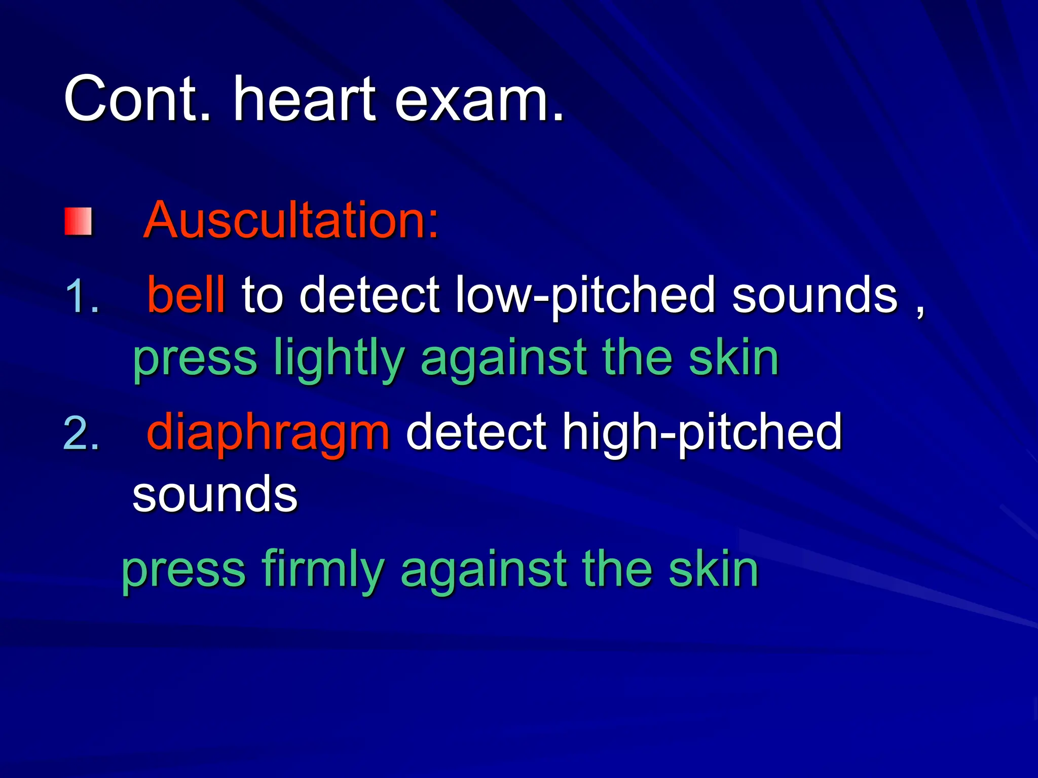
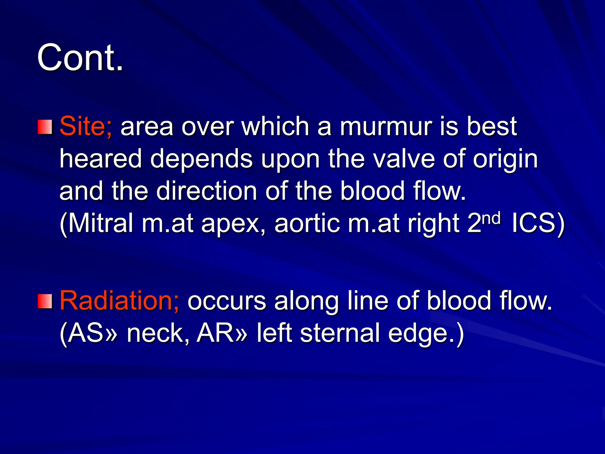
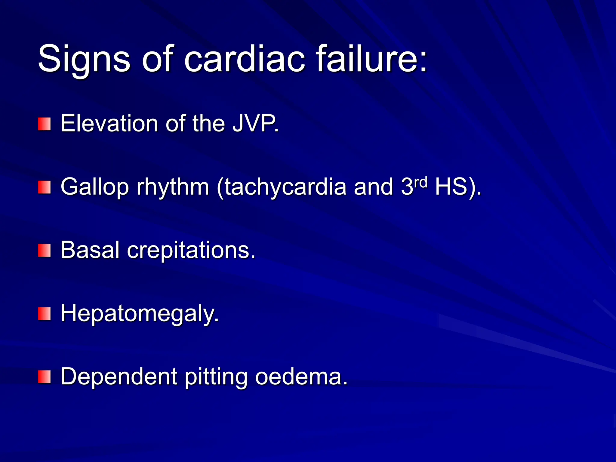
Cardiovascular Examination
Dr. Aisha AL-Ghamdi
Associate professor
Consultant internist
Cardiovascular Examination
General examination.
Heart examination.
Cont.
Blood pressure (supine & erect, why?).
Signs of heart failure in other organs
(lungs, liver, lower limbs )
Fundus examination (arterial changes,
haemorrhages, exudates and
papilloedema) in arterial hypertension.
Radial arterial puls.
1. Rate: average =70/min (range,60-90).
increased & decreased in some conditions
what are they ?
2. Rhythm.
3. Form of pulse wave.
4. Volume.
5. Vessel wall.
6. Synchronicity with other radial artery delay?
7. Radio femoral delay (aortic coarctation).
Jagular venous pressure(JVP).
Position of the patient at (45), head.
Features of JVP, how to differentiate with
carotid pulsation in the neck.
Hepato-jugular reflex’ .
Waves (a, c, x, v, y)
Heart examination;
Inspection :
1. Apex beat .
2. left parasternal movement due to right
ventricular hypertrophy.
3. pulsation in 2d left ICS 2ry to enlarged
PA.
4. epigastric pulsation 2ry to expanded
abdominal aorta
Cont. heart exam.
Palpation :
1. Apex beat 5th ICS midclavicular line.
2. Left parasternal heave 2nd ICS 2ry to
RVH.
3. Thrill (vibrating sensation indicates
palpable murmur).
Cont. heart exam.
Auscultation:
1. bell to detect low-pitched sounds ,
press lightly against the skin
2. diaphragm detect high-pitched
sounds
press firmly against the skin
Cont. auscultation
Normally audible heart sounds:
1st & 2nd HS
Added sounds: 3rd & 4th HS, pericardial
friction rub (pericarditis), opening snap
(m.s), mitral click(m.v.p)
murmers
Murmurs
Turbulent blood flow caused by diseased
valve or if a large amount of blood flows
through a normal valve.
characteristics of murmurs suggest the
cause of it (site, radiation, pitch, timing and
the intensity) .
Cont.
Site; area over which a murmur is best
heared depends upon the valve of origin
and the direction of the blood flow.
(Mitral m.at apex, aortic m.at right 2nd ICS)
Radiation; occurs along line of blood flow.
(AS» neck, AR» left sternal edge.)
Cont.
Pitch; the greater the pressure gradient the
higher the pitch(MS m.» low-pitched, AR m.»
high-pitched)
Timing; in relation to the1st and the 2nd HS
Systolic; time between 1st and the 2nd HS, could
be mid-systolic (AS), pansystolic (MR).
Diastolic; time between 2nd and the 1st HS, can be
divided into three phases. Early (AR), Mid-
diastole (MS), Presystole.
Cont.
Examples of murmurs;
Systolic; MR,TR, AS, PS.
Diastolic; MS,TS, AR, PS.
Continuous; PDA.
Signs of cardiac failure:
Elevation of the JVP.
Gallop rhythm (tachycardia and 3rd HS).
Basal crepitations.
Hepatomegaly.
Dependent pitting oedema.
Examination of peripheral arteries:
All pulses should be examined . What are
they ??
Look for bruit (indicates arterial stenosis)
Look for signs of arterial insufficiency. 5Ps
Pain, Pallor, Pulselessness, Paresthesias,
,Paralysis. (Pain is the earliest sign) are
the signs of acute leg ischemia.
Examination of peripheral veins:
inflammation or thrombosis of a vein may
cause local discomfort or pain.
Manifestations of venous obstruction
depends on site, extent and adequacy of
collateral vessels
Cyanosis and oedema are signs of venous
obstruction.

Cardiovascular Examination. Cardiovascular Examination.

  • 1.
    Cardiovascular Examination Dr. AishaAL-Ghamdi Associate professor Consultant internist
  • 2.
  • 3.
    Cont. Blood pressure (supine& erect, why?). Signs of heart failure in other organs (lungs, liver, lower limbs ) Fundus examination (arterial changes, haemorrhages, exudates and papilloedema) in arterial hypertension.
  • 4.
    Radial arterial puls. 1.Rate: average =70/min (range,60-90). increased & decreased in some conditions what are they ? 2. Rhythm. 3. Form of pulse wave. 4. Volume. 5. Vessel wall. 6. Synchronicity with other radial artery delay? 7. Radio femoral delay (aortic coarctation).
  • 5.
    Jagular venous pressure(JVP). Positionof the patient at (45), head. Features of JVP, how to differentiate with carotid pulsation in the neck. Hepato-jugular reflex’ . Waves (a, c, x, v, y)
  • 6.
    Heart examination; Inspection : 1.Apex beat . 2. left parasternal movement due to right ventricular hypertrophy. 3. pulsation in 2d left ICS 2ry to enlarged PA. 4. epigastric pulsation 2ry to expanded abdominal aorta
  • 7.
    Cont. heart exam. Palpation: 1. Apex beat 5th ICS midclavicular line. 2. Left parasternal heave 2nd ICS 2ry to RVH. 3. Thrill (vibrating sensation indicates palpable murmur).
  • 8.
    Cont. heart exam. Auscultation: 1.bell to detect low-pitched sounds , press lightly against the skin 2. diaphragm detect high-pitched sounds press firmly against the skin
  • 9.
    Cont. auscultation Normally audibleheart sounds: 1st & 2nd HS Added sounds: 3rd & 4th HS, pericardial friction rub (pericarditis), opening snap (m.s), mitral click(m.v.p) murmers
  • 10.
    Murmurs Turbulent blood flowcaused by diseased valve or if a large amount of blood flows through a normal valve. characteristics of murmurs suggest the cause of it (site, radiation, pitch, timing and the intensity) .
  • 11.
    Cont. Site; area overwhich a murmur is best heared depends upon the valve of origin and the direction of the blood flow. (Mitral m.at apex, aortic m.at right 2nd ICS) Radiation; occurs along line of blood flow. (AS» neck, AR» left sternal edge.)
  • 12.
    Cont. Pitch; the greaterthe pressure gradient the higher the pitch(MS m.» low-pitched, AR m.» high-pitched) Timing; in relation to the1st and the 2nd HS Systolic; time between 1st and the 2nd HS, could be mid-systolic (AS), pansystolic (MR). Diastolic; time between 2nd and the 1st HS, can be divided into three phases. Early (AR), Mid- diastole (MS), Presystole.
  • 13.
    Cont. Examples of murmurs; Systolic;MR,TR, AS, PS. Diastolic; MS,TS, AR, PS. Continuous; PDA.
  • 14.
    Signs of cardiacfailure: Elevation of the JVP. Gallop rhythm (tachycardia and 3rd HS). Basal crepitations. Hepatomegaly. Dependent pitting oedema.
  • 15.
    Examination of peripheralarteries: All pulses should be examined . What are they ?? Look for bruit (indicates arterial stenosis) Look for signs of arterial insufficiency. 5Ps Pain, Pallor, Pulselessness, Paresthesias, ,Paralysis. (Pain is the earliest sign) are the signs of acute leg ischemia.
  • 16.
    Examination of peripheralveins: inflammation or thrombosis of a vein may cause local discomfort or pain. Manifestations of venous obstruction depends on site, extent and adequacy of collateral vessels Cyanosis and oedema are signs of venous obstruction.