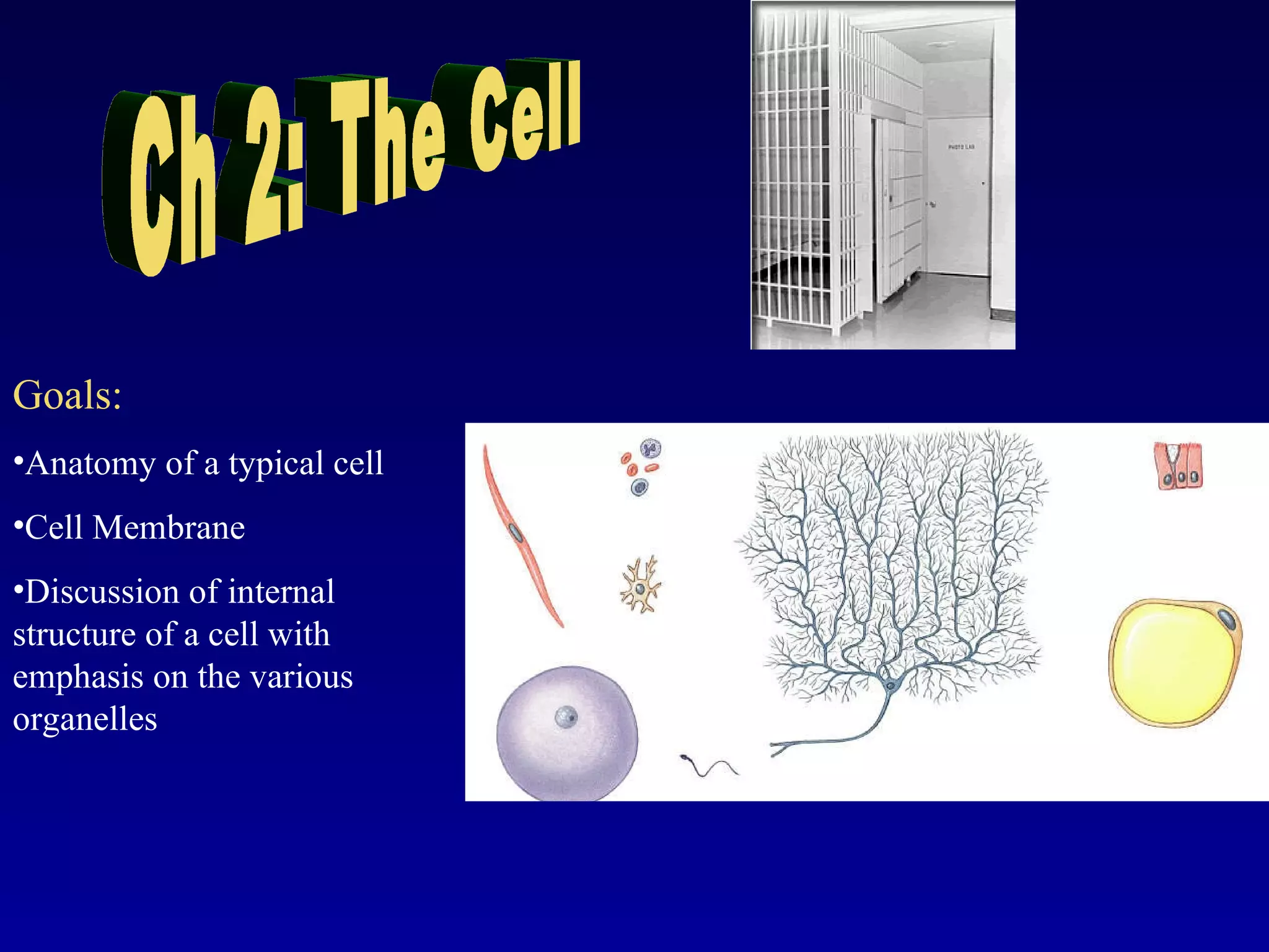
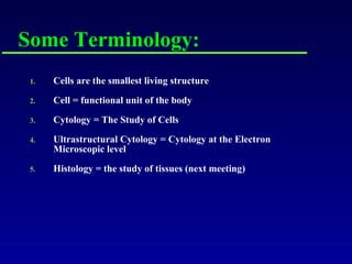
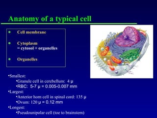
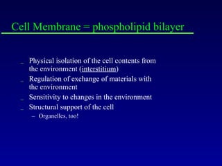
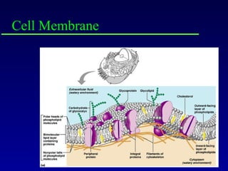
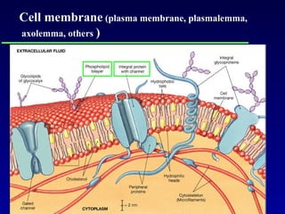
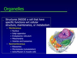
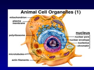
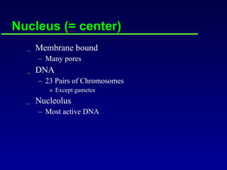
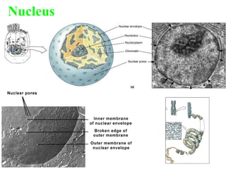
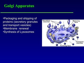
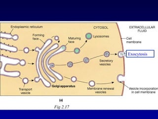
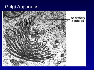
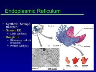
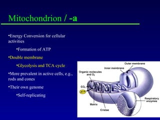
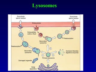
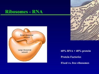
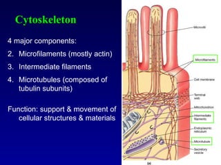
Ch 2: The Cell Goals: Anatomy of a typical cell Cell Membrane Discussion of internal structure of a cell with emphasis on the various organelles
Cells are the smallest living structure Cell = functional unit of the body Cytology = The Study of Cells Ultrastructural Cytology = Cytology at the Electron Microscopic level Histology = the study of tissues (next meeting) Some Terminology:
Anatomy of a typical cell Cell membrane Cytoplasm = cytosol + organelles Organelles Smallest:  Granule cell in cerebellum:  4  μ RBC:  5-7  μ  = 0.005-0.007 mm Largest:  Anterior horn cell in spinal cord: 135  μ   Ovum: 120  μ  = 0.12 mm Longest:  Pseudounipolar cell (toe to brainstem)
Anatomy of a typical cell, cont’d Shapes: Squamous  (scale) - flat, capillaries, lungs Cuboidal  - lines ducts Columnar  - length > width, digestive tract Stratified  - many layers Many others will be covered in histology (next two lectures)
Cell Membrane = phospholipid bilayer Physical isolation of the cell contents from the environment ( interstitium ) Regulation of exchange of materials with the environment Sensitivity to changes in the environment Structural support of the cell Organelles, too!
Cell Membrane
Cell membrane  (plasma membrane, plasmalemma, axolemma, others  )
Membrane Permeability Diffusion Concentration Gradient of Solutes Osmosis Water (solvent) through semipermeable membrane Filtration Hydrostatic Pressure Capillaries! Active Transport  Requires energy (ATP)
Endocytosis = into the cell Pinocytosis Extracellular Fluid Phagocytosis Solid Objects, e.g., bacteria Receptor-mediated Endocytosis Special membrane proteins required
Exocytosis = out of the cell Secretory vesicles  (e.g. hormones) Fluid and waste removal
Cytosol The thick fluid inside any cell Often synonymous with cytoplasm (protoplasm) Cytoplasm = cytosol + organelles Suspends organelles
Organelles Structures INSIDE a cell that have specific functions wrt cellular structure, maintenance, or metabolism Membranous Nucleus Golgi apparatus Endoplasmic reticulum Mitochondria Vesicles and lysosomes Nonmembranous Ribosomes Microtubules (cytoskeleton) Actin/Myosin in muscle cells
 
Nucleus (= center) Membrane bound Many pores DNA 23 Pairs of Chromosomes Except gametes Nucleolus Most active DNA
Nucleus
Golgi Apparatus Packaging and shipping of proteins (secretory granules and transport vesicles) Membrane  renewal Synthesis of Lysosomes
Fig 2.17 Exocytosis
Golgi Apparatus
Endoplasmic Reticulum Synthesis, Storage, transport Smooth ER Lipid synthesis Rough ER Ribosomes make it rough ER Protein synthesis
Mitochondrion  / -a Energy Conversion for cellular activities Formation of ATP Double membrane  Glycolysis and TCA cycle More prevalent in active cells, e.g., rods and cones Their own genome Self-replicating
Lysosomes
Ribosomes - RNA 60% RNA + 40% protein Protein Factories Fixed vs. free ribosomes
Cytoskeleton 4 major components: Microfilaments (mostly actin) Intermediate filaments Microtubules (composed of tubulin subunits) Function: support & movement of cellular structures & materials
Cilium – Cilia (pl.) Compare to microvilli and flagella In 9+2 array
Actin/Myosin The contractile proteins in muscle cells Striations Skeletal muscle
Gap Junctions Tight Junctions Desmosomes Intercellular Attachments Chapter 4, pp 78-80 Fig 4-7 Act as: Seals betw cells Intercellular communication Added strength to resist separation
Channel proteins (connexons) interlock and form pores Abundant in cardiac and smooth muscle Allows efficient intercellular communication 1) Gap Junctions
2) Tight Junctions Interlocking membrane proteins Found near surface of cells lining the digestive tract.  Explain!
3) Desmosomes Proteoglycan layer reinforced by transmembrane proteins (cell adhesion molecules or CAMs) Belt, button and hemidesmosomes Found in superficial layers  of skin
Fig 2.19 a
 
Mitosis (vs. meiosis) Cell Division Interphase – Between mitosis  Prophase – Chromosomes become bunched Metaphase – Chromosomes gather at equator Anaphase – Chromosomes move to poles Telophase – The two new nuclei form Cytokinesis – Actual cell separation Two new  diploid  cells
Mitosis
 
Some cells Fat cells (adipocytes) Cartilage cells (chondrocytes)
More cells Neutrophil Plasma cell
Still more cells Columnar cells Sperm cells (spermatozoa)
River Cullenagh, Ennistymon, Co Clare, Ireland

Chapter2 cellmarieb

  • 1.
    Ch 2: TheCell Goals: Anatomy of a typical cell Cell Membrane Discussion of internal structure of a cell with emphasis on the various organelles
  • 2.
    Cells are thesmallest living structure Cell = functional unit of the body Cytology = The Study of Cells Ultrastructural Cytology = Cytology at the Electron Microscopic level Histology = the study of tissues (next meeting) Some Terminology:
  • 3.
    Anatomy of atypical cell Cell membrane Cytoplasm = cytosol + organelles Organelles Smallest: Granule cell in cerebellum: 4 μ RBC: 5-7 μ = 0.005-0.007 mm Largest: Anterior horn cell in spinal cord: 135 μ Ovum: 120 μ = 0.12 mm Longest: Pseudounipolar cell (toe to brainstem)
  • 4.
    Anatomy of atypical cell, cont’d Shapes: Squamous (scale) - flat, capillaries, lungs Cuboidal - lines ducts Columnar - length > width, digestive tract Stratified - many layers Many others will be covered in histology (next two lectures)
  • 5.
    Cell Membrane =phospholipid bilayer Physical isolation of the cell contents from the environment ( interstitium ) Regulation of exchange of materials with the environment Sensitivity to changes in the environment Structural support of the cell Organelles, too!
  • 6.
  • 7.
    Cell membrane (plasma membrane, plasmalemma, axolemma, others )
  • 8.
    Membrane Permeability DiffusionConcentration Gradient of Solutes Osmosis Water (solvent) through semipermeable membrane Filtration Hydrostatic Pressure Capillaries! Active Transport Requires energy (ATP)
  • 9.
    Endocytosis = intothe cell Pinocytosis Extracellular Fluid Phagocytosis Solid Objects, e.g., bacteria Receptor-mediated Endocytosis Special membrane proteins required
  • 10.
    Exocytosis = outof the cell Secretory vesicles (e.g. hormones) Fluid and waste removal
  • 11.
    Cytosol The thickfluid inside any cell Often synonymous with cytoplasm (protoplasm) Cytoplasm = cytosol + organelles Suspends organelles
  • 12.
    Organelles Structures INSIDEa cell that have specific functions wrt cellular structure, maintenance, or metabolism Membranous Nucleus Golgi apparatus Endoplasmic reticulum Mitochondria Vesicles and lysosomes Nonmembranous Ribosomes Microtubules (cytoskeleton) Actin/Myosin in muscle cells
  • 13.
  • 14.
    Nucleus (= center)Membrane bound Many pores DNA 23 Pairs of Chromosomes Except gametes Nucleolus Most active DNA
  • 15.
  • 16.
    Golgi Apparatus Packagingand shipping of proteins (secretory granules and transport vesicles) Membrane renewal Synthesis of Lysosomes
  • 17.
  • 18.
  • 19.
    Endoplasmic Reticulum Synthesis,Storage, transport Smooth ER Lipid synthesis Rough ER Ribosomes make it rough ER Protein synthesis
  • 20.
    Mitochondrion /-a Energy Conversion for cellular activities Formation of ATP Double membrane Glycolysis and TCA cycle More prevalent in active cells, e.g., rods and cones Their own genome Self-replicating
  • 21.
  • 22.
    Ribosomes - RNA60% RNA + 40% protein Protein Factories Fixed vs. free ribosomes
  • 23.
    Cytoskeleton 4 majorcomponents: Microfilaments (mostly actin) Intermediate filaments Microtubules (composed of tubulin subunits) Function: support & movement of cellular structures & materials
  • 24.
    Cilium – Cilia(pl.) Compare to microvilli and flagella In 9+2 array
  • 25.
    Actin/Myosin The contractileproteins in muscle cells Striations Skeletal muscle
  • 26.
    Gap Junctions TightJunctions Desmosomes Intercellular Attachments Chapter 4, pp 78-80 Fig 4-7 Act as: Seals betw cells Intercellular communication Added strength to resist separation
  • 27.
    Channel proteins (connexons)interlock and form pores Abundant in cardiac and smooth muscle Allows efficient intercellular communication 1) Gap Junctions
  • 28.
    2) Tight JunctionsInterlocking membrane proteins Found near surface of cells lining the digestive tract. Explain!
  • 29.
    3) Desmosomes Proteoglycanlayer reinforced by transmembrane proteins (cell adhesion molecules or CAMs) Belt, button and hemidesmosomes Found in superficial layers of skin
  • 30.
  • 31.
  • 32.
    Mitosis (vs. meiosis)Cell Division Interphase – Between mitosis Prophase – Chromosomes become bunched Metaphase – Chromosomes gather at equator Anaphase – Chromosomes move to poles Telophase – The two new nuclei form Cytokinesis – Actual cell separation Two new diploid cells
  • 33.
  • 34.
  • 35.
    Some cells Fatcells (adipocytes) Cartilage cells (chondrocytes)
  • 36.
  • 37.
    Still more cellsColumnar cells Sperm cells (spermatozoa)
  • 38.

Editor's Notes

  • #8 Membrane permeability covered in physiology