This document summarizes four cardiovascular procedures:
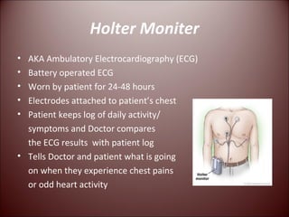
1) Holter monitoring involves wearing portable ECG electrodes for 1-2 days to monitor heart activity and compare to a patient's daily log of symptoms.
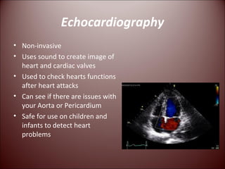
2) Echocardiography uses ultrasound to create images of the heart and valves to assess function and detect issues.
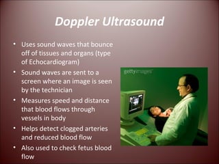
3) Doppler ultrasound uses sound waves to detect blood flow speed and identify clogged arteries or reduced flow.
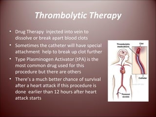
4) Thrombolytic therapy involves injecting clot-busting drugs into veins to dissolve blood clots, with better outcomes if administered within 12 hours of a heart attack.