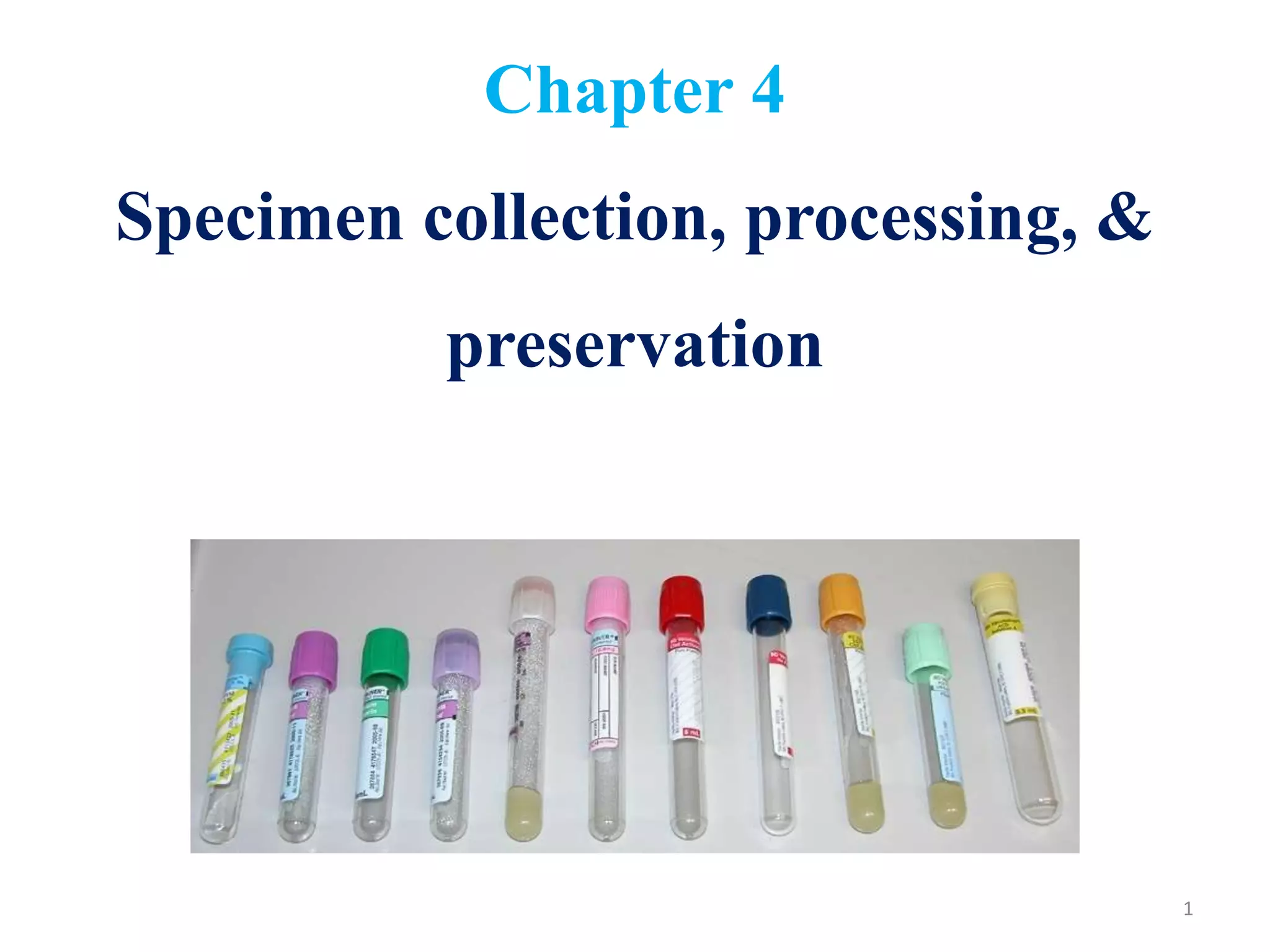
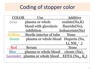
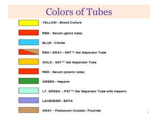
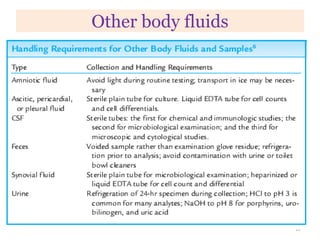
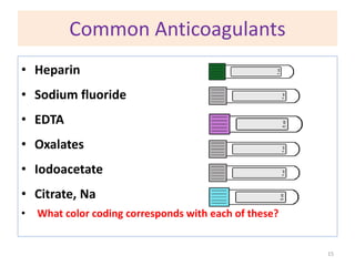
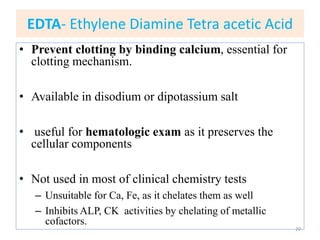
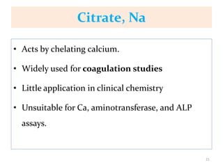
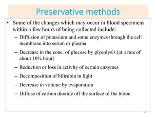
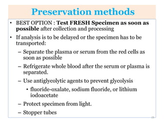
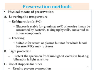
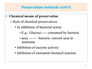
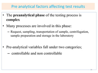
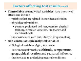
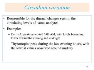
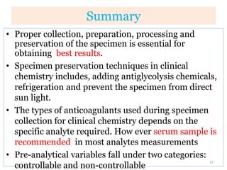
Chapter 4
Specimen collection, processing, &
preservation
1
Objectives
At the end of the chapter, the student will be able to
• Explain about different kind of specimens
• Discuss about factors affect blood composition
• Discuss about preservation, storage, and
transportation of specimens.
• Discuss different preanalytical factors affecting test
results
2
Specimen for Biochemical analysis
• Proper collection, preparation, processing and
preservation of the specimen is essential for obtaining
best results.
• A properly filled test requisition slip should
accompany each specimen.
• Specimen must be labeled at least with the patient’s
name, hospital or identification number , ward, and
date and time of collection.
3
Specimen types
• Biological specimens include:
– Whole blood, serum, plasma, urine, saliva, body
fluids (spinal, synovial, amniotic, pleural,
pericardial, ascetic fluid), and various types of
solid tissue, including specific cell types
• Majority of clinical chemistry tests are done on
blood : serum , whole blood, or plasma.
4
Specimen types
• Random specimen
– collected any time
• Fasting specimen
– collected after no food has been eaten for
more than 6 hours and usually in the morning
• timed specimens
– collected at a particular time after an event
such as eating or taking a medication.
5
Blood
• Blood for analysis may be obtained from veins,
arteries or capillaries.
• Venous blood is usually the specimen of choice and
venipuncture is the method for obtaining this
specimen
• Skin puncture is frequently used to obtain what is
mostly capillary blood
• Arterial puncture is used mainly for blood gas
analysis
6
Venipuncture
• Venous blood is usually collected with
evacuated blood tubes
– Evacuated blood tubes are less expensive, more
convenient
– Different types: vary by the type of additive
present
– The color of the stopper used identifies the
additive present
7
Coding of stopper color
COLOR Use Additive
Gray plasma or whole oxalate(Na,K)
blood with glycolysis fluoride(Na),
inhibition Iodoacetate(Na)
Yellow Sterile interior of tube None .
Green plasma or whole blood Heparin (Na,
Li, NH4
+ )
Red Serum None .
Blue plasma or whole blood citrate (Na) .
Lavender plasma or whole blood EDTA (Na2, K2)
8
Colors of Tubes
9
Serum and plasma
• Serum
– Whole blood is allowed to clot and centrifuged to
obtain the serum
– The most commonly used specimen in clinical
chemistry
• Plasma
– Clotting is preserved by allowing the whole blood to
mix an anticoagulant and centrifuged to obtain the
plasma
10
Urine
• Urine is frequently requested specimen for assessment of
renal function and metabolic conditions
• The required type of urine depends upon the analyte to
be measured
– Eg, a timed 24 hout urine is required for clearance test
• If analysis is to be delayed, the urine specimen can be
retained at 20c or 40c for up to a week or at -20 oC if
longer delay is needed
11
Other body fluids
12
Anticoagulants for blood
• Anticoagulant: chemical substances added to
fresh blood to prevent clotting
• Used when whole blood or plasma is desired
for testing
• The right type of anticoagulant in correct
amounts should be used
13
Anticoagulants for blood cont’d
• Precautions in the selection and use of anticoagulants:
– It should neither remove nor add the substance to
be measured;
• Eg: sodium salts of anticoagulants for sodium
determination, EDTA for calcium determination.
– Use neither excess nor less amount of an
anticoagulant.
– It should not be an enzyme inhibitor if sample is to
be used for enzyme assays.
• Eg: Sodium fluoride and chelating anticoagulants are
enzyme poisons
14
Common Anticoagulants
• Heparin
• Sodium fluoride
• EDTA
• Oxalates
• Iodoacetate
• Citrate, Na
• What color coding corresponds with each of these?
15
Heparin
• Available as Na, K, Li, and NH4
+ salts.
• Acts as antithrombin, preventing the transformation
of prothrombin into thrombin and thus the formation
of fibrin into fibrinogen.
• Most widely used anticoagulant for chemical
analyses.
• It causes the least interference with tests.
• Disadvantage: high cost and temporary action.
16
Sodium fluoride
• Is usually considered as a preservative for blood
glucose, it also acts as a weak anticoagulant.
• Exerts preservative action by inhibiting
glycolytic enzymes
• Potent inhibitor to urease as well.
17
Oxalates, Na, K, Li, NH+
4
• Inhibit blood coagulation by forming insoluble
complexes with calcium ions.
• Used for glucose assay along with fluoride- as
fluoride-oxalate.
• Oxalate inhibits most enzymes- ALP, AMS, LDH,
CK and may also cause precipitation of calcium.
18
Iodoacetate
• Sodium iodoacetate at a concentration of 2g/L is an
effective antiglycolytic agent and a substitute for
NaF.
• Since it has no effect on urease, it can be used when
glucose and urea are done on a single specimen.
• It inhibits CK but appears to have no significant
effect on other clinical chemistry tests.
19
EDTA- Ethylene Diamine Tetra acetic Acid
• Prevent clotting by binding calcium, essential for
clotting mechanism.
• Available in disodium or dipotassium salt
• useful for hematologic exam as it preserves the
cellular components
• Not used in most of clinical chemistry tests
– Unsuitable for Ca, Fe, as it chelates them as well
– Inhibits ALP, CK activities by chelating of metallic
cofactors.
20
Citrate, Na
• Acts by chelating calcium.
• Widely used for coagulation studies
• Little application in clinical chemistry
• Unsuitable for Ca, aminotransferase, and ALP
assays.
21
Preservative methods
• Some of the changes which may occur in blood specimens
within a few hours of being collected include:
– Diffusion of potassium and some enzymes through the cell
membrane into serum or plasma
– Decrease in the conc. of glucose by glycolysis (at a rate of
about 10% hour)
– Reduction or loss in activity of certain enzymes
– Decomposition of bilirubin in light
– Decrease in volume by evaporation
– Diffuse of carbon dioxide off the surface of the blood
22
Preservation methods
• BEST OPTION : Test FRESH Specimen as soon as
possible after collection and processing
• If analysis is to be delayed or the specimen has to be
transported:
– Separate the plasma or serum from the red cells as
soon as possible
– Refrigerate whole blood after the serum or plasma is
separated.
– Use antiglycolytic agents to prevent glycolysis
• fluoride-oxalate, sodium fluoride, or lithium
iodoacetate
– Protect specimen from light.
– Stopper tubes
23
Preservation methods
• Physical means of preservation
A. Lowering the temperature
– Refrigeration(4-80C)
• Glucose is stable for 30 min at 200C otherwise it may be
consumed by bacteria, taking up by cells, converted to
others compounds
– Freezing
• Suitable for serum or plasma but not for whole blood
because RBCs may ruptures
B. Light protection
– Protect the specimen from sun light & excessive heat e.g.
bilurubin is light sensitive
C. Use of stoppers for tubes
– Used to prevent evaporation
24
Preservation methods cont’d
• Chemical means of preservation
– Role of chemical preservatives
• In inhibition of bacterial action
–E.g.. Glucose----- consumed by bacteria
–urea ------ bacteria convert urea to
ammonia
• Inhibition of enzyme activity
• Inhibition of unwanted chemical reaction
25
Pre analytical factors affecting test results
• The preanalytical phase of the testing process is
complex
• Many processes are involved in this phase:
– Request, sampling, transportation of sample, centrifugation,
sample preparation and storage in the laboratory
• Pre-analytical variables fall under two categories;
– controllable and non controllable
26
Factors affecting test results cont’d
• Controllable preanalytical variables: have short-lived
effects and include:
– variables that are related to specimen collection
– physiological variables:
• posture, prolonged bed rest, exercise, physical
training, circadian variation, Pregnancy and
menstrual cycle
– those associated with diet, lifestyle, drugs smoking
• Non-controllable preanalytical variables
– Biological variables: Age , sex , race
– Environmental variables :Altitude, temperature,
geographical location and seasonal influence
– those related to underlying medical conditions 27
Factors affecting test results cont’d
• Specimen related variables It include variables related
to:
– specimen type and collection
– use of anticoagulant
– specimen handling and processing: storage,
transport and preparation
– duration of tourniquet application
– Specimen contamination from Infusion
– Hemolysis
28
Duration of Tourniquet Application
• cause short-term venous occlusion, allowing palpation
of the veins
• > 1 min-hemoconcentration occurs due to fluid
changes
– Low molecular weight compounds (K+) leave the
capillaries and pool in the interstitial fluid region
– increase in the conc of large molecules(proteins) in
the remaining plasma.
29
Duration of…
• Venous stasis from excessive fist clenching
– promotes anaerobic glycolysis with the accumulation
of plasma lactate and a reduction in blood pH
• Reduction in blood pH
– alter drug-protein or hormone-protein binding→
increased free drug or free hormone conc
– Levels of Ca2+, Mg2+ increase since they are released
from albumin
30
Duration of Tourniquet Application…
Specimen Contamination from Fluid Infusion
• Dextrose(sugar) IV infusion→ extremely high glucose
results
• Total parenteral nutrition infusion:
– creates gross turbidity along with elevated lipid and
glucose values
• saline IV infusion: Na+ and Cl- falsely elevated
• blood transfusion: the extent of hemolysis may increase
and result in increased values for potassium, LDH
31
Effects of Hemolysis
• disruption of the RBC membrane and release of
hemoglobin and other cellular components
• It is graded as mild, moderate, or gross hemolysis
based on visible presence of hemoglobin
• Serum shows visual evidence of hemolysis when
hemoglobin conc exceeds 20 mg/dl.
32
Effect of hemolysis
• Caused by a variety of conditions during the
collection and processing steps
– poor placement of the needle into the vein, pulling
back the plunger on a syringe too quickly, and
allowing air leakage due to a poorly fitted needle
– Prolonged tourniquet application with leak of
interstitial fluid into tissue, Tissue trauma during
collection,
– Using a small-bore needle when compared to the size
of the evacuated tube or forcing blood through a
stopper into the evacuated tube
– Exposure of cells to excessive heat during storage or
transportation
33
Effect of hemolysis…
• Hemolysis can be avoided by:
– Checking that the syringe and needle are dry and
that the barrel and plunger of the syringe fit well.
– Not withdrawing the blood too rapidly or moving
the needle once it is in the vein.
– Removing the needle from the syringe before
dispensing the blood into the specimen container.
– Allow the blood to run gently down the inside wall
of the container.
34
• Effect of menstruation
– the reference intervals for estradiol, FSH, LH and
progesterone are influenced by the stages of menstrual
cycle
• Pregnancy
– A dilutional effect is observed due to an increase in the
mean plasma volume, which causes hemodilution
– heat-stable ALP increased (placental origin)
– Increased levels of hormones, such as estrogen,
progesterone, and human placental lactogen
35
Circadian variation
• Responsible for the diurnal changes seen in the
circulating levels of some analytes
• Example:
– Cortisol: peaks at around 6:00 AM, with levels becoming
lower toward the evening and midnight
– Thyrotropin: peak during the late evening hours, with
the lowest values observed around midday
36
Summary
• Proper collection, preparation, processing and
preservation of the specimen is essential for
obtaining best results.
• Specimen preservation techniques in clinical
chemistry includes, adding antiglycolysis chemicals,
refrigeration and prevent the specimen from direct
sun light.
• The types of anticoagulants used during specimen
collection for clinical chemistry depends on the
specific analyte required. How ever serum sample is
recommended in most analytes measurements
• Pre-analytical variables fall under two categories:
controllable and non-controllable
37
Thank you !
38

Chapter 4 Specimen collection, handling, and processing.ppt

  • 1.
    Chapter 4 Specimen collection,processing, & preservation 1
  • 2.
    Objectives At the endof the chapter, the student will be able to • Explain about different kind of specimens • Discuss about factors affect blood composition • Discuss about preservation, storage, and transportation of specimens. • Discuss different preanalytical factors affecting test results 2
  • 3.
    Specimen for Biochemicalanalysis • Proper collection, preparation, processing and preservation of the specimen is essential for obtaining best results. • A properly filled test requisition slip should accompany each specimen. • Specimen must be labeled at least with the patient’s name, hospital or identification number , ward, and date and time of collection. 3
  • 4.
    Specimen types • Biologicalspecimens include: – Whole blood, serum, plasma, urine, saliva, body fluids (spinal, synovial, amniotic, pleural, pericardial, ascetic fluid), and various types of solid tissue, including specific cell types • Majority of clinical chemistry tests are done on blood : serum , whole blood, or plasma. 4
  • 5.
    Specimen types • Randomspecimen – collected any time • Fasting specimen – collected after no food has been eaten for more than 6 hours and usually in the morning • timed specimens – collected at a particular time after an event such as eating or taking a medication. 5
  • 6.
    Blood • Blood foranalysis may be obtained from veins, arteries or capillaries. • Venous blood is usually the specimen of choice and venipuncture is the method for obtaining this specimen • Skin puncture is frequently used to obtain what is mostly capillary blood • Arterial puncture is used mainly for blood gas analysis 6
  • 7.
    Venipuncture • Venous bloodis usually collected with evacuated blood tubes – Evacuated blood tubes are less expensive, more convenient – Different types: vary by the type of additive present – The color of the stopper used identifies the additive present 7
  • 8.
    Coding of stoppercolor COLOR Use Additive Gray plasma or whole oxalate(Na,K) blood with glycolysis fluoride(Na), inhibition Iodoacetate(Na) Yellow Sterile interior of tube None . Green plasma or whole blood Heparin (Na, Li, NH4 + ) Red Serum None . Blue plasma or whole blood citrate (Na) . Lavender plasma or whole blood EDTA (Na2, K2) 8
  • 9.
  • 10.
    Serum and plasma •Serum – Whole blood is allowed to clot and centrifuged to obtain the serum – The most commonly used specimen in clinical chemistry • Plasma – Clotting is preserved by allowing the whole blood to mix an anticoagulant and centrifuged to obtain the plasma 10
  • 11.
    Urine • Urine isfrequently requested specimen for assessment of renal function and metabolic conditions • The required type of urine depends upon the analyte to be measured – Eg, a timed 24 hout urine is required for clearance test • If analysis is to be delayed, the urine specimen can be retained at 20c or 40c for up to a week or at -20 oC if longer delay is needed 11
  • 12.
  • 13.
    Anticoagulants for blood •Anticoagulant: chemical substances added to fresh blood to prevent clotting • Used when whole blood or plasma is desired for testing • The right type of anticoagulant in correct amounts should be used 13
  • 14.
    Anticoagulants for bloodcont’d • Precautions in the selection and use of anticoagulants: – It should neither remove nor add the substance to be measured; • Eg: sodium salts of anticoagulants for sodium determination, EDTA for calcium determination. – Use neither excess nor less amount of an anticoagulant. – It should not be an enzyme inhibitor if sample is to be used for enzyme assays. • Eg: Sodium fluoride and chelating anticoagulants are enzyme poisons 14
  • 15.
    Common Anticoagulants • Heparin •Sodium fluoride • EDTA • Oxalates • Iodoacetate • Citrate, Na • What color coding corresponds with each of these? 15
  • 16.
    Heparin • Available asNa, K, Li, and NH4 + salts. • Acts as antithrombin, preventing the transformation of prothrombin into thrombin and thus the formation of fibrin into fibrinogen. • Most widely used anticoagulant for chemical analyses. • It causes the least interference with tests. • Disadvantage: high cost and temporary action. 16
  • 17.
    Sodium fluoride • Isusually considered as a preservative for blood glucose, it also acts as a weak anticoagulant. • Exerts preservative action by inhibiting glycolytic enzymes • Potent inhibitor to urease as well. 17
  • 18.
    Oxalates, Na, K,Li, NH+ 4 • Inhibit blood coagulation by forming insoluble complexes with calcium ions. • Used for glucose assay along with fluoride- as fluoride-oxalate. • Oxalate inhibits most enzymes- ALP, AMS, LDH, CK and may also cause precipitation of calcium. 18
  • 19.
    Iodoacetate • Sodium iodoacetateat a concentration of 2g/L is an effective antiglycolytic agent and a substitute for NaF. • Since it has no effect on urease, it can be used when glucose and urea are done on a single specimen. • It inhibits CK but appears to have no significant effect on other clinical chemistry tests. 19
  • 20.
    EDTA- Ethylene DiamineTetra acetic Acid • Prevent clotting by binding calcium, essential for clotting mechanism. • Available in disodium or dipotassium salt • useful for hematologic exam as it preserves the cellular components • Not used in most of clinical chemistry tests – Unsuitable for Ca, Fe, as it chelates them as well – Inhibits ALP, CK activities by chelating of metallic cofactors. 20
  • 21.
    Citrate, Na • Actsby chelating calcium. • Widely used for coagulation studies • Little application in clinical chemistry • Unsuitable for Ca, aminotransferase, and ALP assays. 21
  • 22.
    Preservative methods • Someof the changes which may occur in blood specimens within a few hours of being collected include: – Diffusion of potassium and some enzymes through the cell membrane into serum or plasma – Decrease in the conc. of glucose by glycolysis (at a rate of about 10% hour) – Reduction or loss in activity of certain enzymes – Decomposition of bilirubin in light – Decrease in volume by evaporation – Diffuse of carbon dioxide off the surface of the blood 22
  • 23.
    Preservation methods • BESTOPTION : Test FRESH Specimen as soon as possible after collection and processing • If analysis is to be delayed or the specimen has to be transported: – Separate the plasma or serum from the red cells as soon as possible – Refrigerate whole blood after the serum or plasma is separated. – Use antiglycolytic agents to prevent glycolysis • fluoride-oxalate, sodium fluoride, or lithium iodoacetate – Protect specimen from light. – Stopper tubes 23
  • 24.
    Preservation methods • Physicalmeans of preservation A. Lowering the temperature – Refrigeration(4-80C) • Glucose is stable for 30 min at 200C otherwise it may be consumed by bacteria, taking up by cells, converted to others compounds – Freezing • Suitable for serum or plasma but not for whole blood because RBCs may ruptures B. Light protection – Protect the specimen from sun light & excessive heat e.g. bilurubin is light sensitive C. Use of stoppers for tubes – Used to prevent evaporation 24
  • 25.
    Preservation methods cont’d •Chemical means of preservation – Role of chemical preservatives • In inhibition of bacterial action –E.g.. Glucose----- consumed by bacteria –urea ------ bacteria convert urea to ammonia • Inhibition of enzyme activity • Inhibition of unwanted chemical reaction 25
  • 26.
    Pre analytical factorsaffecting test results • The preanalytical phase of the testing process is complex • Many processes are involved in this phase: – Request, sampling, transportation of sample, centrifugation, sample preparation and storage in the laboratory • Pre-analytical variables fall under two categories; – controllable and non controllable 26
  • 27.
    Factors affecting testresults cont’d • Controllable preanalytical variables: have short-lived effects and include: – variables that are related to specimen collection – physiological variables: • posture, prolonged bed rest, exercise, physical training, circadian variation, Pregnancy and menstrual cycle – those associated with diet, lifestyle, drugs smoking • Non-controllable preanalytical variables – Biological variables: Age , sex , race – Environmental variables :Altitude, temperature, geographical location and seasonal influence – those related to underlying medical conditions 27
  • 28.
    Factors affecting testresults cont’d • Specimen related variables It include variables related to: – specimen type and collection – use of anticoagulant – specimen handling and processing: storage, transport and preparation – duration of tourniquet application – Specimen contamination from Infusion – Hemolysis 28
  • 29.
    Duration of TourniquetApplication • cause short-term venous occlusion, allowing palpation of the veins • > 1 min-hemoconcentration occurs due to fluid changes – Low molecular weight compounds (K+) leave the capillaries and pool in the interstitial fluid region – increase in the conc of large molecules(proteins) in the remaining plasma. 29
  • 30.
    Duration of… • Venousstasis from excessive fist clenching – promotes anaerobic glycolysis with the accumulation of plasma lactate and a reduction in blood pH • Reduction in blood pH – alter drug-protein or hormone-protein binding→ increased free drug or free hormone conc – Levels of Ca2+, Mg2+ increase since they are released from albumin 30 Duration of Tourniquet Application…
  • 31.
    Specimen Contamination fromFluid Infusion • Dextrose(sugar) IV infusion→ extremely high glucose results • Total parenteral nutrition infusion: – creates gross turbidity along with elevated lipid and glucose values • saline IV infusion: Na+ and Cl- falsely elevated • blood transfusion: the extent of hemolysis may increase and result in increased values for potassium, LDH 31
  • 32.
    Effects of Hemolysis •disruption of the RBC membrane and release of hemoglobin and other cellular components • It is graded as mild, moderate, or gross hemolysis based on visible presence of hemoglobin • Serum shows visual evidence of hemolysis when hemoglobin conc exceeds 20 mg/dl. 32
  • 33.
    Effect of hemolysis •Caused by a variety of conditions during the collection and processing steps – poor placement of the needle into the vein, pulling back the plunger on a syringe too quickly, and allowing air leakage due to a poorly fitted needle – Prolonged tourniquet application with leak of interstitial fluid into tissue, Tissue trauma during collection, – Using a small-bore needle when compared to the size of the evacuated tube or forcing blood through a stopper into the evacuated tube – Exposure of cells to excessive heat during storage or transportation 33
  • 34.
    Effect of hemolysis… •Hemolysis can be avoided by: – Checking that the syringe and needle are dry and that the barrel and plunger of the syringe fit well. – Not withdrawing the blood too rapidly or moving the needle once it is in the vein. – Removing the needle from the syringe before dispensing the blood into the specimen container. – Allow the blood to run gently down the inside wall of the container. 34
  • 35.
    • Effect ofmenstruation – the reference intervals for estradiol, FSH, LH and progesterone are influenced by the stages of menstrual cycle • Pregnancy – A dilutional effect is observed due to an increase in the mean plasma volume, which causes hemodilution – heat-stable ALP increased (placental origin) – Increased levels of hormones, such as estrogen, progesterone, and human placental lactogen 35
  • 36.
    Circadian variation • Responsiblefor the diurnal changes seen in the circulating levels of some analytes • Example: – Cortisol: peaks at around 6:00 AM, with levels becoming lower toward the evening and midnight – Thyrotropin: peak during the late evening hours, with the lowest values observed around midday 36
  • 37.
    Summary • Proper collection,preparation, processing and preservation of the specimen is essential for obtaining best results. • Specimen preservation techniques in clinical chemistry includes, adding antiglycolysis chemicals, refrigeration and prevent the specimen from direct sun light. • The types of anticoagulants used during specimen collection for clinical chemistry depends on the specific analyte required. How ever serum sample is recommended in most analytes measurements • Pre-analytical variables fall under two categories: controllable and non-controllable 37
  • 38.

Editor's Notes

  • #4 Specimen for analysis is the most important element in obtaining reliable test results.
  • #8 Evacuated blood tubes are less expensive, more convenient, and easier to use than syringes
  • #16 Color codes: heparin = green, Sodium fluoride = gray, EDTA = lavendar, oxalates = gray, iodoacetate = gray, citrate = light blue
  • #17 Mucoitin polysulfuric acid and is available as Na, K, Li, and NH4+ salts.
  • #19 Tubes may be prepared by drying 30g/dl solution in an oven Potassium oxalate at a concentration of 1-2 mg/ml blood is the most widely used oxalate. concs >3mg/ml may cause hemolysis
  • #23 Diffuse of carbon dioxide off the surface of the blood, leading to a lowering in the conc of bicarbonate with a compensatory increase in plasma chloride(chloride shift)
  • #26 NaF, Iodacetate
  • #27 Many things can go wrong with this processes, which affect the concentration or activities of analytes in the sample
  • #29 patient identification,
  • #32 The effects of contamination from IV fluids would vary with the type of fluid being infused
  • #36 Pregnancy increased mobilization of lipids the serum levels of apolipoproteins, triglycerides, and total cholesterol increased
  • #37 Glucose in oral glucose tolerance test is higher when the test is performed in the afternoon