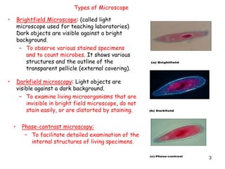
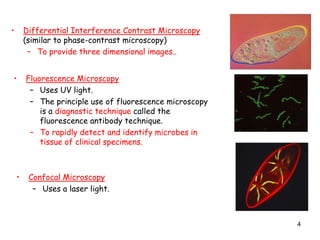
1. The document discusses different types of microscopes used to observe microorganisms, including brightfield, darkfield, phase-contrast, fluorescence, confocal, transmission electron, and scanning electron microscopes.
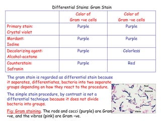
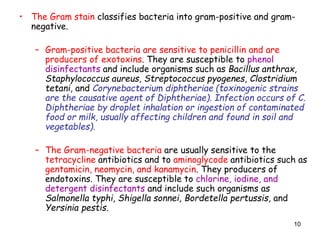
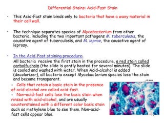
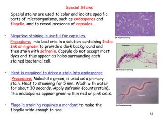
2. It also covers different staining techniques used to prepare specimens for microscopy, including simple stains using a single dye, differential stains like Gram staining and acid-fast staining to classify bacteria, and special stains for structures like capsules, endospores, and flagella.
3. Key points are made about classifying bacteria as gram-positive or gram-negative based on their cell wall composition and how they react to staining, as well as how the acid-fast stain is used to