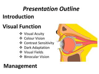
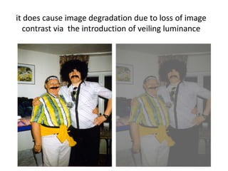
Changes Of Visual Function With Age
Arjun Sapkota
B. Optometry
Maharajgunj Medical Campus
Institute Of Medicine
Presentation Outline
Introduction
Visual Function
 Visual Acuity
 Colour Vision
 Contrast Sensitivity
 Dark Adaptation
 Visual Fields
 Binocular Vision
Management
Aging
The gradual, irreversible biological changes that occur
over the course of time, that do not result from
diseases or other accidents and eventually lead to the
increased risk of death in near future
As a person ages, anatomy and physiology undergo
many changes, that become more apparent with
increasing age
Ageing is a progressive physiological process,
characterized by degeneration of tissues and organ
systems with consequent loss of functional reserves of
these systems
Similarly ageing results anatomical and physiological
changes in eye (as discussed in previous presentation)
These changes result in alteration of visual functions
The term refers to organ(eye) function
Are the parameters of the organ(eye) function such as
VA, CV, CS VF…etc
Can generally be measured psychophysically with fair
accuracy & for each eye separately
Also describe how the eyes and the basic visual system
function
Visual Function
Visual Functions
Describe how the eye &
visual system function
Eg. VA, CV, CS ,VF… etc
For each eye separately
How the person functions in
vision related activities of
daily living
Eg.Reading, Driving, Mobility,
Daily living skills… etc
For the person as a whole
Functional Vision
Driving
ability
Test
Driving
in actual
traffic
Driving
Acuity Test
Color test
Field Test
Many elderly people experience visual problems that
interfere with their daily lives
Decreased visual function is a combination of mainly
aging changes in neuronal elements of visual system,
changes in ocular media, and pupillary miosis
•Ocular disease and vision loss are more prevalent in the
elderly population, the four leading causes of visual
impairment being:
age-related macular degeneration
cataract
glaucoma
diabetic retinopathy
There are several reasons why certain aspects of visual
function deteriorate with increasing age
Optical
vs
Neural factors
Optical Changes
Good vision requires good optics
 Vision is ultimately dependent upon the ability of the
optics of the eye:
= to produce a good quality retinal image
 Age related changes in the optics of the eye can have a
major effect on the visual function of the elderly
patient
 One major change is an increase in the amount of light
that is absorbed by the ocular structures along the visual
axis
Blue light of wavelength (475nm)
 Reduction in the transmission of light and loss of
transparency is greatest in the crystalline lens but also
occurs to a lesser extent in the cornea and vitreous
1. Absorption
The main consequence of increased absorption is a
decrease in the amount of light that reaches the
retina:
i.e.  retinal illumination
The increase in absorption is much greater for
shorter wavelength light
Transmission of Visible Light by Ocular Media
Another major change in the optics of the eye in
later life is increased intraocular light scatter
Although increased scatter may not lead to a
reduction in the amount of retinal illumination BUT
2. Scatter
it does cause image degradation due to loss of image
contrast via the introduction of veiling luminance
Neural Changes
Retinal pigment epithelium (RPE), is vital for integrity
of the rods and cones, shows with age:
 Increased pleomorphism
 Decrease in number of cells in the posterior pole
 Decreased melanin content
 Increased lipofuscin content
 Decreased volume of cytoplasm
loss of rods and cones
Decreased in cone density
Decreased cone photo pigment density
 Structural changes in the outer segments of
photoreceptors
Decreased in the number of ganglionic cells & optic
nerve fibres

Visual acuity
Measurement of the threshold of discrimination of
the two spatially separated targets
Many studies show that acuity decreases with
increasing age
A large proportion of this acuity loss is due to neural
deterioration and cell death in the visual pathway
Factors
Refractive Changes
Age Related Diseases
Decreased retinal luminance
Increased light scattering
Aging neural elements
Refractive Changes
In the normal eye free from cataract and diabetes
Refractive change is hypermetropia at a rate
of little more than 0.25D per decade after 40
Due to increase in axial thickness of lens and
flattening of the anterior surface
1.Spherical Ametropia
 Hirsch found that the median refractive state
increased from +0.18D at ages 45-49 to +1.02 over
the age of 75
 The dispersion in refractive state increased markedly
with age
Myopia
Prevalence of myopia of -1.13D at ages 45 to 64
Increased to 15% (-1.29D) over the age of 75
Decreased in depth & volume of anterior chamber
Nuclear sclerosis
Factors
Hyperopia
Prevalence of hyperopia of +1.13D at ages 45 to 49
Increased to 48% (+1.67D) at ages 70 to 74
Increased refractive index of vitreous
Cortical cataract
Factors
Astigmatism
Against the rule astigmatism increases in older patients
while with the rule astigmatism decreases
Hirsch found that between the age of 40 & 80 the
average change in astigmatism was 1.00D in the ARA
Or an average of 0.25D every 10 years
Changes in astigmatism with age could result
from changes in
 Corneal astigmatism
 Internal astigmatism
 Or both
On the basis of data concerning astigmatism at various
stages of life;
Against the rule astigmatism
With the rule astigmatism
Against the rule astigmatism
 As the upper tarsal plate begins to lose its rigidity in
middle life or beyond, the horizontal meridian is
allowed to steepen so that
 The cornea again assumes an approx. spherical
shape, resulting in a small amount of against-the-
rule astigmatism
Causes
Presbyopia
 Lens becomes more difficult to deform in increasing age
due to lenticular sclerosis and poor elasticity of lens
capsule
 Near point lengthening with age, amplitude of
accommodation decreases with age resulting difficulty
in near vision (decrease in near acuity)
 Generally onset is thought to occur when amplitude
of accommodation has decreased 5D or less
 Amplitude of accommodation decreases, becomes 0
by sixth decade of life
Pathophysiology of presbyopia
Changes in the elastic properties of lens capsule
Sclerosis or hardening of the lens substance
Weakening of the ciliary muscle
Age Related Diseases
1. Cataract
2. Age related macular degeneration
3. Diabetic Retinopathy
 Affects equally persons of either sex usually above
the age of 50 years
Usually bilateral but almost always one eye is
affected earlier than the other
Two forms:
a.The cortical cataract(soft)
b.The nuclear cataract(hard)
Common to find nuclear and cortical senile
cataracts existing in same eye
1. Cataract
 Patient with nuclear sclerosis , distant vision
deteriorates:
 But the near vision remains unaffected(second
sight)
- Due to progressive index myopia
Change in the density of lens nucleus
Compression of older fibers by new fiber formation
Increased refractive index of nucleus
Lens acts as a more conversing optical system
Increased power of the lens(i.e myopic)
2. Age related macular degeneration
 Acquired retinal degeneration caused by
degenerative changes in RPE with subsequent
degeneration of cones and rods
 Causes progressive , irreversible loss of central
vision from fibrous scarring of macular area
 Hypermetropia is most common Ref.. error found in
ARMD
Dry/non-exudative/atrophic ARMD
Responsible for 90% of cases
Causes mild to moderate, gradual loss of vision
Visual acuity ranges from 6/6 –3/60
Patients complain of disorted vision and difficulty in
reading due to central shadowing
Wet/exudative/neo-vascular ARMD
Responsible for only 10% cases of ARMD
Causes rapidly progressive marked loss of vision
VA is worse than 3/60
3. Diabetic Retinopathy
Is a highly specific vascular complication of both
type1 and type 2 diabetes with ageing
Change in refraction with the fluctuation in serum
glucose level
Develops early presbyopia
VA ranges from 6/6 to total blindness and decreased
vision results due to lens changes and retinal
changes(diabetic retinopathy)
Decreased retinal luminance
Young lens absorbs UV light , which damages it
 This damage accumulates ,leading it to absorbs
increasing amount of blue light(475nm)
The increased lenticular absorption reduces retinal
illumination
1.Nuclear sclerosis
As the lens aged
2.Senile Miosis
Pupil becomes smaller & less responsive with age
20 years old man has a diameter of about- 5.3 mm
60 years – 3.2 mm
Senile miosis is due to:
▪ Atrophy of the dilator muscle fibers
▪ Increased rigidity of the iris blood vessels
▪ Or both
Reduction in pupil area, a 60 yr old man receives one
third of the retinal luminance of 20 yr old
Cause decreased retinal blur circles and decreased
retinal luminance
But has advantage of small pupil
It acts as a pinhole, thus increasing the patient’s
depth of field
Patients with senile miosis have often less reliance
on their spectacles, particularly under high
illumination due to reduced pupillary area
Colour Vision
There is a reduction in sensitivity of the short
wavelength sensitive (S) cones in later life
Whether this loss extends to the middle (M) and long
(L) wavelength sensitive cones is still the matter of
debate
 The loss of S-cone sensitivity may be due in part to
optical factors
 since we know that absorption for short wavelength
sensitive light increases in the elderly crystalline lens
 But even when this increased absorption is accounted
for S-cone sensitivity is still reduced in the older eye,
suggesting that neural age changes must also play
some part
Some researchers argue that the S-cone system is
more susceptible to damage by the ageing process
than either the L or M-cone systems
 As a consequence of this reduced S-cone sensitivity
elderly patients tend to exhibit more tritan like
performance in colour vision tasks
 This was demonstrated by Knoblauch et al. (1987) who
used the Farnsworth-Munsell 100 hue test to assess the
variations of colour vision with age as well as luminance
They showed increases in the error score with age
with scores reaching a maximum along the near
vertical axis indicating tritanopia
Tritanopia Color Spectrum
Contrast sensitivity
Studies have shown that with advancing age,
contrast sensitivity declines for gratings of
intermediate and high spatial frequency
 There is little or no loss at low spatial frequencies
Age-related loss of contrast sensitivity can be
explained through changes in:
– Optical properties of the eye
– Pupillary miosis
– Lens opacification
– Increased intraocular light scatter
– Contribution of neural factors
Older individuals are only 1/3 as sensitive to low spatial
frequencies(below 4 cycles/degree)as younger people
Sensitivity loss occur even in absence of
diseases/degenerations even at intermediate and high
frequencies
Spatial Contrast sensitivity
Generally losses at intermediate and high spatial
frequencies only
The magnitude of this loss increases with increasing
spatial frequency
There is greater sensitivity loss when the surrounding
light levels decrease
1
10
100
1000
0.1 1 10 100
ContrastSensitivity
SpatialFrequency (cycles/deg)
71 yrs
21 yrs
Temporal contrast sensitivity
It measures the sensitivity of the visual system to stimuli
which change as a function of time ( flickering /moving )
Studies have shown that there is a loss in temporal
resolution for luminance modulated uniform fields
With drifting sinusoidal grating stimuli there is a loss of
contrast sensitivity with ageing even at low temporal
frequencies, for both colour and luminance stimuli
Critical flicker frequency
The rate at which consecutive visual stimuli are
presented and still be perceived as separate is called
critical flicker frequency
Decline in CFF with ageing from a value of
40Hz(cycles/sec) during the fifth decade to about 30Hz
in the eighth decade
The persistent miosis in the elderly contribute to this
decline
Visual Field Changes with Age
Visual sensitivity across the whole visual field is
adverse affected by the processes of ageing
Studies have indicated isopter constriction in older
adults as well as a generalised loss in sensitivity
throughout the whole of the visual field
A loss in size of visual field ranges from 3% to 3.5% in
middle age to two and four times as much at 60 and
above 65 years respectively
Another approach to examining the visual field is to
assess the ‘functional’ or ‘useful’ field of view
This involves the localisation and identification of
complex stimuli in the periphery
Age related macular degeneration
Central & Para central Scotoma with normal
peripheral finding
Diabetic Retinopathy
In proliferative disease retinal ischemia laser scars &
RD can cause corresponding field loss
Glaucoma
Early glaucomatous field defects include
paracentral scotoma
Arcuate, scotoma, nasal steps & temporal wedges
Progressive VF loss occurs as the disease worsen
Sensitivity and visual threshold
Visual threshold is the minimum amount of light
necessary to see an object
Increases with age
Factors responsible are:
a. Decrease in pupillary aperture results in less light input
b. Decline in the number of photoreceptor cells(rods)
c. Other ageing changes in the retina results in reduced
availability and regeneration capacity of the
photoreceptor pigment(rhodopsin):
- leading to reduce light utilization in aged eye
This is tested by measuring change in visual threshold as
a function of time spent in darkness(dark adaptation)
Dark Adaptation
Is the time dependent increase in visual sensitivity that
occurs in darkness following exposure to bright
illumination levels
 Due to miosis and lens growth (Weale, 1962)
Numerous studies have shown that the elderly have
elevated thresholds( i.e. decreased sensitivity)
throughout the entire time course of dark adaptation
It reveals fundamental information about the function
of rods and cones
 Both the rod & cone portions of the function are
affected(rods are affected before cones )
The mechanisms that underlie these changes in
adaptation in the elderly may be both neural and
optical components
Scotopic sensitivity
Scotopic sensitivity shows o.5 log unit decrease with
age
The loss is enhanced in the perimacular region
Glare function
Scattering of external light within the eye that cast a hazy
veil over the retina:
- Reduces image quality
Elderly patients are more sensitive to glare than younger
An increase in reaction and redetection time in the
presence of a glare source
Disability Glare
Caused by scatter of ocular media
Reduces contrasts
Eg. person at the beach(sun, sand & light reflected off
the water
Discomfort Glare
 When the illumination in visual field is much greater
than the level of illumination for which retina is
adapted
Eg. person driving at night
Specular Glare
When patches of bright light are reflected from smooth,
shiny surface into eye
eg. Light reflected off snow & ice interferes with visibility
Tested by Photostress tests
Binocular vision and ocular
motor system
27 per cent of adults in their sixties have binocular
vision or eye movement disorder. This number rises
to 38 per cent for those over age 80.
Under scotopic conditions, aging people have
difficulty with fixation
–  exotropia with age
 tonic vergence with distance heterophoria
‫۔‬ positive fusional vergence but same negative fusional
vergence
‫۔‬ accommodation with  AC/A ratio
Stereopsis: studies have suggested a decline in stereoacuity
thresholds
The range of voluntary eye movements becomes limited
– supraduction es with age
Dynamic VA and motion perception
Sensitivity to temporal modulation decreases with age
for all temporal frequencies
The ability to detect motion as measured by the
minimum displacement threshold diminishes with age
Decline in Dynamic VA with target velocity increases
with increasing age
 During saccadic eye movements older subjects
show an increased latency of onset
 To a lesser extent saccade duration and velocity
may decrease with age
Older people also exhibit significantly slower smooth
pursuit eye movements for targets moving at speeds
greater than 10º/s.
 There is also a decrease in the ability to resolve the
detail of moving stimuli (Dynamic VA)
Management
Best optical correction and home vision therapy
 Progressives glasses
 Bifocals and trifocals glasses
 Single-vision reading glasses
 Multifocal contact lenses
 Tinted and coated lenses
To reduce reflected glare to the eye from viewed
surfaces
Also enhances the contrasts
AR coating
Yellow tint(amber)
 Blue light blocker
 Block out the blue color
 Also enhances the green color
Photochromatic
Absorb UV radiation in their darkened state
- absorbs 100% UV-B(320 nm – 290 nm)
- and 98% of UV-A(380 nm – 320 nm)
provide the correct amount of protection for the
varying light condition
absorb UV radiation that can otherwise damage
eyes
 Will prevent UV light from reaching the eyes
 Reducing the risk of certain eye disorders such as
the formation of cataracts and macular
degeneration
UV Tint
Advise the ways and means of improving their
visual functions:
– Using more light
– Substitute incandescent for fluorescent light
– Reducing driving speeds
– Avoiding looking directly into the headlights of
oncoming vehicles at night
– Closing one eye in the presence of momentary glare
References
 Internet
 Visual perception, 2nd edition
- Stephen J Schwartz
 Primary Care Optometry, Theodore Grosvenor
 Geriatric ophthalmology
- Andrew G. Lee/Beaver A. Hilary
 Previous Presentations
Thank you

Changes of visual function with age.ppt

  • 1.
    Changes Of VisualFunction With Age Arjun Sapkota B. Optometry Maharajgunj Medical Campus Institute Of Medicine
  • 2.
    Presentation Outline Introduction Visual Function Visual Acuity  Colour Vision  Contrast Sensitivity  Dark Adaptation  Visual Fields  Binocular Vision Management
  • 3.
    Aging The gradual, irreversiblebiological changes that occur over the course of time, that do not result from diseases or other accidents and eventually lead to the increased risk of death in near future
  • 4.
    As a personages, anatomy and physiology undergo many changes, that become more apparent with increasing age Ageing is a progressive physiological process, characterized by degeneration of tissues and organ systems with consequent loss of functional reserves of these systems
  • 5.
    Similarly ageing resultsanatomical and physiological changes in eye (as discussed in previous presentation) These changes result in alteration of visual functions
  • 6.
    The term refersto organ(eye) function Are the parameters of the organ(eye) function such as VA, CV, CS VF…etc Can generally be measured psychophysically with fair accuracy & for each eye separately Also describe how the eyes and the basic visual system function Visual Function
  • 7.
    Visual Functions Describe howthe eye & visual system function Eg. VA, CV, CS ,VF… etc For each eye separately How the person functions in vision related activities of daily living Eg.Reading, Driving, Mobility, Daily living skills… etc For the person as a whole Functional Vision Driving ability Test Driving in actual traffic Driving Acuity Test Color test Field Test
  • 8.
    Many elderly peopleexperience visual problems that interfere with their daily lives Decreased visual function is a combination of mainly aging changes in neuronal elements of visual system, changes in ocular media, and pupillary miosis
  • 9.
    •Ocular disease andvision loss are more prevalent in the elderly population, the four leading causes of visual impairment being: age-related macular degeneration cataract glaucoma diabetic retinopathy
  • 10.
    There are severalreasons why certain aspects of visual function deteriorate with increasing age Optical vs Neural factors
  • 11.
    Optical Changes Good visionrequires good optics  Vision is ultimately dependent upon the ability of the optics of the eye: = to produce a good quality retinal image  Age related changes in the optics of the eye can have a major effect on the visual function of the elderly patient
  • 12.
     One majorchange is an increase in the amount of light that is absorbed by the ocular structures along the visual axis Blue light of wavelength (475nm)  Reduction in the transmission of light and loss of transparency is greatest in the crystalline lens but also occurs to a lesser extent in the cornea and vitreous 1. Absorption
  • 13.
    The main consequenceof increased absorption is a decrease in the amount of light that reaches the retina: i.e.  retinal illumination The increase in absorption is much greater for shorter wavelength light
  • 14.
    Transmission of VisibleLight by Ocular Media
  • 15.
    Another major changein the optics of the eye in later life is increased intraocular light scatter Although increased scatter may not lead to a reduction in the amount of retinal illumination BUT 2. Scatter
  • 16.
    it does causeimage degradation due to loss of image contrast via the introduction of veiling luminance
  • 17.
    Neural Changes Retinal pigmentepithelium (RPE), is vital for integrity of the rods and cones, shows with age:  Increased pleomorphism  Decrease in number of cells in the posterior pole  Decreased melanin content  Increased lipofuscin content  Decreased volume of cytoplasm
  • 18.
    loss of rodsand cones Decreased in cone density Decreased cone photo pigment density  Structural changes in the outer segments of photoreceptors Decreased in the number of ganglionic cells & optic nerve fibres 
  • 19.
    Visual acuity Measurement ofthe threshold of discrimination of the two spatially separated targets Many studies show that acuity decreases with increasing age A large proportion of this acuity loss is due to neural deterioration and cell death in the visual pathway
  • 20.
    Factors Refractive Changes Age RelatedDiseases Decreased retinal luminance Increased light scattering Aging neural elements
  • 21.
    Refractive Changes In thenormal eye free from cataract and diabetes Refractive change is hypermetropia at a rate of little more than 0.25D per decade after 40 Due to increase in axial thickness of lens and flattening of the anterior surface
  • 22.
    1.Spherical Ametropia  Hirschfound that the median refractive state increased from +0.18D at ages 45-49 to +1.02 over the age of 75  The dispersion in refractive state increased markedly with age
  • 23.
    Myopia Prevalence of myopiaof -1.13D at ages 45 to 64 Increased to 15% (-1.29D) over the age of 75 Decreased in depth & volume of anterior chamber Nuclear sclerosis Factors
  • 24.
    Hyperopia Prevalence of hyperopiaof +1.13D at ages 45 to 49 Increased to 48% (+1.67D) at ages 70 to 74 Increased refractive index of vitreous Cortical cataract Factors
  • 25.
    Astigmatism Against the ruleastigmatism increases in older patients while with the rule astigmatism decreases Hirsch found that between the age of 40 & 80 the average change in astigmatism was 1.00D in the ARA Or an average of 0.25D every 10 years
  • 26.
    Changes in astigmatismwith age could result from changes in  Corneal astigmatism  Internal astigmatism  Or both
  • 27.
    On the basisof data concerning astigmatism at various stages of life; Against the rule astigmatism With the rule astigmatism Against the rule astigmatism
  • 28.
     As theupper tarsal plate begins to lose its rigidity in middle life or beyond, the horizontal meridian is allowed to steepen so that  The cornea again assumes an approx. spherical shape, resulting in a small amount of against-the- rule astigmatism Causes
  • 29.
    Presbyopia  Lens becomesmore difficult to deform in increasing age due to lenticular sclerosis and poor elasticity of lens capsule  Near point lengthening with age, amplitude of accommodation decreases with age resulting difficulty in near vision (decrease in near acuity)
  • 30.
     Generally onsetis thought to occur when amplitude of accommodation has decreased 5D or less  Amplitude of accommodation decreases, becomes 0 by sixth decade of life
  • 31.
    Pathophysiology of presbyopia Changesin the elastic properties of lens capsule Sclerosis or hardening of the lens substance Weakening of the ciliary muscle
  • 32.
    Age Related Diseases 1.Cataract 2. Age related macular degeneration 3. Diabetic Retinopathy
  • 33.
     Affects equallypersons of either sex usually above the age of 50 years Usually bilateral but almost always one eye is affected earlier than the other Two forms: a.The cortical cataract(soft) b.The nuclear cataract(hard) Common to find nuclear and cortical senile cataracts existing in same eye 1. Cataract
  • 34.
     Patient withnuclear sclerosis , distant vision deteriorates:  But the near vision remains unaffected(second sight) - Due to progressive index myopia
  • 35.
    Change in thedensity of lens nucleus Compression of older fibers by new fiber formation Increased refractive index of nucleus Lens acts as a more conversing optical system Increased power of the lens(i.e myopic)
  • 36.
    2. Age relatedmacular degeneration  Acquired retinal degeneration caused by degenerative changes in RPE with subsequent degeneration of cones and rods  Causes progressive , irreversible loss of central vision from fibrous scarring of macular area  Hypermetropia is most common Ref.. error found in ARMD
  • 37.
    Dry/non-exudative/atrophic ARMD Responsible for90% of cases Causes mild to moderate, gradual loss of vision Visual acuity ranges from 6/6 –3/60 Patients complain of disorted vision and difficulty in reading due to central shadowing
  • 38.
    Wet/exudative/neo-vascular ARMD Responsible foronly 10% cases of ARMD Causes rapidly progressive marked loss of vision VA is worse than 3/60
  • 39.
    3. Diabetic Retinopathy Isa highly specific vascular complication of both type1 and type 2 diabetes with ageing Change in refraction with the fluctuation in serum glucose level Develops early presbyopia VA ranges from 6/6 to total blindness and decreased vision results due to lens changes and retinal changes(diabetic retinopathy)
  • 40.
    Decreased retinal luminance Younglens absorbs UV light , which damages it  This damage accumulates ,leading it to absorbs increasing amount of blue light(475nm) The increased lenticular absorption reduces retinal illumination 1.Nuclear sclerosis As the lens aged
  • 41.
    2.Senile Miosis Pupil becomessmaller & less responsive with age 20 years old man has a diameter of about- 5.3 mm 60 years – 3.2 mm Senile miosis is due to: ▪ Atrophy of the dilator muscle fibers ▪ Increased rigidity of the iris blood vessels ▪ Or both
  • 42.
    Reduction in pupilarea, a 60 yr old man receives one third of the retinal luminance of 20 yr old Cause decreased retinal blur circles and decreased retinal luminance
  • 43.
    But has advantageof small pupil It acts as a pinhole, thus increasing the patient’s depth of field Patients with senile miosis have often less reliance on their spectacles, particularly under high illumination due to reduced pupillary area
  • 44.
    Colour Vision There isa reduction in sensitivity of the short wavelength sensitive (S) cones in later life Whether this loss extends to the middle (M) and long (L) wavelength sensitive cones is still the matter of debate
  • 45.
     The lossof S-cone sensitivity may be due in part to optical factors  since we know that absorption for short wavelength sensitive light increases in the elderly crystalline lens  But even when this increased absorption is accounted for S-cone sensitivity is still reduced in the older eye, suggesting that neural age changes must also play some part
  • 46.
    Some researchers arguethat the S-cone system is more susceptible to damage by the ageing process than either the L or M-cone systems
  • 47.
     As aconsequence of this reduced S-cone sensitivity elderly patients tend to exhibit more tritan like performance in colour vision tasks  This was demonstrated by Knoblauch et al. (1987) who used the Farnsworth-Munsell 100 hue test to assess the variations of colour vision with age as well as luminance
  • 48.
    They showed increasesin the error score with age with scores reaching a maximum along the near vertical axis indicating tritanopia Tritanopia Color Spectrum
  • 49.
    Contrast sensitivity Studies haveshown that with advancing age, contrast sensitivity declines for gratings of intermediate and high spatial frequency  There is little or no loss at low spatial frequencies Age-related loss of contrast sensitivity can be explained through changes in: – Optical properties of the eye – Pupillary miosis – Lens opacification – Increased intraocular light scatter – Contribution of neural factors
  • 50.
    Older individuals areonly 1/3 as sensitive to low spatial frequencies(below 4 cycles/degree)as younger people Sensitivity loss occur even in absence of diseases/degenerations even at intermediate and high frequencies
  • 51.
    Spatial Contrast sensitivity Generallylosses at intermediate and high spatial frequencies only The magnitude of this loss increases with increasing spatial frequency There is greater sensitivity loss when the surrounding light levels decrease
  • 52.
    1 10 100 1000 0.1 1 10100 ContrastSensitivity SpatialFrequency (cycles/deg) 71 yrs 21 yrs
  • 53.
    Temporal contrast sensitivity Itmeasures the sensitivity of the visual system to stimuli which change as a function of time ( flickering /moving ) Studies have shown that there is a loss in temporal resolution for luminance modulated uniform fields With drifting sinusoidal grating stimuli there is a loss of contrast sensitivity with ageing even at low temporal frequencies, for both colour and luminance stimuli
  • 55.
    Critical flicker frequency Therate at which consecutive visual stimuli are presented and still be perceived as separate is called critical flicker frequency Decline in CFF with ageing from a value of 40Hz(cycles/sec) during the fifth decade to about 30Hz in the eighth decade The persistent miosis in the elderly contribute to this decline
  • 56.
    Visual Field Changeswith Age Visual sensitivity across the whole visual field is adverse affected by the processes of ageing Studies have indicated isopter constriction in older adults as well as a generalised loss in sensitivity throughout the whole of the visual field A loss in size of visual field ranges from 3% to 3.5% in middle age to two and four times as much at 60 and above 65 years respectively
  • 57.
    Another approach toexamining the visual field is to assess the ‘functional’ or ‘useful’ field of view This involves the localisation and identification of complex stimuli in the periphery
  • 58.
    Age related maculardegeneration Central & Para central Scotoma with normal peripheral finding Diabetic Retinopathy In proliferative disease retinal ischemia laser scars & RD can cause corresponding field loss
  • 59.
    Glaucoma Early glaucomatous fielddefects include paracentral scotoma Arcuate, scotoma, nasal steps & temporal wedges Progressive VF loss occurs as the disease worsen
  • 60.
    Sensitivity and visualthreshold Visual threshold is the minimum amount of light necessary to see an object Increases with age Factors responsible are: a. Decrease in pupillary aperture results in less light input b. Decline in the number of photoreceptor cells(rods)
  • 61.
    c. Other ageingchanges in the retina results in reduced availability and regeneration capacity of the photoreceptor pigment(rhodopsin): - leading to reduce light utilization in aged eye This is tested by measuring change in visual threshold as a function of time spent in darkness(dark adaptation)
  • 62.
    Dark Adaptation Is thetime dependent increase in visual sensitivity that occurs in darkness following exposure to bright illumination levels  Due to miosis and lens growth (Weale, 1962)
  • 63.
    Numerous studies haveshown that the elderly have elevated thresholds( i.e. decreased sensitivity) throughout the entire time course of dark adaptation It reveals fundamental information about the function of rods and cones
  • 64.
     Both therod & cone portions of the function are affected(rods are affected before cones ) The mechanisms that underlie these changes in adaptation in the elderly may be both neural and optical components
  • 65.
    Scotopic sensitivity Scotopic sensitivityshows o.5 log unit decrease with age The loss is enhanced in the perimacular region
  • 66.
    Glare function Scattering ofexternal light within the eye that cast a hazy veil over the retina: - Reduces image quality Elderly patients are more sensitive to glare than younger An increase in reaction and redetection time in the presence of a glare source
  • 67.
    Disability Glare Caused byscatter of ocular media Reduces contrasts Eg. person at the beach(sun, sand & light reflected off the water Discomfort Glare  When the illumination in visual field is much greater than the level of illumination for which retina is adapted Eg. person driving at night
  • 68.
    Specular Glare When patchesof bright light are reflected from smooth, shiny surface into eye eg. Light reflected off snow & ice interferes with visibility Tested by Photostress tests
  • 69.
    Binocular vision andocular motor system 27 per cent of adults in their sixties have binocular vision or eye movement disorder. This number rises to 38 per cent for those over age 80. Under scotopic conditions, aging people have difficulty with fixation –  exotropia with age
  • 70.
     tonic vergencewith distance heterophoria ‫۔‬ positive fusional vergence but same negative fusional vergence ‫۔‬ accommodation with  AC/A ratio Stereopsis: studies have suggested a decline in stereoacuity thresholds The range of voluntary eye movements becomes limited – supraduction es with age
  • 71.
    Dynamic VA andmotion perception Sensitivity to temporal modulation decreases with age for all temporal frequencies The ability to detect motion as measured by the minimum displacement threshold diminishes with age
  • 72.
    Decline in DynamicVA with target velocity increases with increasing age  During saccadic eye movements older subjects show an increased latency of onset  To a lesser extent saccade duration and velocity may decrease with age
  • 73.
    Older people alsoexhibit significantly slower smooth pursuit eye movements for targets moving at speeds greater than 10º/s.  There is also a decrease in the ability to resolve the detail of moving stimuli (Dynamic VA)
  • 74.
    Management Best optical correctionand home vision therapy  Progressives glasses  Bifocals and trifocals glasses  Single-vision reading glasses  Multifocal contact lenses  Tinted and coated lenses
  • 75.
    To reduce reflectedglare to the eye from viewed surfaces Also enhances the contrasts AR coating Yellow tint(amber)  Blue light blocker  Block out the blue color  Also enhances the green color
  • 76.
    Photochromatic Absorb UV radiationin their darkened state - absorbs 100% UV-B(320 nm – 290 nm) - and 98% of UV-A(380 nm – 320 nm) provide the correct amount of protection for the varying light condition absorb UV radiation that can otherwise damage eyes
  • 77.
     Will preventUV light from reaching the eyes  Reducing the risk of certain eye disorders such as the formation of cataracts and macular degeneration UV Tint
  • 78.
    Advise the waysand means of improving their visual functions: – Using more light – Substitute incandescent for fluorescent light – Reducing driving speeds – Avoiding looking directly into the headlights of oncoming vehicles at night – Closing one eye in the presence of momentary glare
  • 79.
    References  Internet  Visualperception, 2nd edition - Stephen J Schwartz  Primary Care Optometry, Theodore Grosvenor  Geriatric ophthalmology - Andrew G. Lee/Beaver A. Hilary  Previous Presentations
  • 80.