Downloaded 1,421 times
![Ischium
SKELETAL SYSTEM
Dorsal View
Met atarsals
Astragalus]Tarsals
Calcaneum](https://image.slidesharecdn.com/chancofrogatlasinc-130623210447-phpapp02/75/Atlas-of-the-From-3-2048.jpg)
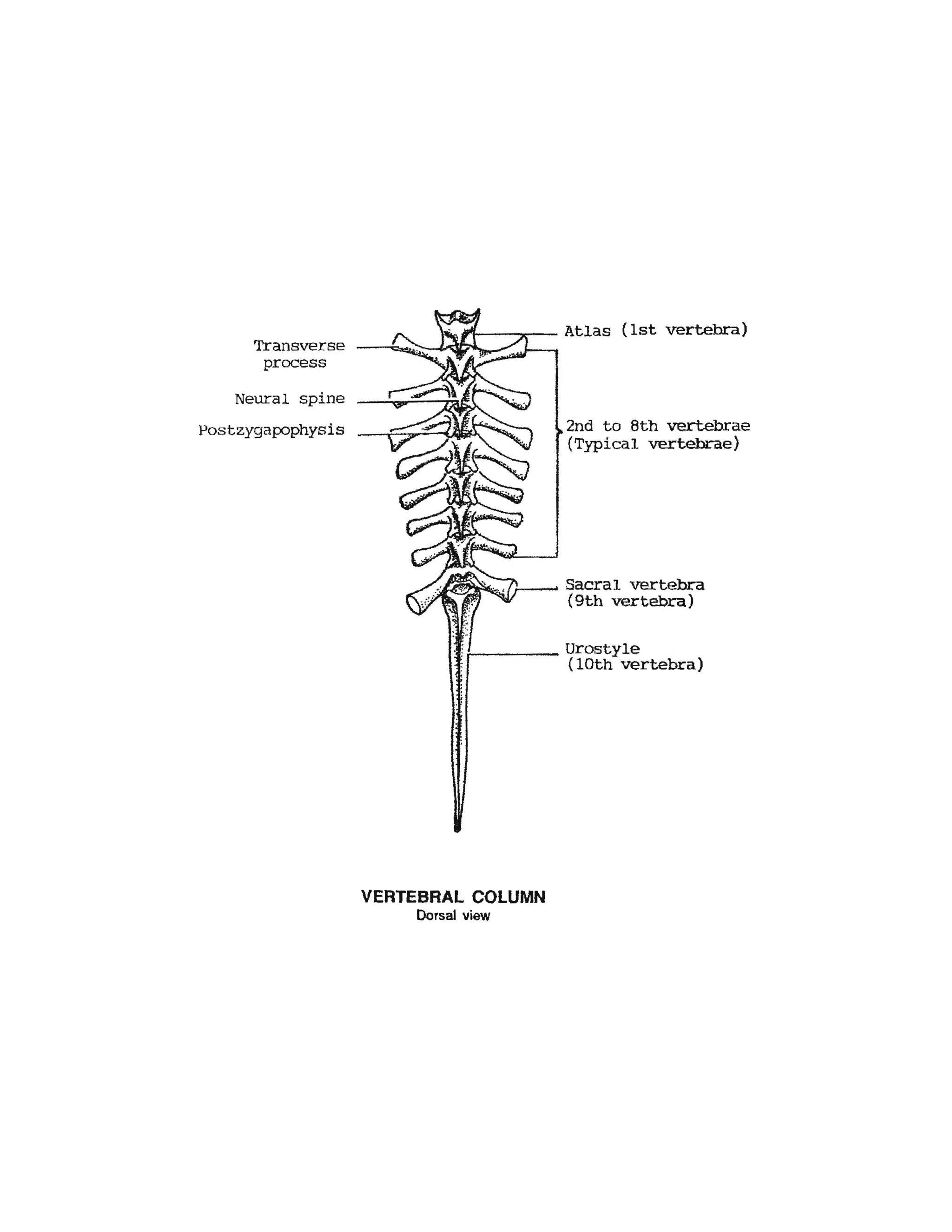
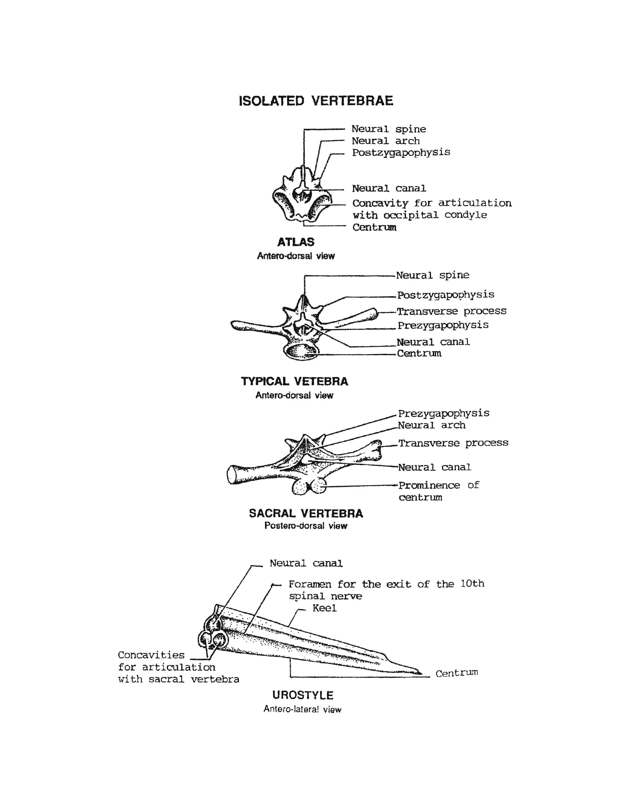

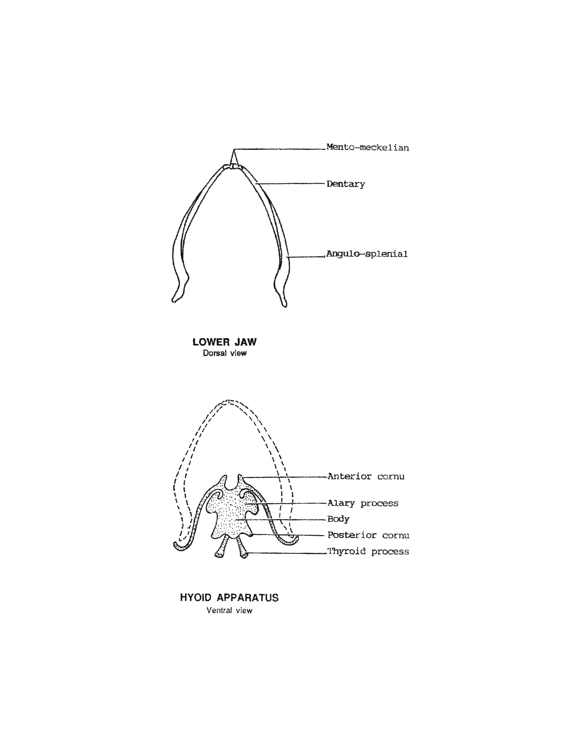


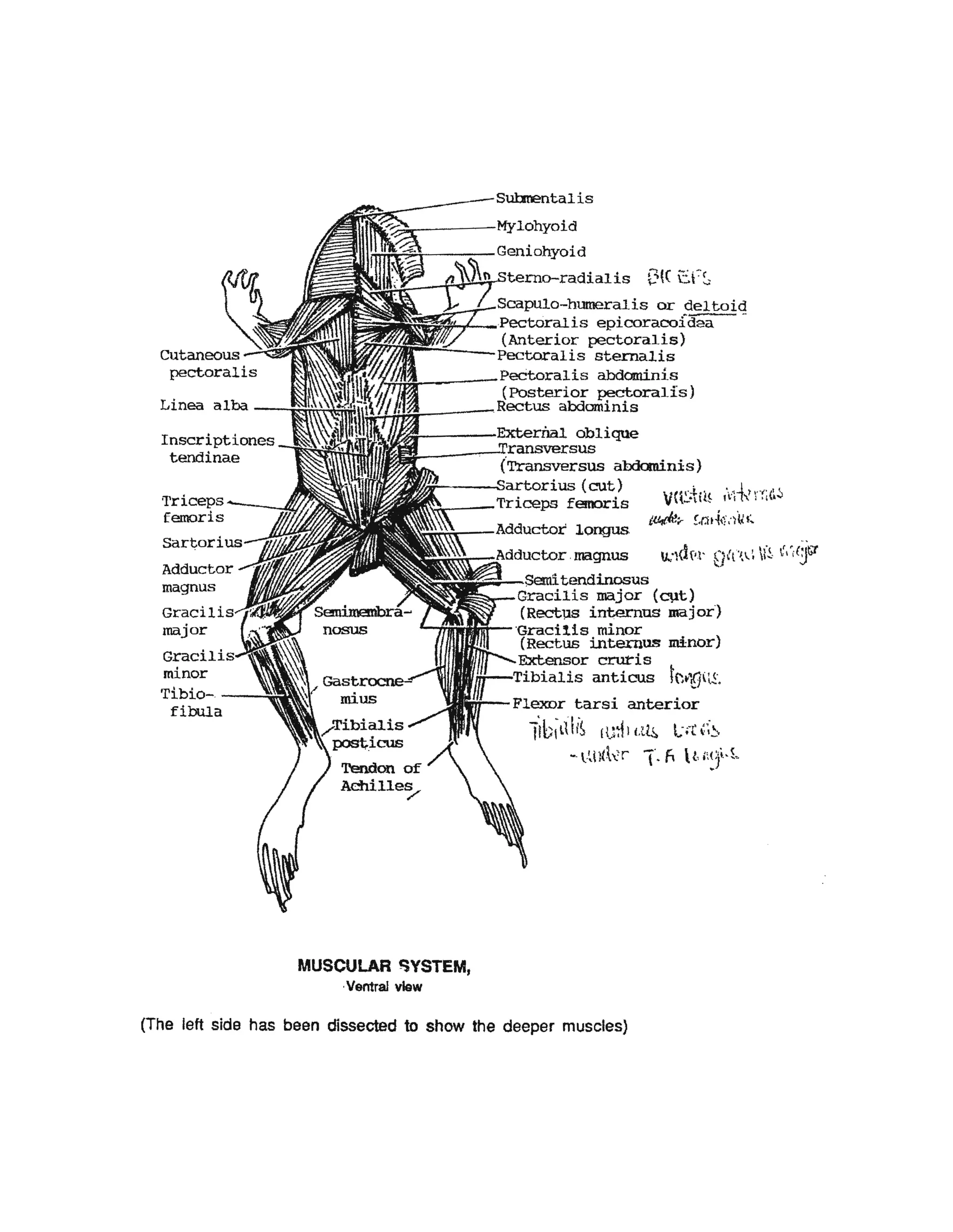

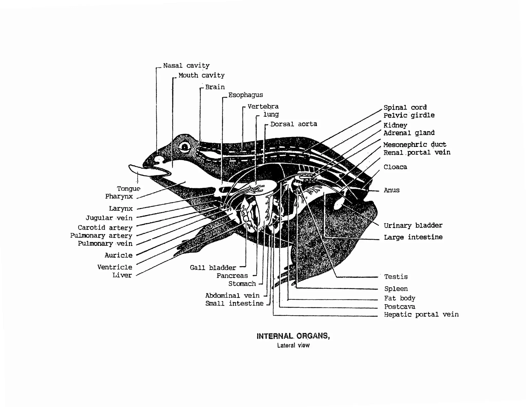
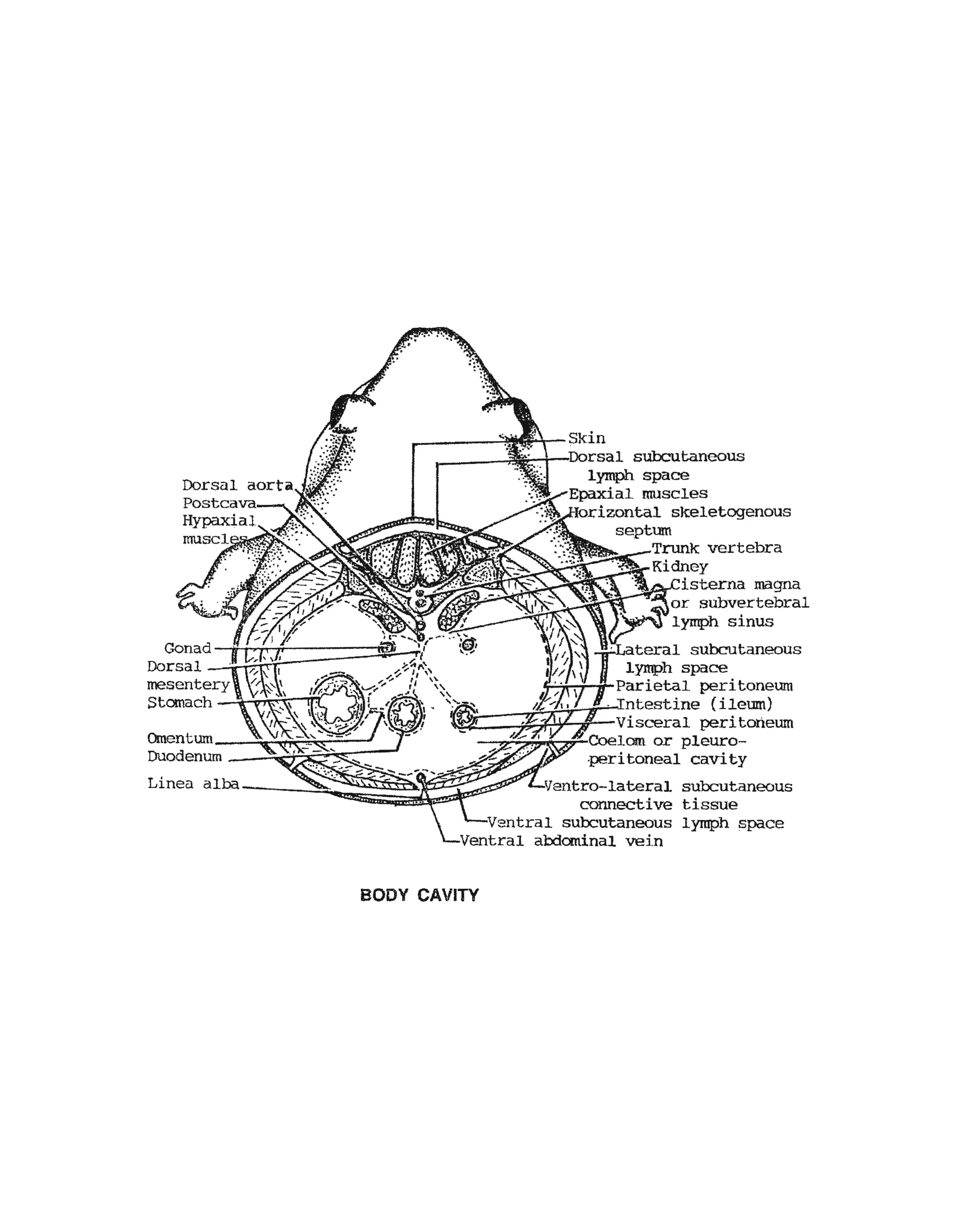


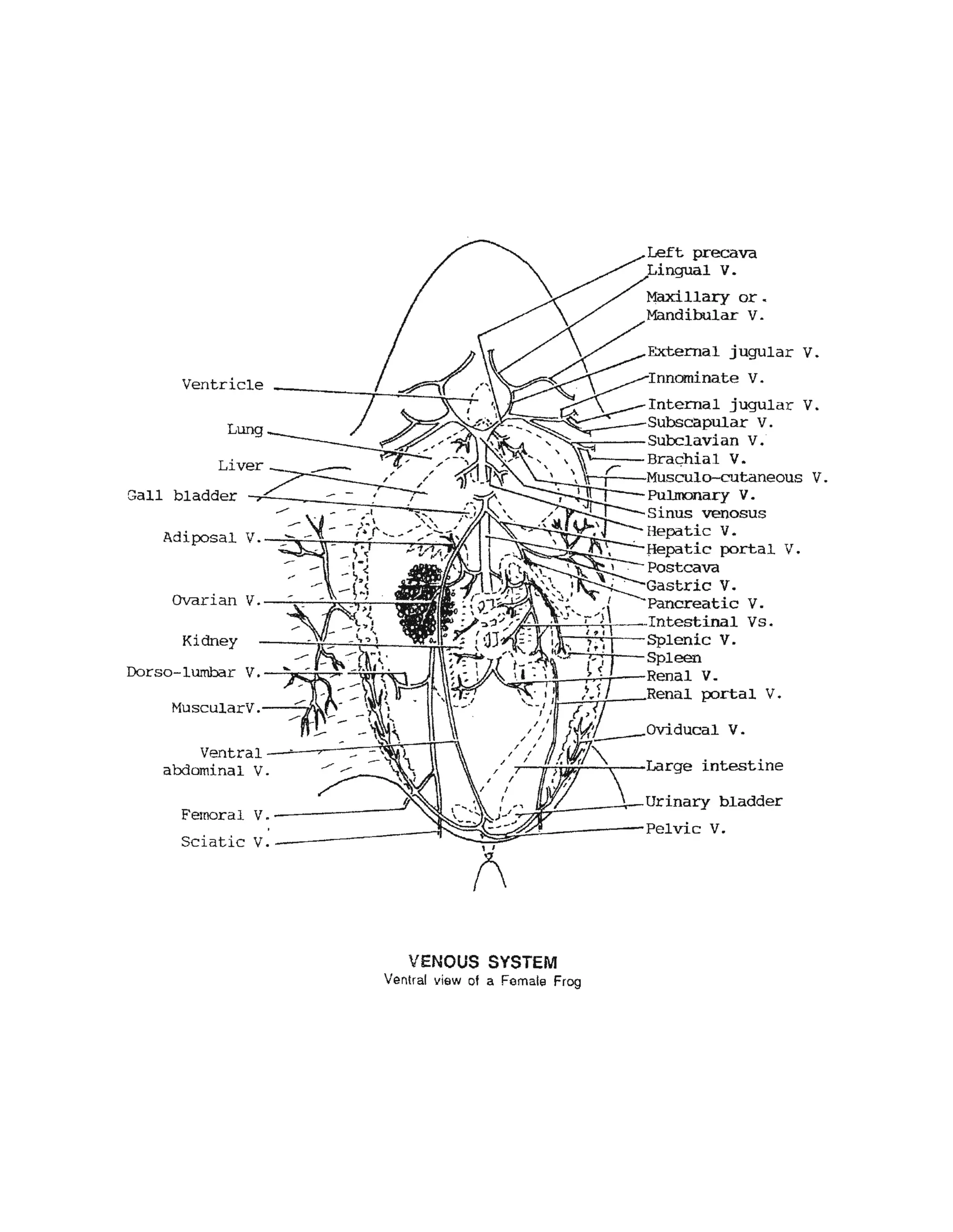


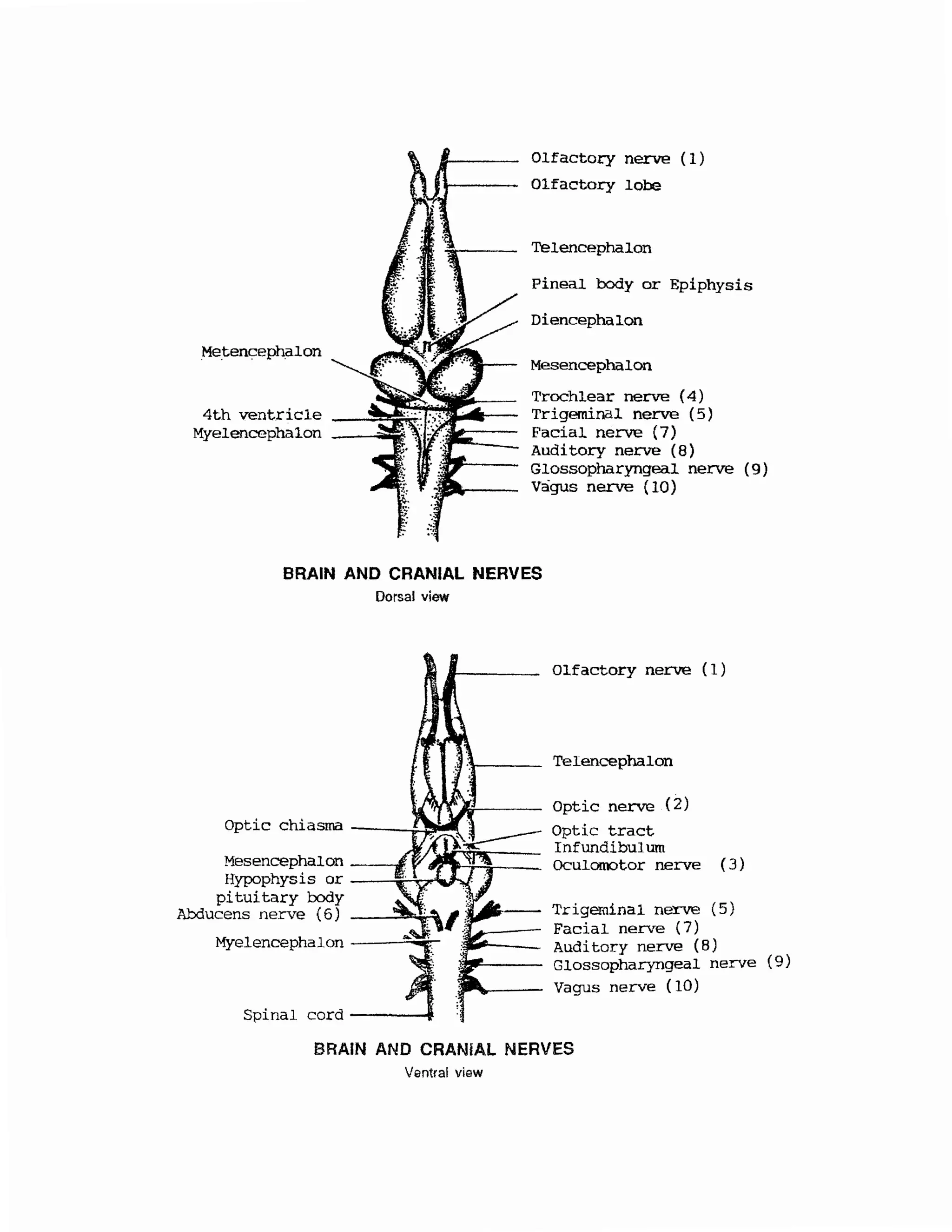


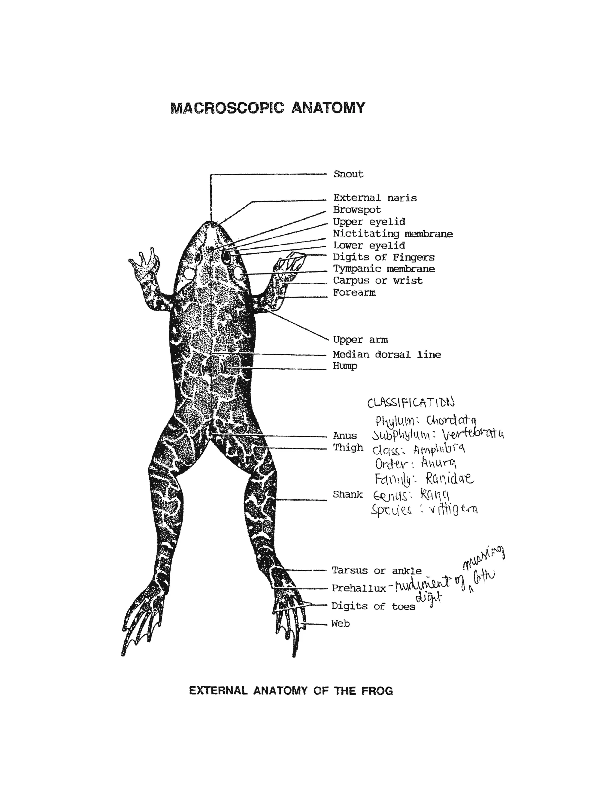
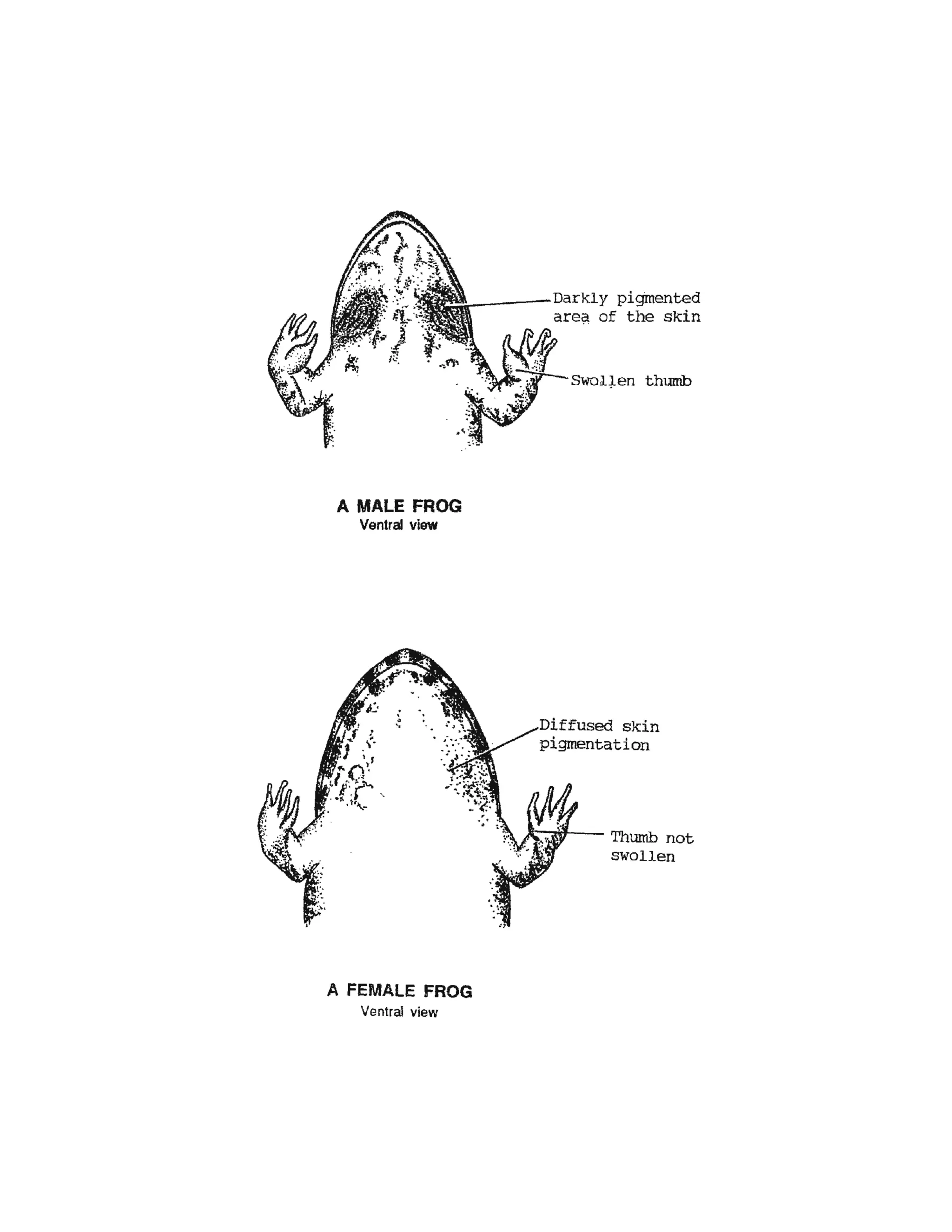
The document provides detailed descriptions and diagrams of the external and internal anatomy of frogs, including: 1) Descriptions and diagrams of the skeletal system, muscular system, digestive system, circulatory system, urogenital system, nervous system, and sense organs of frogs. 2) Labels and explanations of the major bones, muscles, organs, blood vessels, sections of the brain and spinal cord, and cranial nerves. 3) Annotations of the external features of male and female frogs including distinguishing characteristics.


