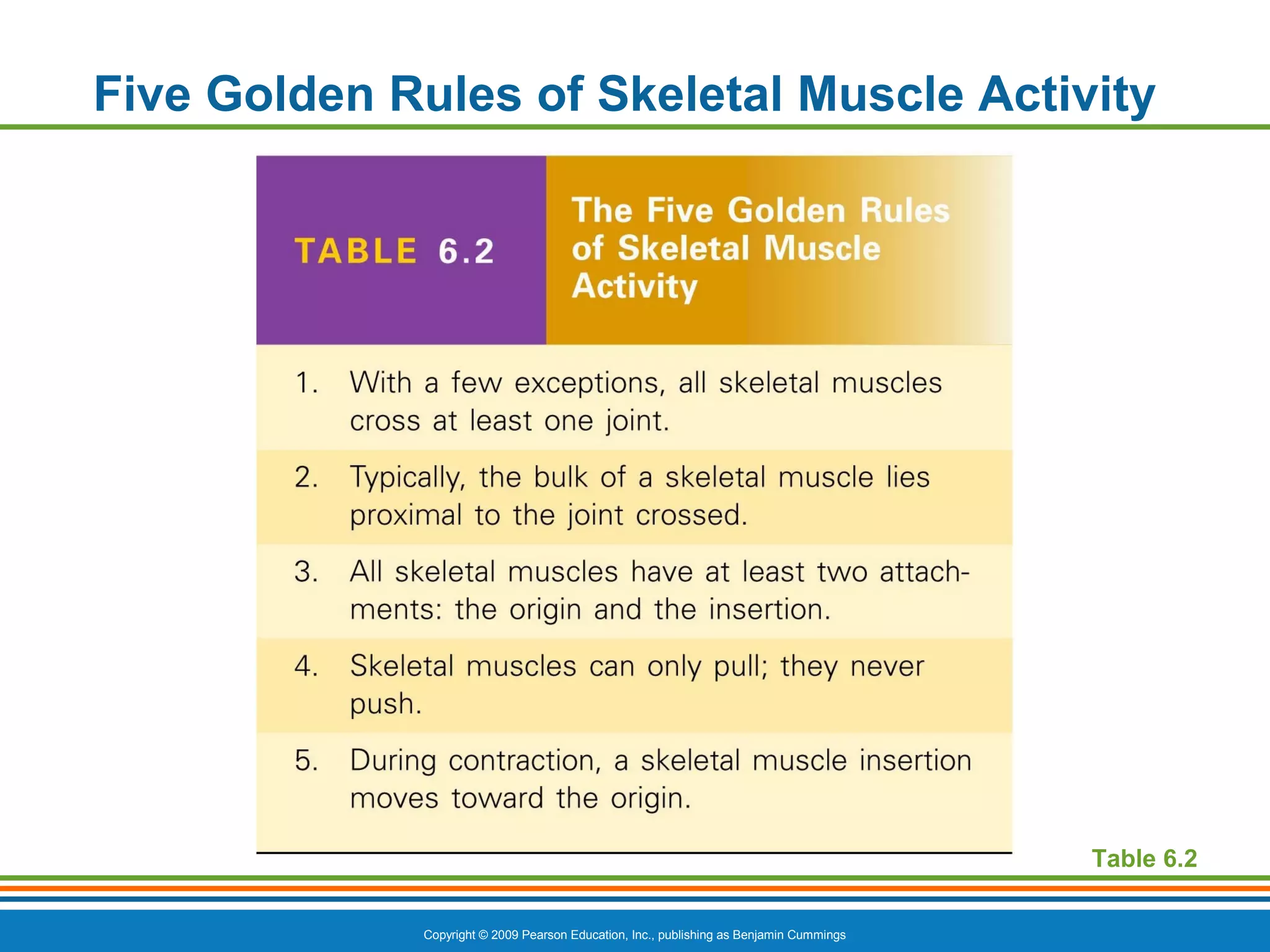
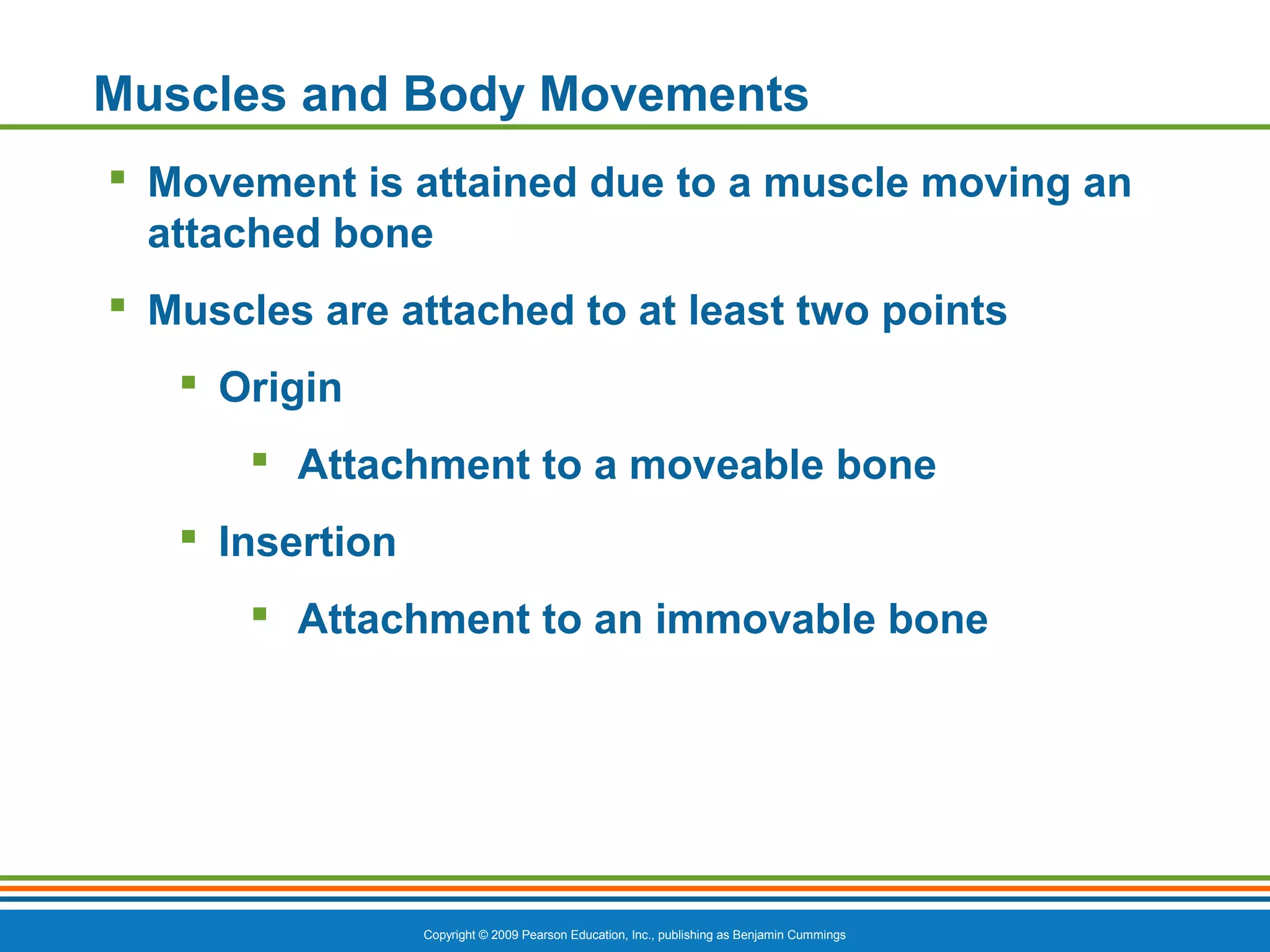
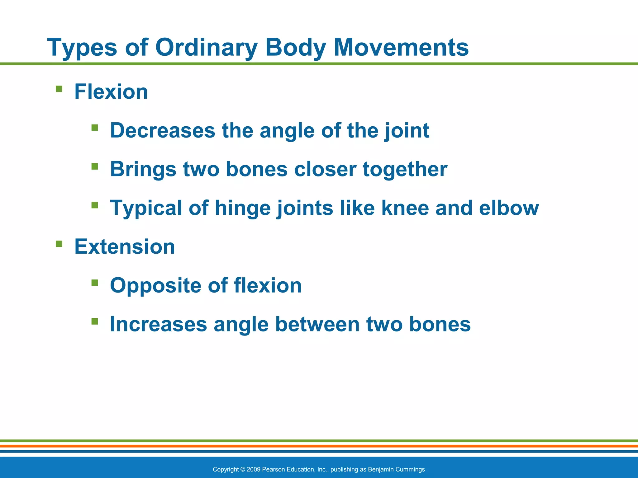
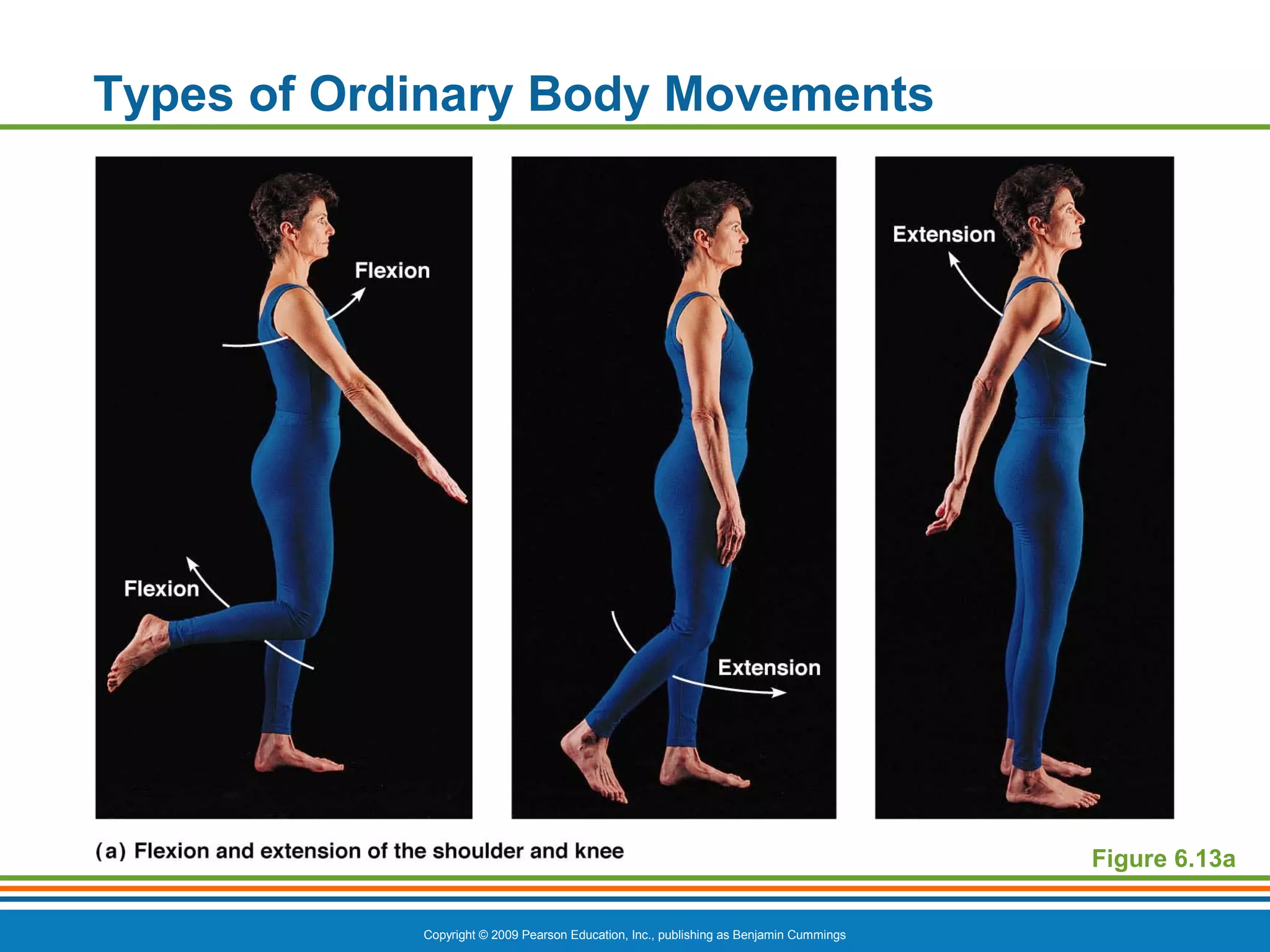
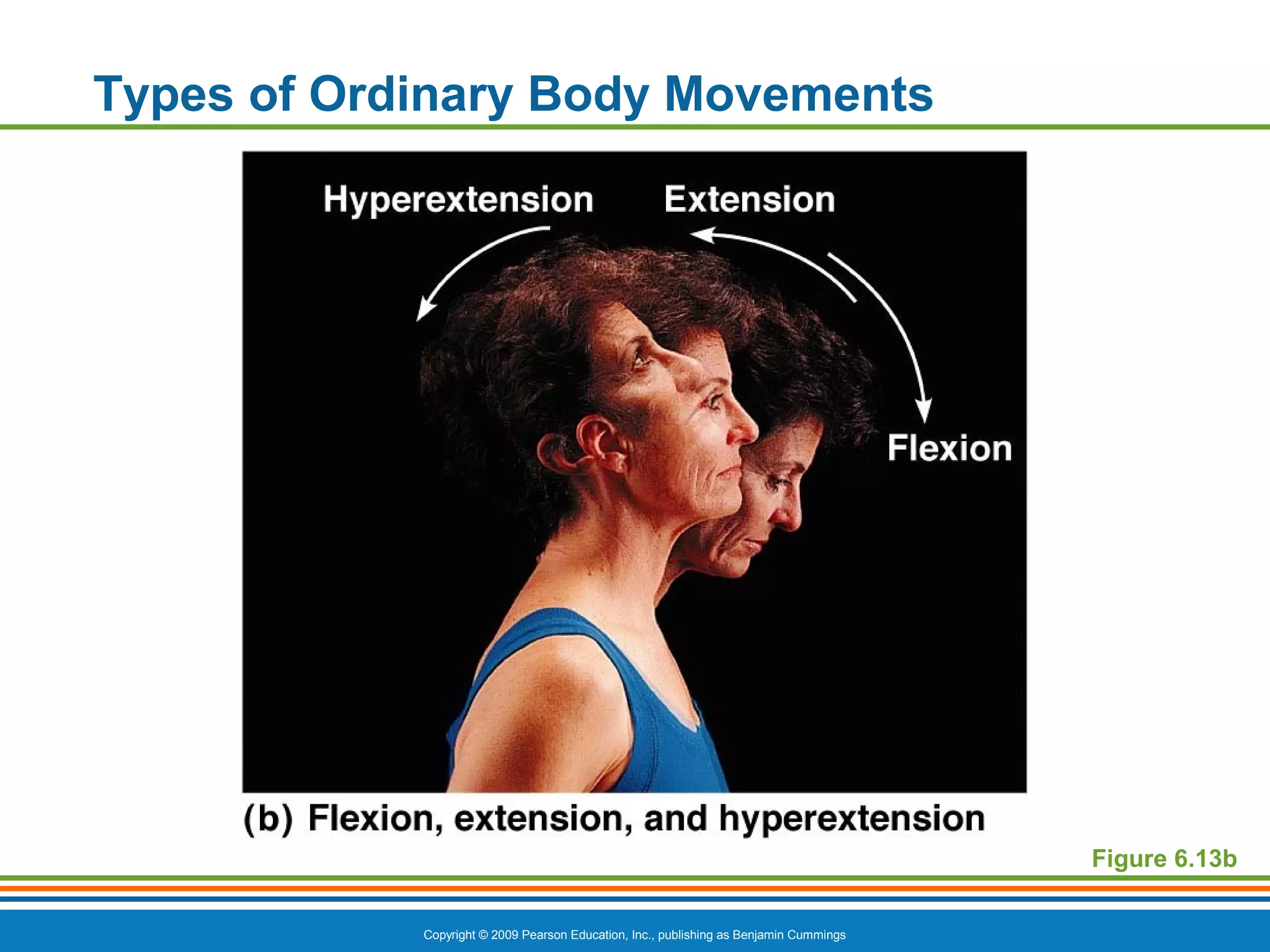
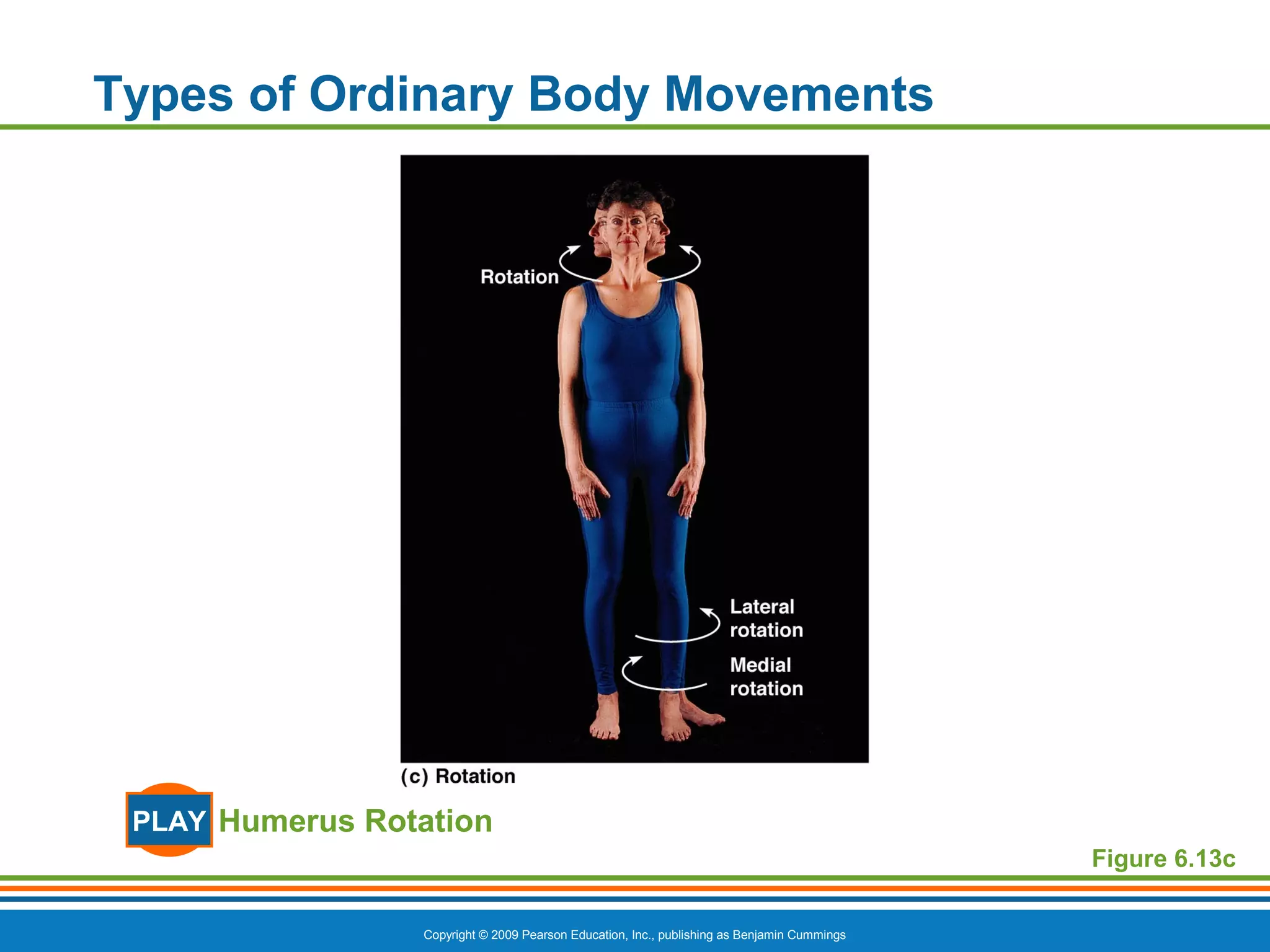
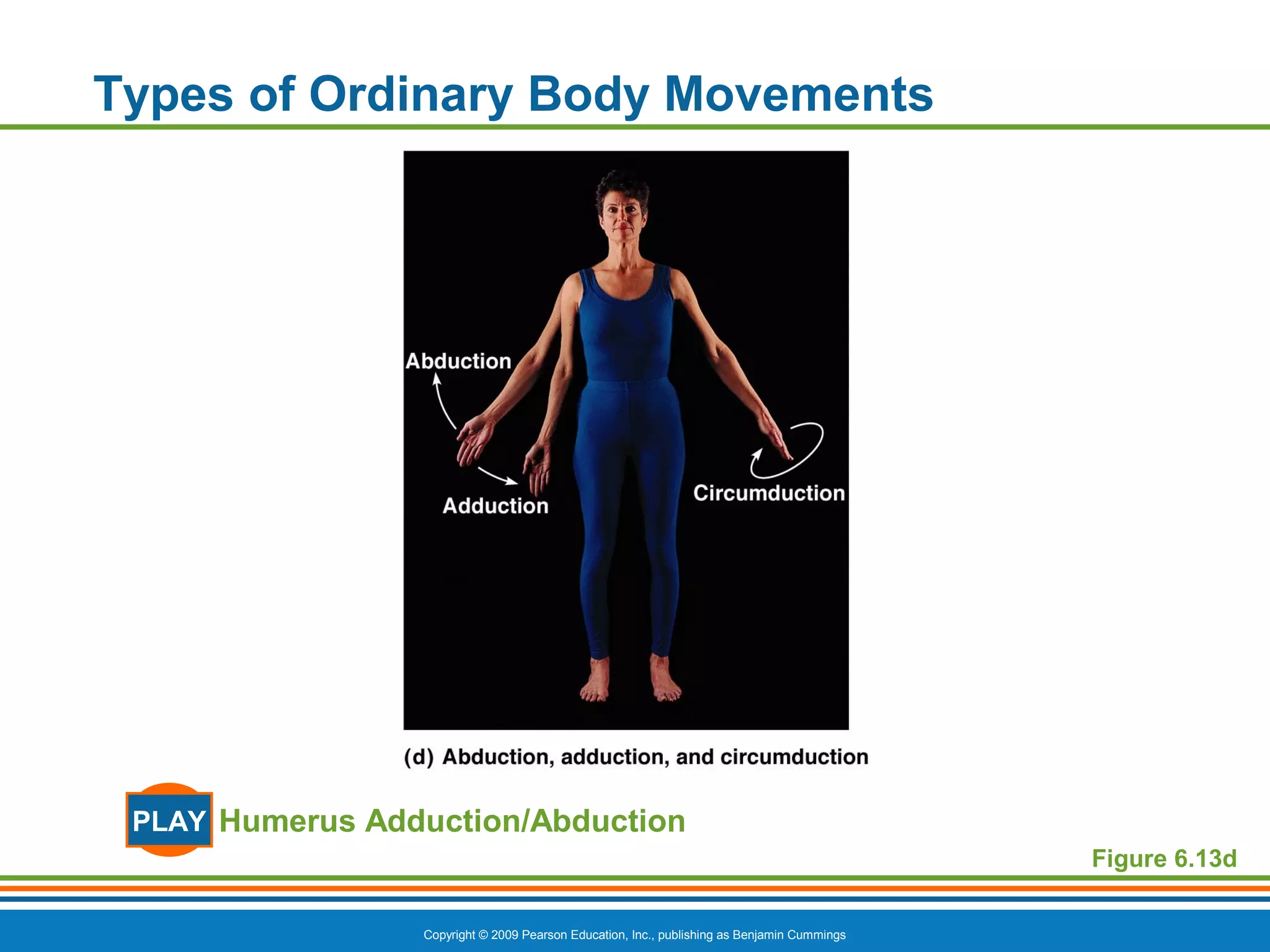
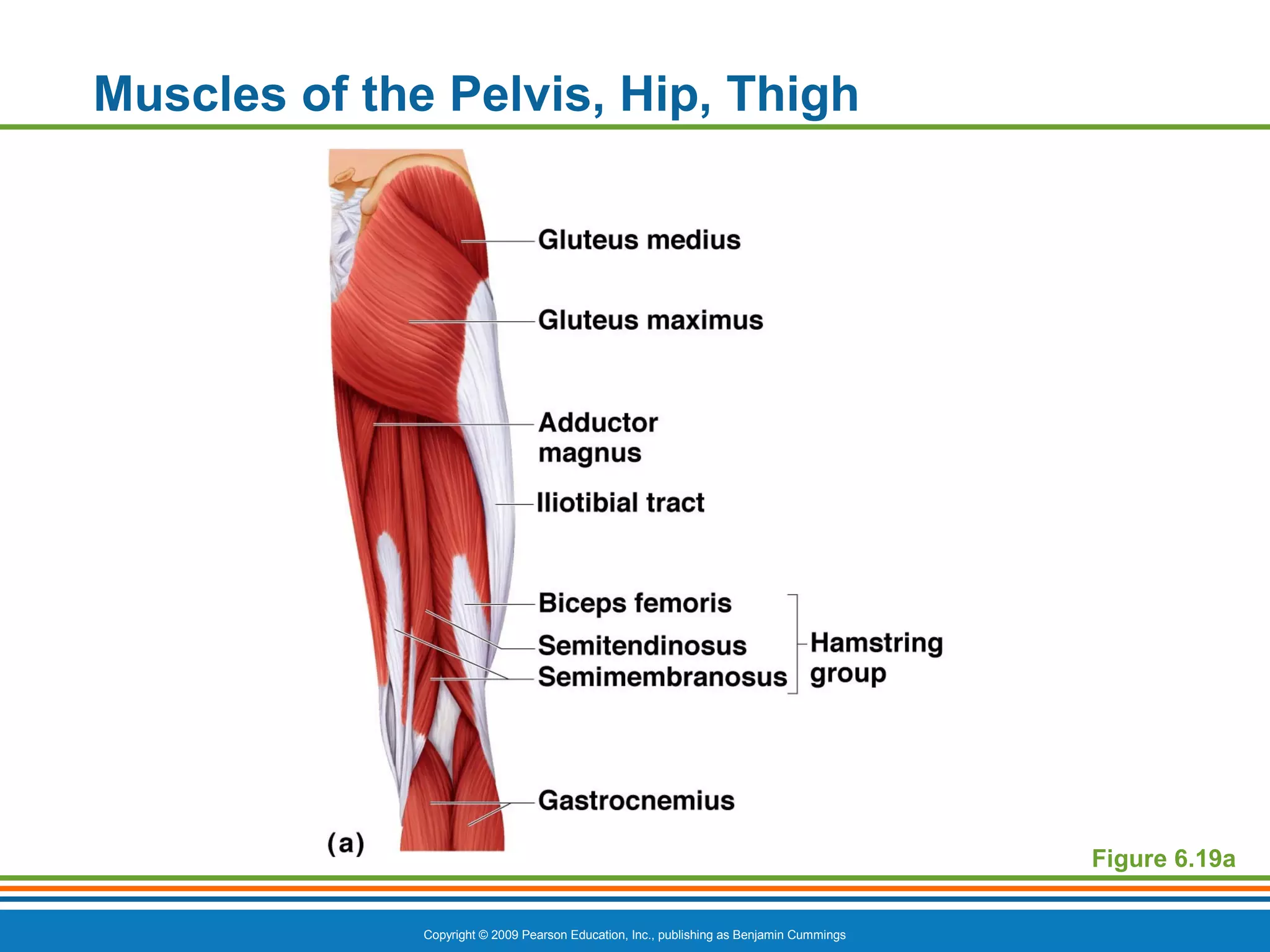
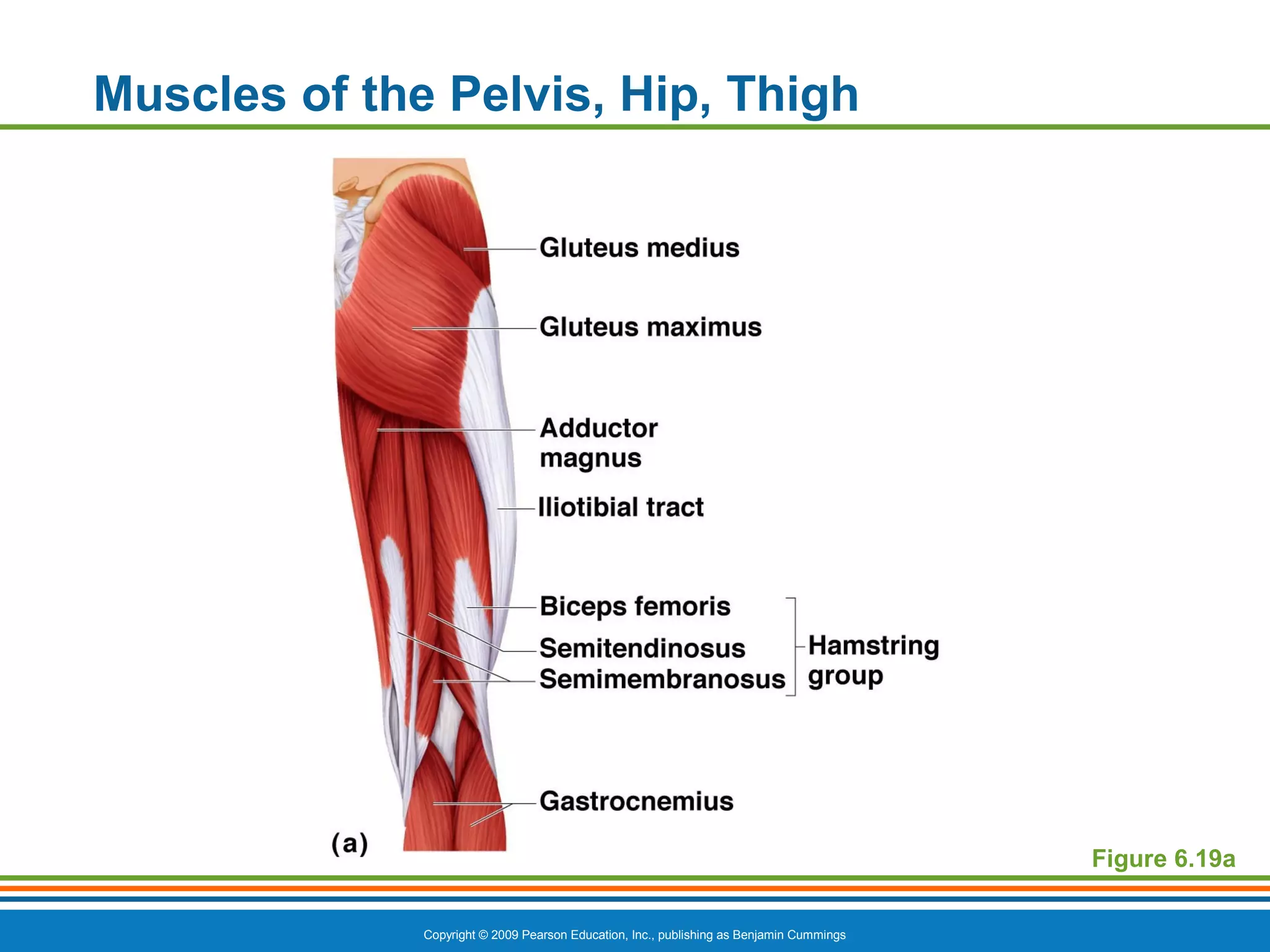
PowerPoint®
Lecture Slide Presentation
by Patty Bostwick-Taylor,
Florence-Darlington Technical College
Copyright © 2009 Pearson Education, Inc., publishing as Benjamin Cummings
PART C
6
The Muscular
System
Copyright © 2009 Pearson Education, Inc., publishing as Benjamin Cummings
Five Golden Rules of Skeletal Muscle Activity
Table 6.2
Copyright © 2009 Pearson Education, Inc., publishing as Benjamin Cummings
Muscles and Body Movements
 Movement is attained due to a muscle moving an
attached bone
 Muscles are attached to at least two points
 Origin
 Attachment to a moveable bone
 Insertion
 Attachment to an immovable bone
Copyright © 2009 Pearson Education, Inc., publishing as Benjamin Cummings
Muscles and Body Movements
Figure 6.12
Copyright © 2009 Pearson Education, Inc., publishing as Benjamin Cummings
Types of Ordinary Body Movements
 Flexion
 Decreases the angle of the joint
 Brings two bones closer together
 Typical of hinge joints like knee and elbow
 Extension
 Opposite of flexion
 Increases angle between two bones
Copyright © 2009 Pearson Education, Inc., publishing as Benjamin Cummings
Types of Ordinary Body Movements
Figure 6.13a
Copyright © 2009 Pearson Education, Inc., publishing as Benjamin Cummings
Types of Ordinary Body Movements
Figure 6.13b
Copyright © 2009 Pearson Education, Inc., publishing as Benjamin Cummings
Types of Ordinary Body Movements
 Rotation
 Movement of a bone around its longitudinal
axis
 Common in ball-and-socket joints
 Example is when you move atlas around the
dens of axis (shake your head “no”)
Copyright © 2009 Pearson Education, Inc., publishing as Benjamin Cummings
Types of Ordinary Body Movements
Humerus RotationPLAY
Figure 6.13c
Copyright © 2009 Pearson Education, Inc., publishing as Benjamin Cummings
Types of Ordinary Body Movements
 Abduction
 Movement of a limb away from the midline
 Adduction
 Opposite of abduction
 Movement of a limb toward the midline
Copyright © 2009 Pearson Education, Inc., publishing as Benjamin Cummings
Types of Ordinary Body Movements
Figure 6.13d
Humerus Adduction/AbductionPLAY
Copyright © 2009 Pearson Education, Inc., publishing as Benjamin Cummings
Types of Ordinary Body Movements
 Circumduction
 Combination of flexion, extension, abduction,
and adduction
 Common in ball-and-socket joints
Copyright © 2009 Pearson Education, Inc., publishing as Benjamin Cummings
Types of Ordinary Body Movements
Wrist CircumductionPLAY
Humerus CircumductionPLAY
Figure 6.13d
Copyright © 2009 Pearson Education, Inc., publishing as Benjamin Cummings
Special Movements
 Dorsiflexion
 Lifting the foot so that the superior surface
approaches the shin
 Plantar flexion
 Depressing the foot (pointing the toes)
Copyright © 2009 Pearson Education, Inc., publishing as Benjamin Cummings
Special Movements
Figure 6.13e
Ankle Dorsiflexion/Plantar FlexionPLAY
Copyright © 2009 Pearson Education, Inc., publishing as Benjamin Cummings
Special Movements
 Inversion
 Turn sole of foot medially
 Eversion
 Turn sole of foot laterally
Copyright © 2009 Pearson Education, Inc., publishing as Benjamin Cummings
Special Movements
Ankle Inversion/EversionPLAY
Figure 6.13f
Copyright © 2009 Pearson Education, Inc., publishing as Benjamin Cummings
Special Movements
 Supination
 Forearm rotates laterally so palm faces
anteriorly
 Pronation
 Forearm rotates medially so palm faces
posteriorly
Copyright © 2009 Pearson Education, Inc., publishing as Benjamin Cummings
Special Movements
Figure 6.13g
Elbow Pronation/SupinationPLAY
Copyright © 2009 Pearson Education, Inc., publishing as Benjamin Cummings
Special Movements
 Opposition
 Move thumb to touch the tips of other fingers
on the same hand
Copyright © 2009 Pearson Education, Inc., publishing as Benjamin Cummings
Special Movements
Hand OppositionPLAY
Figure 6.13h
Copyright © 2009 Pearson Education, Inc., publishing as Benjamin Cummings
Types of Muscles
 Prime mover—muscle with the major
responsibility for a certain movement
 Antagonist—muscle that opposes or reverses a
prime mover
 Synergist—muscle that aids a prime mover in a
movement and helps prevent rotation
 Fixator—stabilizes the origin of a prime mover
Copyright © 2009 Pearson Education, Inc., publishing as Benjamin Cummings
Naming Skeletal Muscles
 By direction of muscle fibers
 Example: Rectus (straight)
 By relative size of the muscle
 Example: Maximus (largest)
Copyright © 2009 Pearson Education, Inc., publishing as Benjamin Cummings
Naming Skeletal Muscles
 By location of the muscle
 Example: Temporalis (temporal bone)
 By number of origins
 Example: Triceps (three heads)
Copyright © 2009 Pearson Education, Inc., publishing as Benjamin Cummings
Naming Skeletal Muscles
 By location of the muscle’s origin and insertion
 Example: Sterno (on the sternum)
 By shape of the muscle
 Example: Deltoid (triangular)
 By action of the muscle
 Example: Flexor and extensor (flexes or
extends a bone)
Copyright © 2009 Pearson Education, Inc., publishing as Benjamin Cummings
Head and Neck Muscles
 Facial muscles
 Frontalis—raises eyebrows
 Orbicularis oculi—closes eyes, squints,
blinks, winks
 Orbicularis oris—closes mouth and protrudes
the lips
 Buccinator—flattens the cheek, chews
 Zygomaticus—raises corners of the mouth
 Chewing muscles
 Masseter—closes the jaw and elevates
mandible
 Temporalis—synergist of the masseter, closes
jaw
Copyright © 2009 Pearson Education, Inc., publishing as Benjamin Cummings
Head and Neck Muscles
 Neck muscles
 Platysma—pulls the corners of the mouth
inferiorly
 Sternocleidomastoid—flexes the neck, rotates
the head
Copyright © 2009 Pearson Education, Inc., publishing as Benjamin Cummings
Head and Neck Muscles
Figure 6.15
Copyright © 2009 Pearson Education, Inc., publishing as Benjamin Cummings
Muscles of Trunk, Shoulder, Arm
 Anterior muscles
 Pectoralis major—adducts and flexes the
humerus
 Intercostal muscles
 External intercostals—raise rib cage
during inhalation
 Internal intercostals—depress the rib cage
to move air out of the lungs when you
exhale forcibly
Copyright © 2009 Pearson Education, Inc., publishing as Benjamin Cummings
Anterior Muscles of Trunk, Shoulder, Arm
Figure 6.16a
Copyright © 2009 Pearson Education, Inc., publishing as Benjamin Cummings
Muscles of Trunk, Shoulder, Arm
 Muscles of the abdominal girdle
 Rectus abdominis—flexes vertebral column
and compresses abdominal contents
(defecation, childbirth, forced breathing)
 External and internal obliques—flex vertebral
column; rotate trunk and bend it laterally
 Transversus abdominis—compresses
abdominal contents
Copyright © 2009 Pearson Education, Inc., publishing as Benjamin Cummings
Anterior Muscles of Trunk, Shoulder, Arm
Figure 6.16b
Copyright © 2009 Pearson Education, Inc., publishing as Benjamin Cummings
Muscles of Trunk, Shoulder, Arm
 Posterior muscles
 Trapezius—elevates, depresses, adducts, and
stabilizes the scapula
 Latissimus dorsi—extends and adducts the
humerus
 Deltoid—arm abduction
Copyright © 2009 Pearson Education, Inc., publishing as Benjamin Cummings
Muscles of Posterior Neck, Trunk, Arm
Figure 6.17a
Copyright © 2009 Pearson Education, Inc., publishing as Benjamin Cummings
Muscles of the Upper Limb
 Biceps brachii—supinates forearm, flexes elbow
 Brachialis—elbow flexion
 Brachioradialis—weak muscle
 Triceps brachii—elbow extension (antagonist to
biceps brachii)
Copyright © 2009 Pearson Education, Inc., publishing as Benjamin Cummings
Anterior Muscles of Trunk, Shoulder, Arm
Figure 6.16a
Copyright © 2009 Pearson Education, Inc., publishing as Benjamin Cummings
Muscles of Posterior Neck, Trunk, Arm
Figure 6.17a
Copyright © 2009 Pearson Education, Inc., publishing as Benjamin Cummings
Muscles of the Lower Limb
 Gluteus maximus—hip extension
 Gluteus medius—hip abduction, steadies pelvis
when walking
 Iliopsoas—hip flexion, keeps the upper body from
falling backward when standing erect
 Adductor muscles—adduct the thighs
Copyright © 2009 Pearson Education, Inc., publishing as Benjamin Cummings
Muscles of the Pelvis, Hip, Thigh
Figure 6.19a
Copyright © 2009 Pearson Education, Inc., publishing as Benjamin Cummings
Muscles of the Pelvis, Hip, Thigh
Figure 6.19c
Copyright © 2009 Pearson Education, Inc., publishing as Benjamin Cummings
Muscles of the Lower Limb
 Muscles causing movement at the knee joint
 Hamstring group—thigh extension and knee
flexion
 Biceps femoris
 Semimembranosus
 Semitendinosus
Copyright © 2009 Pearson Education, Inc., publishing as Benjamin Cummings
Muscles of the Pelvis, Hip, Thigh
Figure 6.19a
Copyright © 2009 Pearson Education, Inc., publishing as Benjamin Cummings
Muscles of the Lower Limb
 Muscles causing movement at the knee joint
 Sartorius—flexes the thigh
 Quadriceps group—extends the knee
 Rectus femoris
 Vastus muscles (three)
Copyright © 2009 Pearson Education, Inc., publishing as Benjamin Cummings
Muscles of the Pelvis, Hip, Thigh
Figure 6.19c
Copyright © 2009 Pearson Education, Inc., publishing as Benjamin Cummings
Muscles of the Lower Limb
 Muscles causing movement at ankle and foot
 Tibialis anterior—dorsiflexion and foot
inversion
 Extensor digitorum longus—toe extension and
dorsiflexion of the foot
 Fibularis muscles—plantar flexion, everts the
foot
 Soleus—plantar flexion
Copyright © 2009 Pearson Education, Inc., publishing as Benjamin Cummings
Muscles of the Lower Leg
Figure 6.20a
Copyright © 2009 Pearson Education, Inc., publishing as Benjamin Cummings
Muscles of the Lower Leg
Figure 6.20b
Copyright © 2009 Pearson Education, Inc., publishing as Benjamin Cummings
Superficial Muscles: Anterior
Figure 6.21
Copyright © 2009 Pearson Education, Inc., publishing as Benjamin Cummings
Superficial Muscles: Posterior
Figure 6.22
Copyright © 2009 Pearson Education, Inc., publishing as Benjamin Cummings
Intramuscular Injection Sites
Figure 6.18, 6.19b, d

CVA A&P - Chapter 6c: Muscle Movements and Major Muscles

  • 1.
    PowerPoint® Lecture Slide Presentation byPatty Bostwick-Taylor, Florence-Darlington Technical College Copyright © 2009 Pearson Education, Inc., publishing as Benjamin Cummings PART C 6 The Muscular System
  • 2.
    Copyright © 2009Pearson Education, Inc., publishing as Benjamin Cummings Five Golden Rules of Skeletal Muscle Activity Table 6.2
  • 3.
    Copyright © 2009Pearson Education, Inc., publishing as Benjamin Cummings Muscles and Body Movements  Movement is attained due to a muscle moving an attached bone  Muscles are attached to at least two points  Origin  Attachment to a moveable bone  Insertion  Attachment to an immovable bone
  • 4.
    Copyright © 2009Pearson Education, Inc., publishing as Benjamin Cummings Muscles and Body Movements Figure 6.12
  • 5.
    Copyright © 2009Pearson Education, Inc., publishing as Benjamin Cummings Types of Ordinary Body Movements  Flexion  Decreases the angle of the joint  Brings two bones closer together  Typical of hinge joints like knee and elbow  Extension  Opposite of flexion  Increases angle between two bones
  • 6.
    Copyright © 2009Pearson Education, Inc., publishing as Benjamin Cummings Types of Ordinary Body Movements Figure 6.13a
  • 7.
    Copyright © 2009Pearson Education, Inc., publishing as Benjamin Cummings Types of Ordinary Body Movements Figure 6.13b
  • 8.
    Copyright © 2009Pearson Education, Inc., publishing as Benjamin Cummings Types of Ordinary Body Movements  Rotation  Movement of a bone around its longitudinal axis  Common in ball-and-socket joints  Example is when you move atlas around the dens of axis (shake your head “no”)
  • 9.
    Copyright © 2009Pearson Education, Inc., publishing as Benjamin Cummings Types of Ordinary Body Movements Humerus RotationPLAY Figure 6.13c
  • 10.
    Copyright © 2009Pearson Education, Inc., publishing as Benjamin Cummings Types of Ordinary Body Movements  Abduction  Movement of a limb away from the midline  Adduction  Opposite of abduction  Movement of a limb toward the midline
  • 11.
    Copyright © 2009Pearson Education, Inc., publishing as Benjamin Cummings Types of Ordinary Body Movements Figure 6.13d Humerus Adduction/AbductionPLAY
  • 12.
    Copyright © 2009Pearson Education, Inc., publishing as Benjamin Cummings Types of Ordinary Body Movements  Circumduction  Combination of flexion, extension, abduction, and adduction  Common in ball-and-socket joints
  • 13.
    Copyright © 2009Pearson Education, Inc., publishing as Benjamin Cummings Types of Ordinary Body Movements Wrist CircumductionPLAY Humerus CircumductionPLAY Figure 6.13d
  • 14.
    Copyright © 2009Pearson Education, Inc., publishing as Benjamin Cummings Special Movements  Dorsiflexion  Lifting the foot so that the superior surface approaches the shin  Plantar flexion  Depressing the foot (pointing the toes)
  • 15.
    Copyright © 2009Pearson Education, Inc., publishing as Benjamin Cummings Special Movements Figure 6.13e Ankle Dorsiflexion/Plantar FlexionPLAY
  • 16.
    Copyright © 2009Pearson Education, Inc., publishing as Benjamin Cummings Special Movements  Inversion  Turn sole of foot medially  Eversion  Turn sole of foot laterally
  • 17.
    Copyright © 2009Pearson Education, Inc., publishing as Benjamin Cummings Special Movements Ankle Inversion/EversionPLAY Figure 6.13f
  • 18.
    Copyright © 2009Pearson Education, Inc., publishing as Benjamin Cummings Special Movements  Supination  Forearm rotates laterally so palm faces anteriorly  Pronation  Forearm rotates medially so palm faces posteriorly
  • 19.
    Copyright © 2009Pearson Education, Inc., publishing as Benjamin Cummings Special Movements Figure 6.13g Elbow Pronation/SupinationPLAY
  • 20.
    Copyright © 2009Pearson Education, Inc., publishing as Benjamin Cummings Special Movements  Opposition  Move thumb to touch the tips of other fingers on the same hand
  • 21.
    Copyright © 2009Pearson Education, Inc., publishing as Benjamin Cummings Special Movements Hand OppositionPLAY Figure 6.13h
  • 22.
    Copyright © 2009Pearson Education, Inc., publishing as Benjamin Cummings Types of Muscles  Prime mover—muscle with the major responsibility for a certain movement  Antagonist—muscle that opposes or reverses a prime mover  Synergist—muscle that aids a prime mover in a movement and helps prevent rotation  Fixator—stabilizes the origin of a prime mover
  • 23.
    Copyright © 2009Pearson Education, Inc., publishing as Benjamin Cummings Naming Skeletal Muscles  By direction of muscle fibers  Example: Rectus (straight)  By relative size of the muscle  Example: Maximus (largest)
  • 24.
    Copyright © 2009Pearson Education, Inc., publishing as Benjamin Cummings Naming Skeletal Muscles  By location of the muscle  Example: Temporalis (temporal bone)  By number of origins  Example: Triceps (three heads)
  • 25.
    Copyright © 2009Pearson Education, Inc., publishing as Benjamin Cummings Naming Skeletal Muscles  By location of the muscle’s origin and insertion  Example: Sterno (on the sternum)  By shape of the muscle  Example: Deltoid (triangular)  By action of the muscle  Example: Flexor and extensor (flexes or extends a bone)
  • 26.
    Copyright © 2009Pearson Education, Inc., publishing as Benjamin Cummings Head and Neck Muscles  Facial muscles  Frontalis—raises eyebrows  Orbicularis oculi—closes eyes, squints, blinks, winks  Orbicularis oris—closes mouth and protrudes the lips  Buccinator—flattens the cheek, chews  Zygomaticus—raises corners of the mouth  Chewing muscles  Masseter—closes the jaw and elevates mandible  Temporalis—synergist of the masseter, closes jaw
  • 27.
    Copyright © 2009Pearson Education, Inc., publishing as Benjamin Cummings Head and Neck Muscles  Neck muscles  Platysma—pulls the corners of the mouth inferiorly  Sternocleidomastoid—flexes the neck, rotates the head
  • 28.
    Copyright © 2009Pearson Education, Inc., publishing as Benjamin Cummings Head and Neck Muscles Figure 6.15
  • 29.
    Copyright © 2009Pearson Education, Inc., publishing as Benjamin Cummings Muscles of Trunk, Shoulder, Arm  Anterior muscles  Pectoralis major—adducts and flexes the humerus  Intercostal muscles  External intercostals—raise rib cage during inhalation  Internal intercostals—depress the rib cage to move air out of the lungs when you exhale forcibly
  • 30.
    Copyright © 2009Pearson Education, Inc., publishing as Benjamin Cummings Anterior Muscles of Trunk, Shoulder, Arm Figure 6.16a
  • 31.
    Copyright © 2009Pearson Education, Inc., publishing as Benjamin Cummings Muscles of Trunk, Shoulder, Arm  Muscles of the abdominal girdle  Rectus abdominis—flexes vertebral column and compresses abdominal contents (defecation, childbirth, forced breathing)  External and internal obliques—flex vertebral column; rotate trunk and bend it laterally  Transversus abdominis—compresses abdominal contents
  • 32.
    Copyright © 2009Pearson Education, Inc., publishing as Benjamin Cummings Anterior Muscles of Trunk, Shoulder, Arm Figure 6.16b
  • 33.
    Copyright © 2009Pearson Education, Inc., publishing as Benjamin Cummings Muscles of Trunk, Shoulder, Arm  Posterior muscles  Trapezius—elevates, depresses, adducts, and stabilizes the scapula  Latissimus dorsi—extends and adducts the humerus  Deltoid—arm abduction
  • 34.
    Copyright © 2009Pearson Education, Inc., publishing as Benjamin Cummings Muscles of Posterior Neck, Trunk, Arm Figure 6.17a
  • 35.
    Copyright © 2009Pearson Education, Inc., publishing as Benjamin Cummings Muscles of the Upper Limb  Biceps brachii—supinates forearm, flexes elbow  Brachialis—elbow flexion  Brachioradialis—weak muscle  Triceps brachii—elbow extension (antagonist to biceps brachii)
  • 36.
    Copyright © 2009Pearson Education, Inc., publishing as Benjamin Cummings Anterior Muscles of Trunk, Shoulder, Arm Figure 6.16a
  • 37.
    Copyright © 2009Pearson Education, Inc., publishing as Benjamin Cummings Muscles of Posterior Neck, Trunk, Arm Figure 6.17a
  • 38.
    Copyright © 2009Pearson Education, Inc., publishing as Benjamin Cummings Muscles of the Lower Limb  Gluteus maximus—hip extension  Gluteus medius—hip abduction, steadies pelvis when walking  Iliopsoas—hip flexion, keeps the upper body from falling backward when standing erect  Adductor muscles—adduct the thighs
  • 39.
    Copyright © 2009Pearson Education, Inc., publishing as Benjamin Cummings Muscles of the Pelvis, Hip, Thigh Figure 6.19a
  • 40.
    Copyright © 2009Pearson Education, Inc., publishing as Benjamin Cummings Muscles of the Pelvis, Hip, Thigh Figure 6.19c
  • 41.
    Copyright © 2009Pearson Education, Inc., publishing as Benjamin Cummings Muscles of the Lower Limb  Muscles causing movement at the knee joint  Hamstring group—thigh extension and knee flexion  Biceps femoris  Semimembranosus  Semitendinosus
  • 42.
    Copyright © 2009Pearson Education, Inc., publishing as Benjamin Cummings Muscles of the Pelvis, Hip, Thigh Figure 6.19a
  • 43.
    Copyright © 2009Pearson Education, Inc., publishing as Benjamin Cummings Muscles of the Lower Limb  Muscles causing movement at the knee joint  Sartorius—flexes the thigh  Quadriceps group—extends the knee  Rectus femoris  Vastus muscles (three)
  • 44.
    Copyright © 2009Pearson Education, Inc., publishing as Benjamin Cummings Muscles of the Pelvis, Hip, Thigh Figure 6.19c
  • 45.
    Copyright © 2009Pearson Education, Inc., publishing as Benjamin Cummings Muscles of the Lower Limb  Muscles causing movement at ankle and foot  Tibialis anterior—dorsiflexion and foot inversion  Extensor digitorum longus—toe extension and dorsiflexion of the foot  Fibularis muscles—plantar flexion, everts the foot  Soleus—plantar flexion
  • 46.
    Copyright © 2009Pearson Education, Inc., publishing as Benjamin Cummings Muscles of the Lower Leg Figure 6.20a
  • 47.
    Copyright © 2009Pearson Education, Inc., publishing as Benjamin Cummings Muscles of the Lower Leg Figure 6.20b
  • 48.
    Copyright © 2009Pearson Education, Inc., publishing as Benjamin Cummings Superficial Muscles: Anterior Figure 6.21
  • 49.
    Copyright © 2009Pearson Education, Inc., publishing as Benjamin Cummings Superficial Muscles: Posterior Figure 6.22
  • 50.
    Copyright © 2009Pearson Education, Inc., publishing as Benjamin Cummings Intramuscular Injection Sites Figure 6.18, 6.19b, d