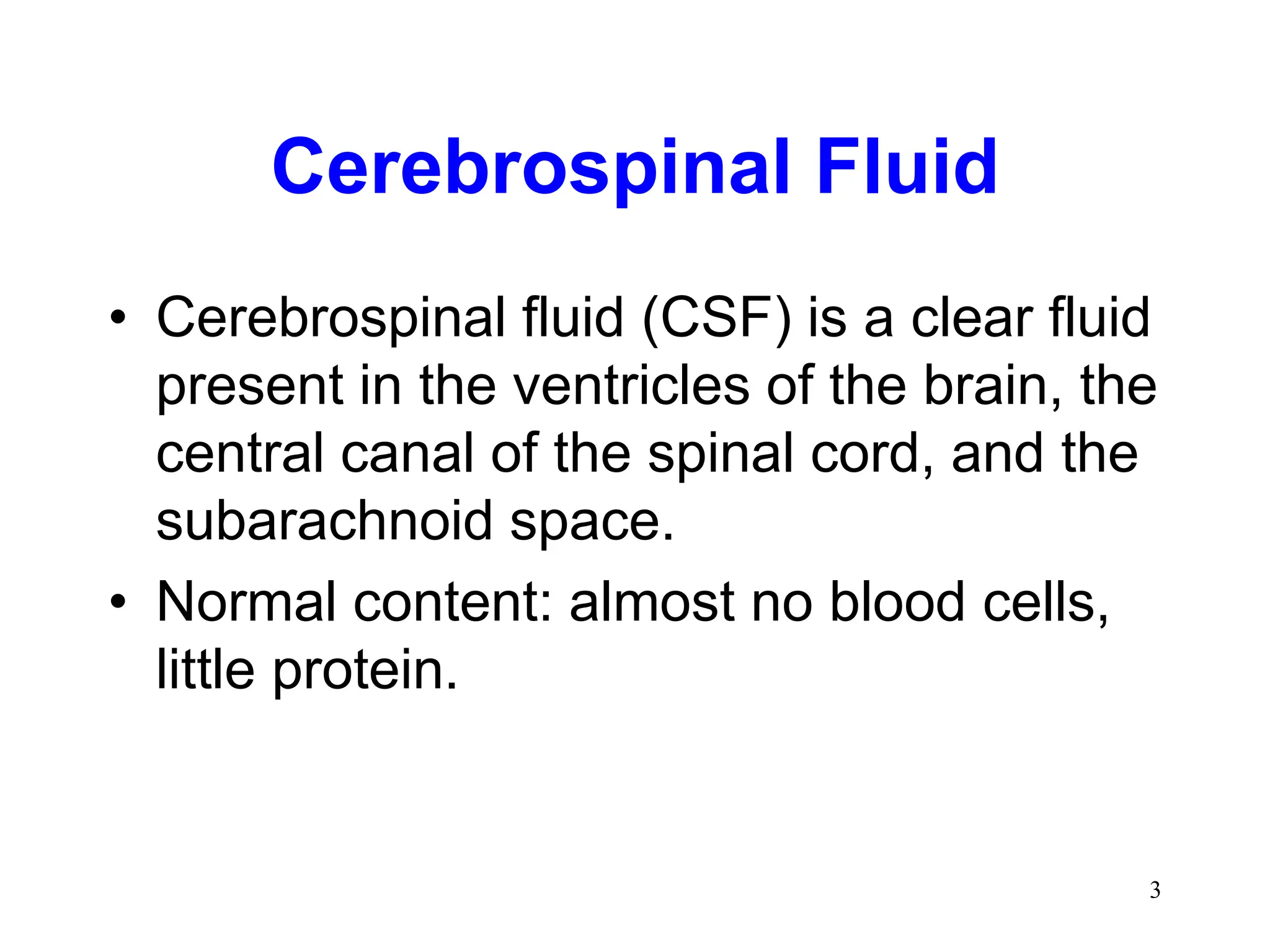
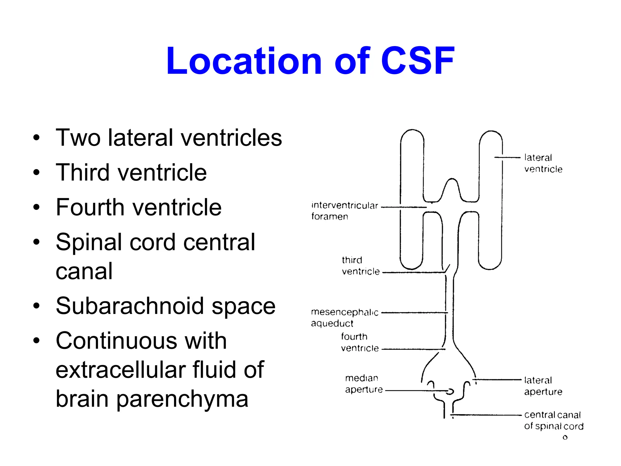
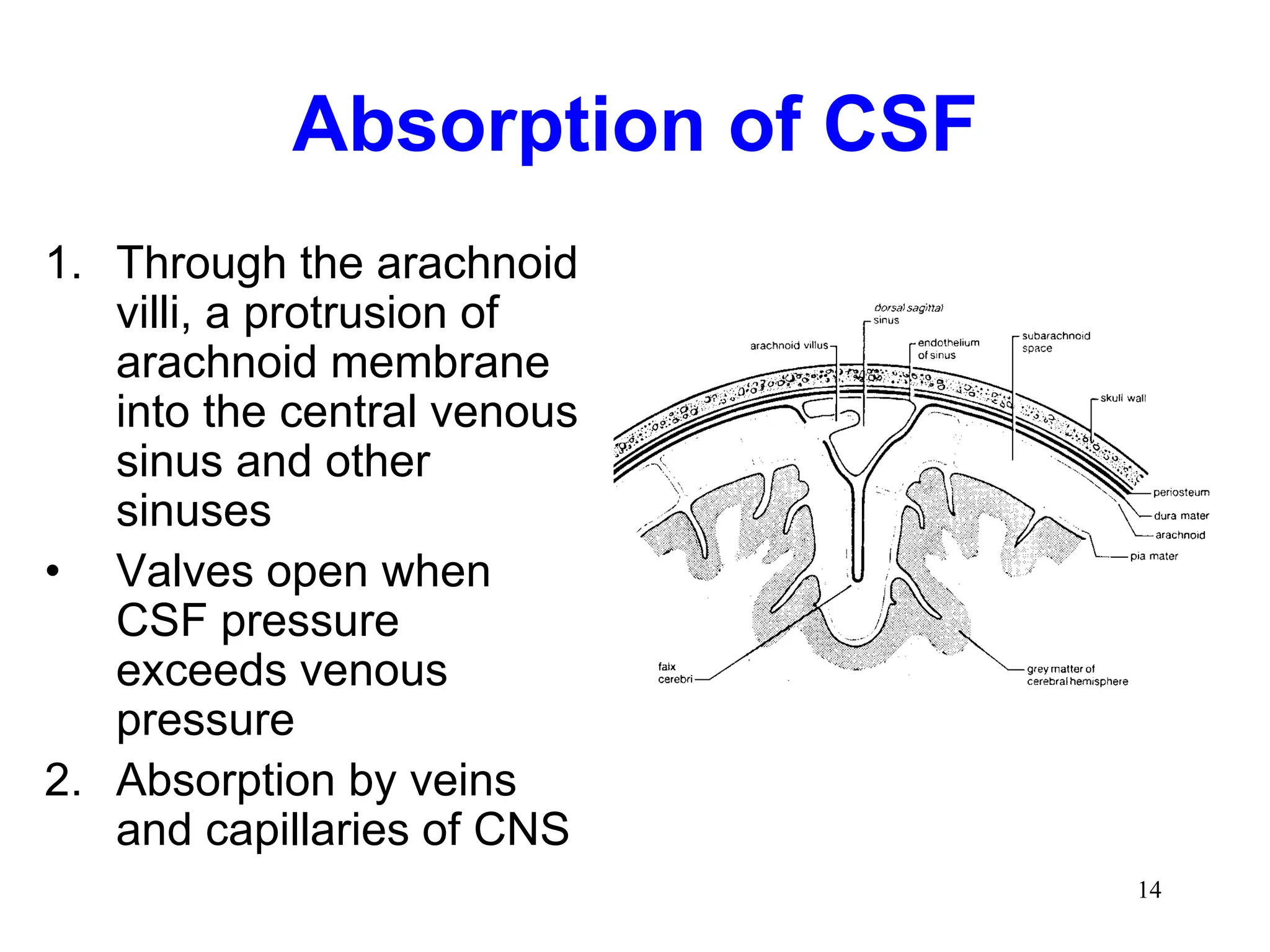
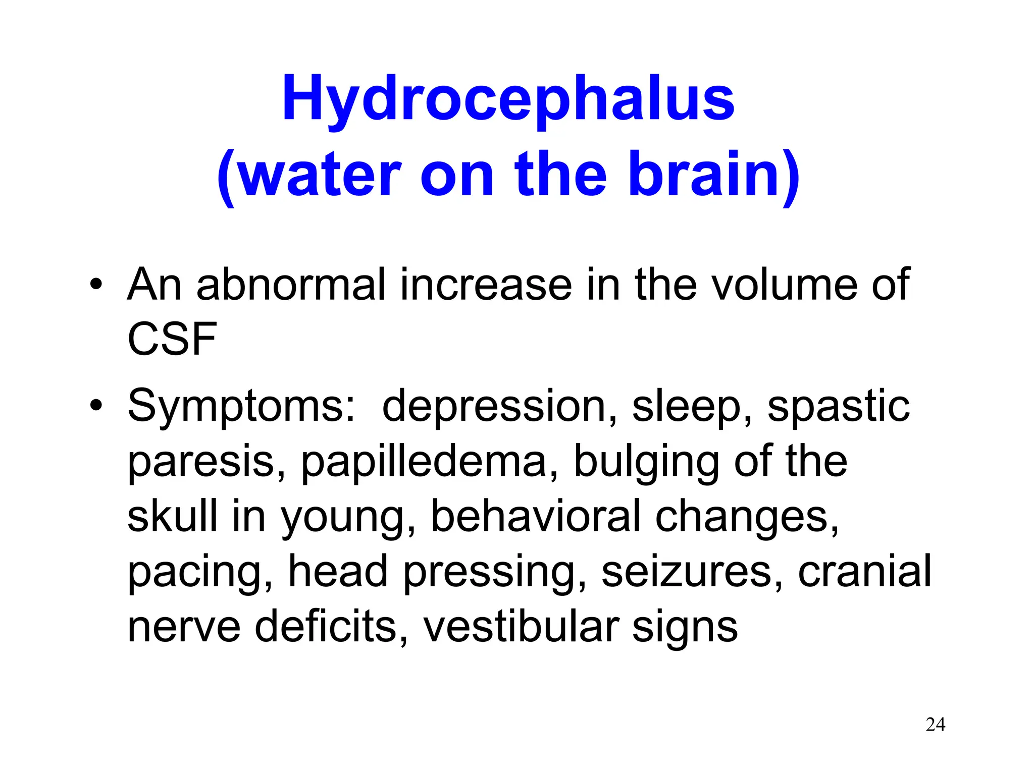
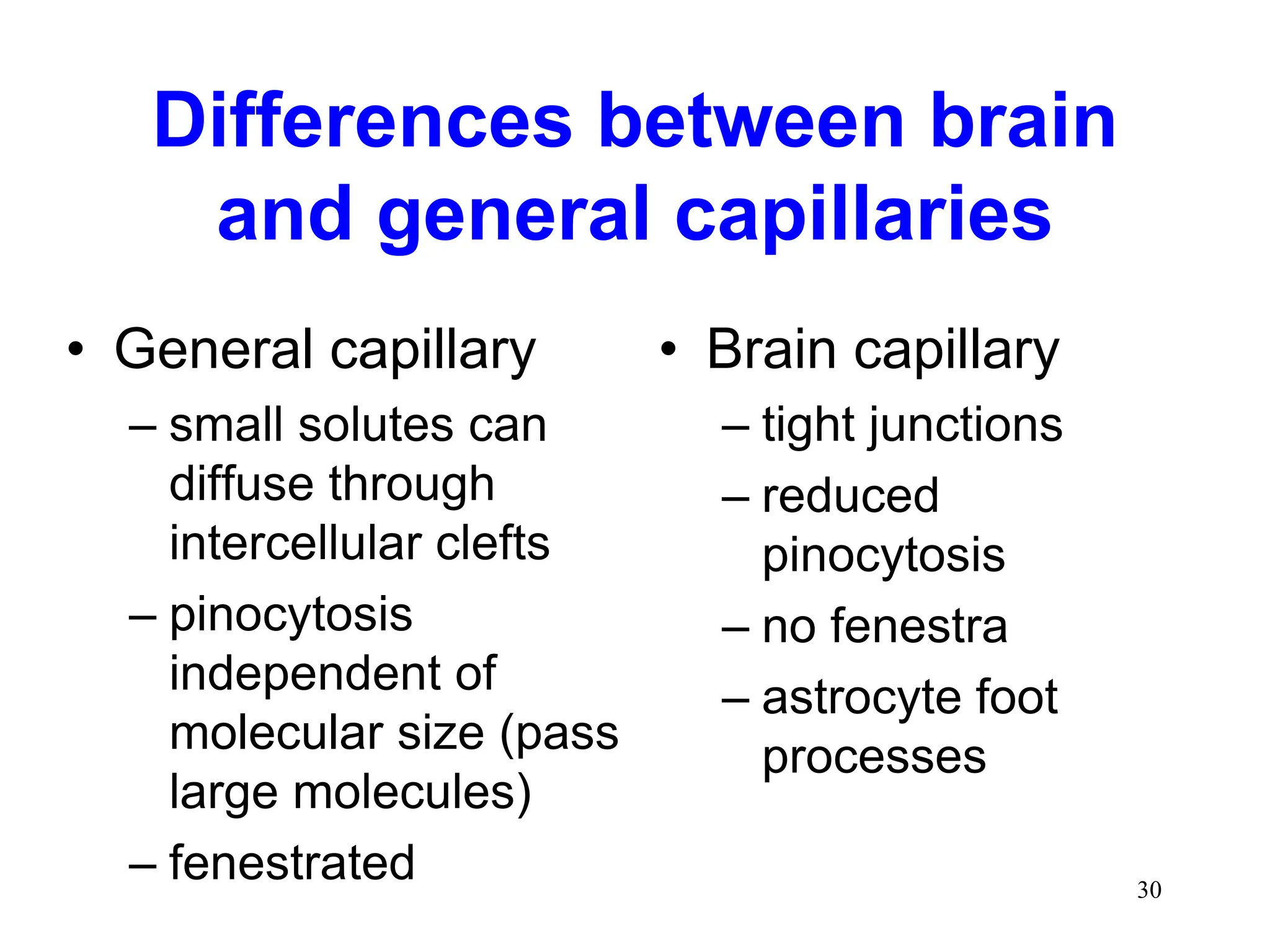
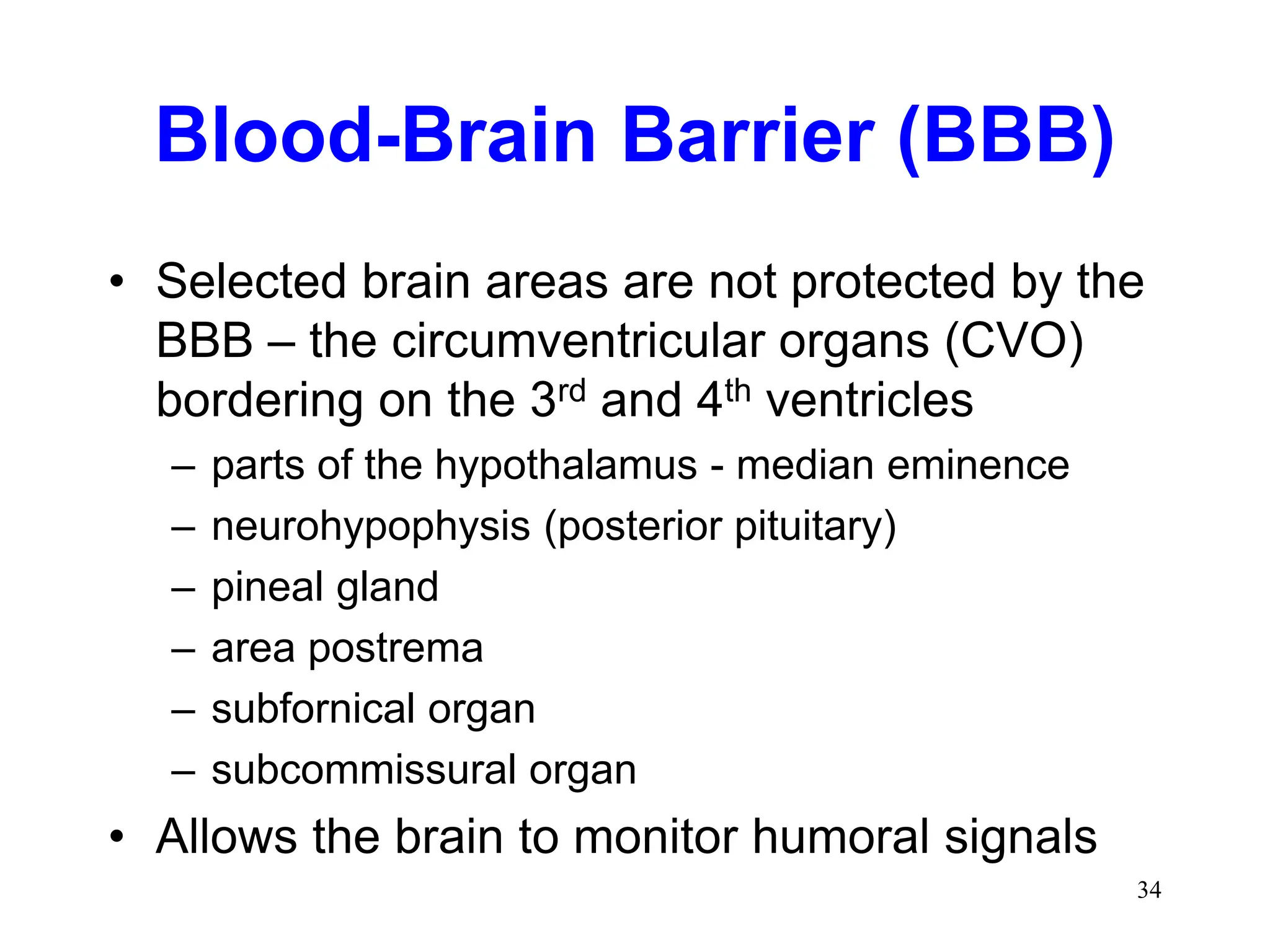
1
CEREBROSPINAL FLUID (CSF)
AND THE BLOOD-BRAIN
BARRIER (BBB)
2
Student Preparation
Textbook of Medical Physiology,
Guyton and Hall, Ch. 61
3
Cerebrospinal Fluid
• Cerebrospinal fluid (CSF) is a clear fluid
present in the ventricles of the brain, the
central canal of the spinal cord, and the
subarachnoid space.
• Normal content: almost no blood cells,
little protein.
4
CSF Cont.
• Its rate of formation, flow, and
absorption is sufficiently high to cause
its replacement several times daily.
Measuring its pressure, performing cell
counts, and analyzing levels of various
biochemical constituents is
accomplished with a spinal tap.
5
CSF Cont.
• Injecting radiopaque dyes into the
subarachnoid space is the basis of a
common neuroradiologic technique
called a myelogram.
• Obstruction of flow of CSF is a common
cause of hydrocephalus.
6
CSF Cont.
• An understanding of the formation, flow,
and absorption of CSF is essential to an
understanding of these diagnostic
procedures and the pathophysiology of
hydrocephalus.
7
Cerebrospinal Fluid
• A clear, colorless fluid that surrounds and
permeates the CNS. Offers support,
protection and nourishment.
• Functions:
– Protection of cranial contents
– Modulates pressure changes (has the same
specific gravity as brain tissues)
– Serves as a chemical buffer to maintain constant
ionic environment
– Serves as a transport medium for nutrients and
metabolites, endocrine substances and even
neurotransmitters
8
Location of CSF
• Two lateral ventricles
• Third ventricle
• Fourth ventricle
• Spinal cord central
canal
• Subarachnoid space
• Continuous with
extracellular fluid of
brain parenchyma
9
10
Formation of CSF
• Choroid plexuses of
lateral, third and
fourth ventricles
• Ependymal lining of
ventricular system
• Pia-glial membrane
• Blood vessels
11
Formation of CSF
• Cells are believed to actively secrete
Na+ into the ventricular system in
exchange for K+. Sodium ions
electrically attract Cl- and osmotically
draw water from the blood vascular
system to constitute the CSF.
12
Composition of CSF
13
Circulation of CSF
• Due to
– Pulsation of blood in choroid plexus
– Due to pulsation of ependymal cells
• Lateral ventricles interventricular
foramen of Monroe third ventricle
mesencephalic aqueduct (aqueduct of
Sylvius) fourth ventricle
– Spinal cord central canal;
– Also, out the lateral apertures to the subarachnoid
space to the venous system
14
Absorption of CSF
1. Through the arachnoid
villi, a protrusion of
arachnoid membrane
into the central venous
sinus and other
sinuses
• Valves open when
CSF pressure
exceeds venous
pressure
2. Absorption by veins
and capillaries of CNS
15
CSF Pressure
• Dog - 170 mm H2O
• Cow - 200 mm H2O
• Horse - 400 mm H2O
• Subarachnoid
hemorrhage blocks
absorption, raises
pressure
• Congenital excessive
formation or blockage to
flow raises pressure
• Regulated by rate of
absorption and rate of
formation.
• Brain tumors lower
absorption & raise
pressure
• Inflamed meninges can
cause excessive
formation & raise
pressure
16
Jugular compression maneuver
(Queckenstedt maneuver)
• Compress jugular veins in neck -
increases CSF pressure at the
cerebellomedullary cistern and the
lumbar cistern (normal)
• Decreased pressure at lumbar cistern
indicates blockage of spinal central
canal
17
Cerebrospinal fluid
collection
18
Cerebrospinal fluid
collection
• Small animals:
atlanto-occipital
(cerebellomedullary)
cistern
neck
19
20
Cerebrospinal fluid
collection
• Large animals
–Cerebellomedullary
cistern
–Lumbar cistern
(L5-L6)
21
22
CSF collection
23
Dog Lumbar Myelogram
24
Hydrocephalus
(water on the brain)
• An abnormal increase in the volume of
CSF
• Symptoms: depression, sleep, spastic
paresis, papilledema, bulging of the
skull in young, behavioral changes,
pacing, head pressing, seizures, cranial
nerve deficits, vestibular signs
25
Types of Hydrocephalus
• Communicating –
due to excessive
formation of CSF
or lack of
absorption
(nothing blocking
flow)
• Non-communicating –
due to obstruction of
flow through ventricle
system
26
Ventriculoperitoneal Shunt
27
Coup – Coup-Contrecoup
Closed Head Injuries
Coup Coup-contrecoup
With a blow to the
back of the
head, the forehead
strikes the surface
(coup), then the
brain bounces
back against
rear skull
(contrecoup)
28
Blood-Brain Barrier (BBB)
• Direct injection of large molecules (e.g.,
a dye) into the venous system will often
penetrate all tissues except the brain.
This observation led to the notion of a
physiological barrier to flow of
substances from blood to brain tissue –
the blood brain barrier.
• The BBB functions to preserve a stable
environment for neurons and glia of the
CNS
• The human brain consists of 2% of total
body weight, but receives 16% of the
cardiac output and 25% of the oxygen
consumed by the body.
• The CNS requires both protection as
well as a sustained energy supply.
• The BBB provides the protection, and
special nutrient transporters (i.e. for
glucose) provide the energy sources.
29
30
Differences between brain
and general capillaries
• General capillary
– small solutes can
diffuse through
intercellular clefts
– pinocytosis
independent of
molecular size (pass
large molecules)
– fenestrated
• Brain capillary
– tight junctions
– reduced
pinocytosis
– no fenestra
– astrocyte foot
processes
31
General vs. Brain Capillaries
32
Brain capillary
• Tight junctions
• Surrounded by
astrocytes
• Few vesicles
• Carrier mediated
transport of glucose
and amino acids
33
Blood-Brain Barrier
• Lipid soluble molecules will cross
• Negatively charged molecules cross more
freely than positively charged ones
• High CO2/low O2 produces vasodilation and
decreases resistance of BBB
• Injury or inflammation decreases resistance
of BBB (allows some antibiotics to be used for
treatment)
34
Blood-Brain Barrier (BBB)
• Selected brain areas are not protected by the
BBB – the circumventricular organs (CVO)
bordering on the 3rd and 4th ventricles
– parts of the hypothalamus - median eminence
– neurohypophysis (posterior pituitary)
– pineal gland
– area postrema
– subfornical organ
– subcommissural organ
• Allows the brain to monitor humoral signals
35
Blood – CSF Barrier
• Barrier from blood to the CSF
• Similar structural mechanism to the BBB
Brain Edema
• Vasogenic edema – an increase in brain
volume resulting from a breakdown in
the BBB. Interstitial volume increases,
intracranial pressure (ICP) increases,
ventricles shrink.
• Cytotoxic edema – intracellular swelling
of neurons, glia, endothelia due to drug
poisoning.
36
37
38
39
40
41
42
43

Cerebrospinal Fluid Blood Brain Barrier.ppt

  • 1.
    1 CEREBROSPINAL FLUID (CSF) ANDTHE BLOOD-BRAIN BARRIER (BBB)
  • 2.
    2 Student Preparation Textbook ofMedical Physiology, Guyton and Hall, Ch. 61
  • 3.
    3 Cerebrospinal Fluid • Cerebrospinalfluid (CSF) is a clear fluid present in the ventricles of the brain, the central canal of the spinal cord, and the subarachnoid space. • Normal content: almost no blood cells, little protein.
  • 4.
    4 CSF Cont. • Itsrate of formation, flow, and absorption is sufficiently high to cause its replacement several times daily. Measuring its pressure, performing cell counts, and analyzing levels of various biochemical constituents is accomplished with a spinal tap.
  • 5.
    5 CSF Cont. • Injectingradiopaque dyes into the subarachnoid space is the basis of a common neuroradiologic technique called a myelogram. • Obstruction of flow of CSF is a common cause of hydrocephalus.
  • 6.
    6 CSF Cont. • Anunderstanding of the formation, flow, and absorption of CSF is essential to an understanding of these diagnostic procedures and the pathophysiology of hydrocephalus.
  • 7.
    7 Cerebrospinal Fluid • Aclear, colorless fluid that surrounds and permeates the CNS. Offers support, protection and nourishment. • Functions: – Protection of cranial contents – Modulates pressure changes (has the same specific gravity as brain tissues) – Serves as a chemical buffer to maintain constant ionic environment – Serves as a transport medium for nutrients and metabolites, endocrine substances and even neurotransmitters
  • 8.
    8 Location of CSF •Two lateral ventricles • Third ventricle • Fourth ventricle • Spinal cord central canal • Subarachnoid space • Continuous with extracellular fluid of brain parenchyma
  • 9.
  • 10.
    10 Formation of CSF •Choroid plexuses of lateral, third and fourth ventricles • Ependymal lining of ventricular system • Pia-glial membrane • Blood vessels
  • 11.
    11 Formation of CSF •Cells are believed to actively secrete Na+ into the ventricular system in exchange for K+. Sodium ions electrically attract Cl- and osmotically draw water from the blood vascular system to constitute the CSF.
  • 12.
  • 13.
    13 Circulation of CSF •Due to – Pulsation of blood in choroid plexus – Due to pulsation of ependymal cells • Lateral ventricles interventricular foramen of Monroe third ventricle mesencephalic aqueduct (aqueduct of Sylvius) fourth ventricle – Spinal cord central canal; – Also, out the lateral apertures to the subarachnoid space to the venous system
  • 14.
    14 Absorption of CSF 1.Through the arachnoid villi, a protrusion of arachnoid membrane into the central venous sinus and other sinuses • Valves open when CSF pressure exceeds venous pressure 2. Absorption by veins and capillaries of CNS
  • 15.
    15 CSF Pressure • Dog- 170 mm H2O • Cow - 200 mm H2O • Horse - 400 mm H2O • Subarachnoid hemorrhage blocks absorption, raises pressure • Congenital excessive formation or blockage to flow raises pressure • Regulated by rate of absorption and rate of formation. • Brain tumors lower absorption & raise pressure • Inflamed meninges can cause excessive formation & raise pressure
  • 16.
    16 Jugular compression maneuver (Queckenstedtmaneuver) • Compress jugular veins in neck - increases CSF pressure at the cerebellomedullary cistern and the lumbar cistern (normal) • Decreased pressure at lumbar cistern indicates blockage of spinal central canal
  • 17.
  • 18.
    18 Cerebrospinal fluid collection • Smallanimals: atlanto-occipital (cerebellomedullary) cistern neck
  • 19.
  • 20.
    20 Cerebrospinal fluid collection • Largeanimals –Cerebellomedullary cistern –Lumbar cistern (L5-L6)
  • 21.
  • 22.
  • 23.
  • 24.
    24 Hydrocephalus (water on thebrain) • An abnormal increase in the volume of CSF • Symptoms: depression, sleep, spastic paresis, papilledema, bulging of the skull in young, behavioral changes, pacing, head pressing, seizures, cranial nerve deficits, vestibular signs
  • 25.
    25 Types of Hydrocephalus •Communicating – due to excessive formation of CSF or lack of absorption (nothing blocking flow) • Non-communicating – due to obstruction of flow through ventricle system
  • 26.
  • 27.
    27 Coup – Coup-Contrecoup ClosedHead Injuries Coup Coup-contrecoup With a blow to the back of the head, the forehead strikes the surface (coup), then the brain bounces back against rear skull (contrecoup)
  • 28.
    28 Blood-Brain Barrier (BBB) •Direct injection of large molecules (e.g., a dye) into the venous system will often penetrate all tissues except the brain. This observation led to the notion of a physiological barrier to flow of substances from blood to brain tissue – the blood brain barrier. • The BBB functions to preserve a stable environment for neurons and glia of the CNS
  • 29.
    • The humanbrain consists of 2% of total body weight, but receives 16% of the cardiac output and 25% of the oxygen consumed by the body. • The CNS requires both protection as well as a sustained energy supply. • The BBB provides the protection, and special nutrient transporters (i.e. for glucose) provide the energy sources. 29
  • 30.
    30 Differences between brain andgeneral capillaries • General capillary – small solutes can diffuse through intercellular clefts – pinocytosis independent of molecular size (pass large molecules) – fenestrated • Brain capillary – tight junctions – reduced pinocytosis – no fenestra – astrocyte foot processes
  • 31.
  • 32.
    32 Brain capillary • Tightjunctions • Surrounded by astrocytes • Few vesicles • Carrier mediated transport of glucose and amino acids
  • 33.
    33 Blood-Brain Barrier • Lipidsoluble molecules will cross • Negatively charged molecules cross more freely than positively charged ones • High CO2/low O2 produces vasodilation and decreases resistance of BBB • Injury or inflammation decreases resistance of BBB (allows some antibiotics to be used for treatment)
  • 34.
    34 Blood-Brain Barrier (BBB) •Selected brain areas are not protected by the BBB – the circumventricular organs (CVO) bordering on the 3rd and 4th ventricles – parts of the hypothalamus - median eminence – neurohypophysis (posterior pituitary) – pineal gland – area postrema – subfornical organ – subcommissural organ • Allows the brain to monitor humoral signals
  • 35.
    35 Blood – CSFBarrier • Barrier from blood to the CSF • Similar structural mechanism to the BBB
  • 36.
    Brain Edema • Vasogenicedema – an increase in brain volume resulting from a breakdown in the BBB. Interstitial volume increases, intracranial pressure (ICP) increases, ventricles shrink. • Cytotoxic edema – intracellular swelling of neurons, glia, endothelia due to drug poisoning. 36
  • 37.
  • 38.
  • 39.
  • 40.
  • 41.
  • 42.
  • 43.