Download to read offline

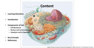


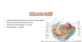
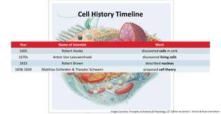
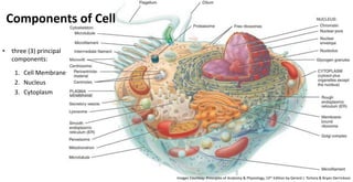


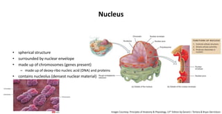


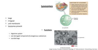
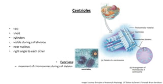

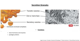


The document provides an overview of the cell, its structure, and function, including key components like the cell membrane, nucleus, and cytoplasm. It discusses historical milestones in cell discovery, cell theory, and details on various organelles, their roles, and importance in cellular processes. Additionally, it highlights essential concepts such as the cellular basis of life and the functions of different cell parts.


![03 [chapter 3 the cellular level of organization]](https://cdn.slidesharecdn.com/ss_thumbnails/03chapter3thecellularleveloforganization-170828035521-thumbnail.jpg?width=640&height=640&fit=bounds)


![20 [chapter 20 the cardiovascular system the heart]](https://cdn.slidesharecdn.com/ss_thumbnails/20chapter20thecardiovascularsystem-theheart-170828133506-thumbnail.jpg?width=640&height=640&fit=bounds)