Download free for 30 days
Sign in
Upload
Language (EN)
Support
Business
Mobile
Social Media
Marketing
Technology
Art & Photos
Career
Design
Education
Presentations & Public Speaking
Government & Nonprofit
Healthcare
Internet
Law
Leadership & Management
Automotive
Engineering
Software
Recruiting & HR
Retail
Sales
Services
Science
Small Business & Entrepreneurship
Food
Environment
Economy & Finance
Data & Analytics
Investor Relations
Sports
Spiritual
News & Politics
Travel
Self Improvement
Real Estate
Entertainment & Humor
Health & Medicine
Devices & Hardware
Lifestyle
Change Language
Language
English
Español
Português
Français
Deutsche
Cancel
Save
Submit search
EN
AS
Uploaded by
Asmita Sodhi
92 views
Methods used to_assess_implant_stability.unlocked
dental implant
Health & Medicine
◦
Read more
1
Save
Share
Embed
Embed presentation
Download
Download to read offline
1
/ 12
2
/ 12
3
/ 12
4
/ 12
5
/ 12
6
/ 12
7
/ 12
8
/ 12
9
/ 12
10
/ 12
11
/ 12
12
/ 12
More Related Content
PDF
Implant Stability: Methods and Recent Advances
by
Abu-Hussein Muhamad
PDF
Implant stability rfa
by
Asmita Sodhi
PDF
Implant stability 2013
by
Asmita Sodhi
PDF
Role of primary stability for osseointegration
by
Asmita Sodhi
PDF
Implant stability the password
by
Asmita Sodhi
PDF
Primary stability a predictable parameter
by
Asmita Sodhi
PDF
Implant stability intech
by
Asmita Sodhi
PPTX
Implant stability1
by
Asmita Sodhi
Implant Stability: Methods and Recent Advances
by
Abu-Hussein Muhamad
Implant stability rfa
by
Asmita Sodhi
Implant stability 2013
by
Asmita Sodhi
Role of primary stability for osseointegration
by
Asmita Sodhi
Implant stability the password
by
Asmita Sodhi
Primary stability a predictable parameter
by
Asmita Sodhi
Implant stability intech
by
Asmita Sodhi
Implant stability1
by
Asmita Sodhi
What's hot
PDF
A Clinical Study Resonance Frequency Analysis of Stability during the Healing...
by
Abu-Hussein Muhamad
PDF
Implant Stability: Methods and Recent Advances
by
Abu-Hussein Muhamad
PDF
Implant stability in immediate implant
by
Asmita Sodhi
PPTX
Christensen prosthesis
by
NikitaChhabariya
PPTX
PROGRESSIVE LOADING IN IMPLANTS
by
shari kurup
PPTX
Implant loading 2
by
bhuvanesh4668
PPTX
Implant loading
by
bhuvanesh4668
PPTX
Implant Loading Protocols Journal Club-Comparative evaluation of the influenc...
by
Partha Sarathi Adhya
PPTX
Loading protocols in implant
by
PiyaliBhattacharya10
PDF
Parameters for a Success Implant Integration Revisited Part I by Oded Bahat
by
Oded Bahat
PDF
68th publication sjmcr - 1st name
by
Dr Rahul Vinay Chandra Tiwari
PPTX
Immediate loading
by
Anuja Gunjal
A Clinical Study Resonance Frequency Analysis of Stability during the Healing...
by
Abu-Hussein Muhamad
Implant Stability: Methods and Recent Advances
by
Abu-Hussein Muhamad
Implant stability in immediate implant
by
Asmita Sodhi
Christensen prosthesis
by
NikitaChhabariya
PROGRESSIVE LOADING IN IMPLANTS
by
shari kurup
Implant loading 2
by
bhuvanesh4668
Implant loading
by
bhuvanesh4668
Implant Loading Protocols Journal Club-Comparative evaluation of the influenc...
by
Partha Sarathi Adhya
Loading protocols in implant
by
PiyaliBhattacharya10
Parameters for a Success Implant Integration Revisited Part I by Oded Bahat
by
Oded Bahat
68th publication sjmcr - 1st name
by
Dr Rahul Vinay Chandra Tiwari
Immediate loading
by
Anuja Gunjal
More from Asmita Sodhi
PDF
Patient Management Common Injuries (2025).pdf
by
Asmita Sodhi
PPTX
Implant lecture
by
Asmita Sodhi
PPT
Case selection & treatment planning
by
Asmita Sodhi
PDF
Minimal invasive drainage
by
Asmita Sodhi
PPT
Ragging a menace
by
Asmita Sodhi
PPTX
Iimportance of keeping records in dental practice
by
Asmita Sodhi
PDF
Methods used to_assess_implant_stability
by
Asmita Sodhi
PDF
Measurement of primary and secondary stability
by
Asmita Sodhi
PDF
Implant stability measurement (2)
by
Asmita Sodhi
PDF
Implant stability measurement (1)
by
Asmita Sodhi
PPTX
Full mouth rehabilitation
by
Asmita Sodhi
PPTX
Calcium metabolism
by
Asmita Sodhi
DOCX
Thumb sucking is a behavior found in humans
by
Asmita Sodhi
Patient Management Common Injuries (2025).pdf
by
Asmita Sodhi
Implant lecture
by
Asmita Sodhi
Case selection & treatment planning
by
Asmita Sodhi
Minimal invasive drainage
by
Asmita Sodhi
Ragging a menace
by
Asmita Sodhi
Iimportance of keeping records in dental practice
by
Asmita Sodhi
Methods used to_assess_implant_stability
by
Asmita Sodhi
Measurement of primary and secondary stability
by
Asmita Sodhi
Implant stability measurement (2)
by
Asmita Sodhi
Implant stability measurement (1)
by
Asmita Sodhi
Full mouth rehabilitation
by
Asmita Sodhi
Calcium metabolism
by
Asmita Sodhi
Thumb sucking is a behavior found in humans
by
Asmita Sodhi
Methods used to_assess_implant_stability.unlocked
1.
The International Journal
of Oral & Maxillofacial Implants 743 Methods Used to Assess Implant Stability: Current Status Mihoko Atsumi, DDS, PhD1/Sang-Hoon Park, DDS, MS2/Hom-Lay Wang, DDS, MSD3 Successful osseointegration is a prerequisite for functional dental implants. Continuous monitoring in an objective and quantitative manner is important to determine the status of implant stability. Histori- cally, the gold standard method used to evaluate degree of osseointegration was microscopic or histo- logic analysis. However, due to the invasiveness of this method and related ethical issues, various other methods of analysis have been proposed: radiographs, cutting torque resistance, reverse torque, modal analysis, and resonance frequency analysis. This review focuses on the methods currently avail- able for the evaluation of implant stability. (More than 50 references.) INT J ORAL MAXILLOFAC IMPLANTS 2007;22:743–754 Key words: cutting resistance analysis, implant stability evaluation, radiographic assessment, resonance frequency analysis, reverse torque test Successful osseointegration has been viewed as a direct structural and functional connection exist- ing between ordered, living bone and the surface of a load-carrying implant1,2 under a light microscope. Histologic appearance resembled a functional anky- losis with no intervention of fibrous or connective tissue between bone and implant surface.3–7 Osseointegration is also a measure of implant sta- bility, which can occur at 2 different stages: primary and secondary.8 Primary stability of an implant mostly comes from mechanical engagement with cortical bone. Secondary stability, on the other hand, offers biological stability through bone regeneration and remodeling.5,9,10 The former is a requirement for successful secondary stability.10 The latter, however, dictates the time of functional loading.11 Degree of implant stability may also depend on the condition of the surrounding tissues. It is, therefore, of an utmost importance to be able to quantify implant stability at various time points and to project a long- term prognosis based upon measured implant stabil- ity. Presently, various diagnostic analyses have been suggested to define implant stability: standardized radiographs, cutting torque resistance analysis, reverse torque test, modal analysis, and resonance frequency analysis (RFA). Therefore, the purpose of this paper was to review methods currently used to evaluate implant stability. An online search for studies in English and Japan- ese was performed using MEDLINE, Pre-MEDLINE, and the Cochrane Oral Health Group trials register. Publications from January 1970 to March 2006 were selected based on the following search terms: “implant mobility,”“Periotest,”“resonance frequency test,” “insertion torque,” “reverse torque,” “cutting resistance,”“implant stability,” and “mobility.” All of the search terms were combined with the term “implant.” A hand search of International Journal of Periodontics and Restorative Dentistry, Journal of Clini- cal Periodontology, International Journal of Oral & Maxillofacial Implants, Clinical Oral Implants Research, Journal of Periodontology, implant-related textbooks, and implant-related journals was also executed. Papers were considered relevant if they included the aforementioned key words and were published in English or Japanese. Articles published in peer- reviewed publications and current publications were 1Assistant Professor, Department of Oral and Maxillofacial Reha- bilitation, Prosthetics and Geriatric Dentistry Division, Kanagawa Dental College, Yokosuka, Japan. 2Assistant Professor, University of Maryland, Department of Peri- odontology, Baltimore, Maryland. 3Professor and Director of Graduate Periodontics, Department of Periodontics and Oral Medicine, School of Dentistry, University of Michigan, Ann Arbor, Michigan. Correspondence to: Dr Hom-Lay Wang, University of Michigan, School of Dentistry, 1011 N. University Avenue, Ann Arbor, MI 48109-1078. Fax: +734 936 0374. E-mail: homlay@umich.edu Atsumi.qxd 9/17/07 3:19 PM Page 743 COPYRIGHT © 2007 BY QUINTESSENCE PUBLISHING CO, INC. PRINTING OF THIS DOCUMENT IS RESTRICTED TO PERSONAL USE ONLY. NO PART OF THIS ARTICLE MAY BE REPRODUCED OR TRANSMITTED IN ANY FORM WITHOUT WRITTEN PERMISSION FROM THE PUBLISHER
2.
744 Volume 22,
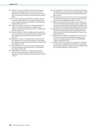
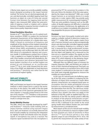
Number 5, 2007 Atsumi et al preferred to non-peer-reviewed and early publica- tions. More than 200 papers matched the inclusion criteria; however, only 114 most relevant articles/chapters were selected and reviewed. IMPLANT STABILITY Implant stability, an indirect indication of osseointe- gration, is a measure of the clinical immobility of an implant.5,9 It is achieved at 2 levels: cortical bone (pri- mary stability) and cancellous bone (secondary sta- bility). Secure primary stability leads to predictable secondary stability.12 Secondary stability has been shown to begin to increase at 4 weeks after implant placement.13 At this time point, the lowest implant stability is expected. Therefore, the original Bråne- mark protocol2 suggested a 3- to 6-month non- loaded healing period to achieve adequate stability before functional loading. Osseointegration is, however, a patient-depen- dent wound healing process affected by various fac- tors (Table 1). Quantification of implant stability at various time points may provide significant informa- tion as to the individualized “optimal healing” time. Raghavendra et al13 proposed that measurement of osseointegration be approached in a quantitative manner, as primary and secondary stability are in an inverse relationship. However, in clinical practice, experience-driven decision still dominates, as objec- tive guidelines have not been established. Table 2 summarizes currently available methods for the objective assessment of implant stability at pre-, intra-, and postsurgical time points. Histologic or histomorphometric analysis, however, is not feasi- ble for daily practice, as this may require unnecessary biopsy. RADIOGRAPHIC ANALYSIS Radiographic evaluation is a noninvasive method that can be performed at any stage of healing. Bitewing view is used to measure crestal bone level, which has been suggested as an important radiographic indica- tor for implant success.14–16 It has been reported that 1.5 mm of radiographic crestal bone loss can be expected in the first year of loading in a stable implant, with 0.1 mm of subsequent annual bone loss.17–20 However, several problems must be addressed. First, 1.5 mm is a mean value. Second, due to a low incidence of implant failure,changes in radio- graphic bone level alone cannot precisely predict implant stability. Third, it is impractical for a clinician to detect changes in radiographic bone loss at 0.1 mm resolution. Fourth, crestal bone changes can only be reliably measured without distortion when the central ray of the x-ray source is perfectly parallel with the structures of interest. This would necessitate a series of standardized radiographs with a customized template for reliable and repeatable measurements, which is impractical. Lastly, conventional periapical or panoramic views do not provide information on a facial bone level, and bone loss at this level precedes mesiodistal bone loss.21 Neither bone quality nor density can be quantified with this method. Even changes in bone mineral cannot be radiographically detected until 40% of demineralization had occurred.22 Numerous limitations exist with the use of a conventional radiograph alone in making an accu- rate, independent assessment of implant stability. Computer-assisted measurements of crestal bone level change may prove to be the most accurate way to use radiographic information, as a standard devia- tion within 0.1 mm (0.01 to 0.51 mm) has been reported.23 However, this method is not convenient for use in clinical practice.24 CUTTING TORQUE RESISTANCE ANALYSIS In cutting resistance analysis (CRA), originally devel- oped by Johansson and Strid25 and later improved by Friberg et al26–29 in in vitro and in vivo human models, the energy (J/mm3) required for a current- fed electric motor in cutting off a unit volume of bone during implant surgery is measured. This energy was shown to be significantly correlated with bone density, which has been suggested as one of factors that significantly influences implant stabil- ity.26,29 To minimize the interoperator variation, hand pressure during drilling was controlled.27 CRA can be used to identify any area of low-density bone (or poor-quality bone) and to quantify bone hardness Table 1 Factors that Influence Implant Stability Factors Affecting Primary Stability Bone quantity and quality Surgical technique, including the skill of the surgeon Implant (eg, geometry, length, diameter, surface characteristics) Factors Affecting Secondary Stability Primary stability Bone modeling and remodeling Implant surface conditions Atsumi.qxd 9/17/07 3:19 PM Page 744 COPYRIGHT © 2007 BY QUINTESSENCE PUBLISHING CO, INC. PRINTING OF THIS DOCUMENT IS RESTRICTED TO PERSONAL USE ONLY. NO PART OF THIS ARTICLE MAY BE REPRODUCED OR TRANSMITTED IN ANY FORM WITHOUT WRITTEN PERMISSION FROM THE PUBLISHER
3.
The International Journal
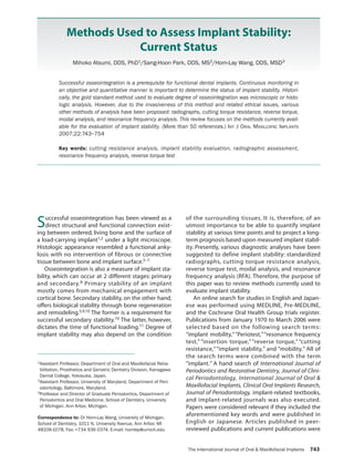
of Oral & Maxillofacial Implants 745 Atsumi et al during the low-speed threading of implant osteotomy sites. A torque gauge incorporated within the drilling unit (eg, Osseocare; Nobel Biocare, Göte- borg, Sweden) can be used to measure implant inser- tion torque in Ncm to indirectly represent J/mm3. Insertion torque values have been used to measure bone quality in various parts of the jaw during implant placement.30 CRA gives a far more objective assessment of bone density than clinician-dependent evaluation of bone quality based on Lekholm and Zarb classification.31 Clinical relevance was demonstrated by studies that showed the highest frequency of implant failures in jaws with advanced resorption and poor bone qual- ity, often seen in maxilla.17,18,32–34 Therefore, cutting resistance value may provide useful information in determining an optimal healing period in a given arch location with a certain bone quality.26 The major limitation of CRA is that it does not give any information on bone quality until the osteotomy site is prepared. CRA also cannot identify the lower “critical” limit of cutting torque value (ie, the value at which an implant would be at risk).29 Furthermore, longitudinal data cannot be collected to assess bone quality changes after implant placement. Its primary use, therefore, lies in estimating the primary stability of an implant. For instance, in Misch’s 6 time-depen- dent stages of implant failures—(1) surgical, (2) osseous healing,(3) early loading, (4) intermediate, (5) late, and (6) long-term35—CRA can only provide information on the first 2 stages. Estimation of implant primary stability alone from CRA is still of value, as high implant failure rates are observed in the first 3 phases.36,37 Nonetheless, long-term evalua- tion of implant stability after implant placement, phases 3 to 7, is desired and should not be over- looked. This limitation has led to development of other diagnostic tests.Table 3 summarizes CRA. REVERSE TORQUE TEST Unlike CRA, which measures the bone density and the resistance to cutting torque, the reverse torque test (RTT), proposed by Roberts et al38 and devel- oped by Johansson and Albrektsson,39–41 measures the “critical” torque threshold where bone-implant contact (BIC) was destroyed. This indirectly provides information on the degree of BIC in a given implant. In the study conducted by Johansson and Albrekts- son,a reverse torque was applied to remove implants placed in the tibiae of rabbits 1, 3, 6, and 12 months postsurgery. Reverse torque value and histologic evaluation showed that greater BIC could be achieved with a longer healing time. Similar observa- tions at the histologic level have been made in other animal studies.42–44 Removal torque value (RTV) as an indirect measurement of BIC or clinical osseointe- gration was later reported to range from 45 to 48 Ncm in 404 clinically osseointegrated implants in humans.45 Sullivan et al further speculated that any RTV greater than 20 Ncm may be acceptable as a cri- terion for a successful osseointegration, since none of the implants in their study45 could be removed during abutment connection at 20 Ncm. It was fur- ther suggested that RTT is, therefore, a reliable diag- nostic method for verification of osseointegration. Table 2 Currently Available Methods to Evaluate Implant Stability and the Time of Use for Each Method Pre Intra Post Noninvasiveness Objectivity Histologic analysis + + + – +++ Percussion test – ++ ++ + + Radiographs ++ ++ ++ ++ – Reverse torque – – ++ – ++ Cutting resistance – +++ – + ++ Vibration analysis Periotest – ++ ++ ++ ++? RFA – +++ +++ +++ ++? +++ = method with highest reliability; ++ = method with certain reliability; + = method with doubtful reliability; – = application is impossible; ? = More information is needed. Table 3 Advantages and Disadvantages of CRA Advantages 1. Detect bone density 2. High correlation between cutting resistance and bone quality 3. Reliable method to assess bone quality 4. Identify bone density during surgery 5. Can be used in daily practice Disadvantages 1. Can only be used during surgery Atsumi.qxd 9/17/07 3:19 PM Page 745 COPYRIGHT © 2007 BY QUINTESSENCE PUBLISHING CO, INC. PRINTING OF THIS DOCUMENT IS RESTRICTED TO PERSONAL USE ONLY. NO PART OF THIS ARTICLE MAY BE REPRODUCED OR TRANSMITTED IN ANY FORM WITHOUT WRITTEN PERMISSION FROM THE PUBLISHER
4.
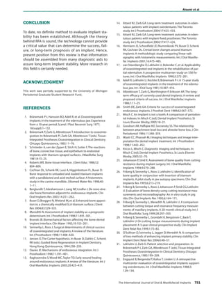
746 Volume 22,
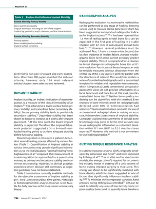
Number 5, 2007 Atsumi et al However, this method has been criticized as being destructive.8 Brånemark et al2 cautioned about the risk of irreversible plastic deformation within peri- implant bone and of implant failure if unnecessary load was applied to an implant that was still under- going osseointegration. Furthermore, a 20-Ncm threshold RTV for successful osseointegration has not yet been supported by scientific data. The threshold limit varies among patients depending on the implant material and the bone quality and quan- tity. A threshold RTV may be lower in type 4 bone than in denser bone, for instance. Hence, subjecting implants placed in this bone type to RTV may result in a shearing of BIC interface and cause implant fail- ure. Furthermore, RTV can only provide information as to “all or none” outcome (osseointegrated or failed); it cannot quantify degree of osseointegration. Hence,RTT is mainly used in experiments. MODAL ANALYSIS Modal analysis measures the natural frequency or displacement signal of a system in resonance, which is initiated by external steady-state waves or a tran- sient impulse force (Table 4). Modal analysis, in other words, is a vibration analysis. It is widely used as an effective test method for structural analysis in engi- neering and the health-care field.46,47 Dental applica- tions include the quantification of osseointegra- tion.48–51 Modal analysis can be performed in 2 models:theoretical and experimental.52 Two or 3-dimensional finite element modeling (FEM) is an example of computer-simulated theoreti- cal modal analysis, which is mathematically con- structed using known biomechanical properties (eg, Young’s modulus [Pa], Poisson ratio, and density in g/cm3) of structures of interest. Theoretical modal analysis such as FEM may be useful in investigation of the vibrational characteristics of objects that may be difficult to excite because of a damping effect from boundary conditions such as the periodontal ligament (PDL) in an in vivo model.49 By altering boundary conditions such as the bone level, FEM can theoretically be used to calculate the anticipated stress and strain in various simulated peri-implant bone levels.50,51 Experimental or dynamic modal analysis, on the other hand, measures structural changes and dynamic characteristics (eg, natural characteristic fre- quency, characteristic mode, and attenuation) of a system that is excited in an in vitro model via vibra- tion testing (eg, impactor or hammer). This in vitro approach provides a more reliable assessment of an object than a theoretical model. This analysis has been applied in dentistry to quantify the degree of osseointegration and implant stability.49 Frequency analysis and mechanical impedance analysis can be used for detecting response waves in modal analysis.52 By combining the vibration and response detecting methods, various kinds of vibration analy- ses can be performed.53 Some techniques derived from these theoretical concepts are being tested for use in evaluating implant mobility. Percussion Test A percussion test is one of the simplest methods that can be used to estimate the level of osseointe- gration.8,54–56 This test is based upon vibrational- acoustic science and impact-response theory. A clini- cal judgment on osseointegration is made based on the sound heard upon percussion with a metallic instrument. A clearly ringing “crystal” sound indicates successful osseointegration, whereas a “dull” sound may indicate no osseointegration. However, this method heavily relies on the clinician’s experience level and subjective belief.Therefore,it cannot be used experimentally as a standardized testing method. Impact Hammer Method Impact hammer method is another example of tran- sient impact as a source of excitement force during experimental modal analysis.53,57 It is an improved version of the percussion test except that sound gen- erated from a contact between a hammer and an object is processed through fast Fourier transform (FFT) for analysis of transfer characteristics. By enhancing the response detection using various devices, such as a microphone, an accelerometer, or a strain gauge, and by processing the detected response with FFT, it becomes possible to quantify and qualify the response wave in the form of disloca- tion, speed, acceleration, stress, distortion, sound, and other physical properties. Periotest (Siemens, Ben- sheim, Germany) and Dental Mobility Checker (DMC; Table 4 Implant Stability Measurement Based on Modal or Vibration Analysis Theoretical Modal Analysis 1. Finite element method Experimental Modal Analysis 1. Percussion test 2. Impact hammer method (Periotest, Siemens, Bensheim, Germany; Dental Mobility Checker, J. Morita, Suita, Japan) 3. RFA (Osstell, Integration Diagnostics, Göteborg, Sweden; Implomate, Bio Tech One, Taipei, Taiwan) 4. Others (pulsed oscillation waveform by Kaneko) Atsumi.qxd 9/17/07 3:19 PM Page 746 COPYRIGHT © 2007 BY QUINTESSENCE PUBLISHING CO, INC. PRINTING OF THIS DOCUMENT IS RESTRICTED TO PERSONAL USE ONLY. NO PART OF THIS ARTICLE MAY BE REPRODUCED OR TRANSMITTED IN ANY FORM WITHOUT WRITTEN PERMISSION FROM THE PUBLISHER
5.
The International Journal
of Oral & Maxillofacial Implants 747 Atsumi et al J. Morita, Suita, Japan) are currently available mobility testers designed according to the impact hammer method. The former has an electromagnetically dri- ven and electronically controlled tapping head that hammers an object at a rate of 4 times per second. Contact time between the tapping head and the object is also measured. DMC utilizes the same prin- ciple of tapping a tooth or implant with a dental hammer. A frequency response function is built-in to detect bone-quality–dependent sound. Pulsed Oscillation Waveform Kaneko et al58,59 described the use of a pulsed oscil- lation waveform (POWF) to analyze the mechanical vibrational characteristics of the implant-bone inter- face using forced excitation of a steady-state wave. POWF is based on estimation of frequency and amplitude of the vibration of the implant induced by a small pulsed force. This system consists of acousto- electric driver (AED), acoustoelectric receiver (AER), pulse generator, and oscilloscope. Both the AED and AER consist of a piezoelectric element and a punc- ture needle. A multifrequency pulsed force of about 1 kHz is applied to an implant by lightly touching it with 2 fine needles connected with piezoelectric ele- ments. Resonance and vibration generated from bone-implant interface of an excited implant are picked up and displayed on an oscilloscope screen.58,59 An in vitro study showed that the sensi- tivity of the POWF test depended on load directions and positions.58 Sensitivity was rather low for the assessment of implant rigidity. IMPLANT STABILITY EVALUATION METHODS DMC and Periotest are based on the impact hammer method, in which impact force is used as the excita- tion force. In this theory, “the width of the first peak on the time axis of the spectrum generated by tran- sient impulse is inversely proportional to the time axis of the impulse.”57,60,61 Therefore, in the presence of impact force, lower rigidity of the tested substance results in a longer time axis. Dental Mobility Checker The DMC, which was originally developed by Aoki60 and Hirakawa,61 measures tooth mobility with an impact hammer method using transient impact force. Aoki and Hirakawa successfully detected the level of tooth mobility by converting the integration (ie, rigidity) of tooth and alveolar bone into acoustic signals. A microphone was used as a receiver. The response signal transferred from the microphone is processed by FFT for conversion for analysis in the time axis. Hence, the duration of the first wave gener- ated by the impact was detected.62 DMC uses a small impact hammer as an excitation device. It is easily used even in molar regions. DMC may provide quite stable measurement for osseointegrated implants.63 There are some problems, however, such as the diffi- culties of double-tapping and difficulty in attaining constant excitation. Furthermore, the application of a small force to an implant immediately after placement may jeopardize the process of osseointegration.2 Periotest Periotest has been thoroughly studied and advo- cated as a reliable method to determine implant sta- bility.8,64–71 Unlike DMC, which applies impact force with a hammer, Periotest uses an electromagnetically driven and electronically controlled tapping metallic rod in a handpiece. Response to a striking or “bark- ing” is measured by a small accelerometer incorpo- rated into the head. Like DMC, contact time between the test object and tapping rod is measured on the time axis as a signal for analysis. The signals are then converted to a unique value called the Periotest value (PTV), which depends on the damping charac- teristics of tissues surrounding teeth or implants.72 Although they use different types of receivers for impulse responses, DMC and Periotest are similar in terms of their theoretical background.They both use a transient impulse as an excitation force, and in both cases analysis is conducted on the time axis. In addi- tion, both were originally developed to measure the mobility of a natural tooth.64,65 In the case of a natural tooth, the buffering capac- ity of the PDL poses a problem in analyzing the dis- tribution of impact force exerted on a tooth. When dynamic characteristics are analyzed based upon an assumption that the whole periodontal structure functions as a mechanical unit, it is difficult to model the attenuation from the PDL. The soft tissue, includ- ing the periosteum, is considered a viscoelastic medium; thus, Hooke’s law does not apply to the behavior of the PDL under an applied load. Thus, vis- coelasticity of the PDL has always posed a difficulty in analysis of the physical characteristics of peri- odontal tissue. By contrast, bone-implant interface with no PDL is believed to be similar to the serial spring model which follows Hooke’s law, and mobil- ity measurement is considered easier. Most reports of the use of a natural tooth mobility detector such as Periotest to measure implant mobil- ity have pointed out a lack of sensitivity in these devices.55,68 Such devices permit a very wide dynamic range (in case of Periotest, PTV is –8 to +50) to permit the measurement of a wide variety of nat- Atsumi.qxd 9/17/07 3:19 PM Page 747 COPYRIGHT © 2007 BY QUINTESSENCE PUBLISHING CO, INC. PRINTING OF THIS DOCUMENT IS RESTRICTED TO PERSONAL USE ONLY. NO PART OF THIS ARTICLE MAY BE REPRODUCED OR TRANSMITTED IN ANY FORM WITHOUT WRITTEN PERMISSION FROM THE PUBLISHER
6.
ural tooth mobility.68
However, the dynamic range used for measuring implant mobility is very limited. Thus, the sensitivity of these devices is insufficient to measure implant mobility. Although many similarities do exist between the tissue structures around an implant and a natural tooth, conclusions from periodontal studies may not be directly applicable to implants.73 In the use of mobility measurement to assess implant stability, the presence or absence of a PDL makes a crucial differ- ence. Similar to impact/vibration testing, values mea- sured with Periotest are significantly influenced by excitation conditions, such as position and direction. The Periotest user’s manual contains clear instruc- tions about striking point position and angle: “The Periotest measurement must be made in a midbuc- cal direction”and“During measurement the Periotest handpiece must always be held perpendicular to the tooth axes.”72 Considering the intraoral environment, and the pen-grip–shaped handpiece of the Periotest, it is clear that it can be used quite easily for the ante- rior region. However, its use for the molar region is extremely difficult because of the presence of buccal mucosa.74 Derhami et al75 used a fixing device to hold a handpiece at the correct angle. This fixing device was used for an in vitro measurement using a cranial bone model, and its clinical application seems difficult. However, Periotest is believed to be an effec- tive evaluation method once the difficulty of control- ling impact force is solved. Long-term data on Periotest have shown that it can be an objective clinical measurement of the sta- bility of bone-implant anchorage.70,71 Aparicio used Periotest to measure implant stability and found a direct correlation between PTV and the degree of ini- tial osseointegration.69 It was further suggested that PTV should be included in the current success crite- ria. Another study with sample size of more than 2,900 implants showed a similar finding.70,71 How- ever, differences with respect to implant design, diameter, length, and bone quality and quantity were not accounted for in that study; analysis in a pattern of changes over time may be more reasonable. A measured bone value only represents its condition at the moment of measurement. Bone is subject to life- long metabolism, which will in turn affect PTV over time. Thus, average value is not a proper way to determine a critical value for implant stability. Even if it could be assumed that PTV precisely reflects the condition of BIC as reported by previous studies,76,77 an average PTV has no importance. Johansson and Albrektsson observed that “implants inserted in different people do not necessarily attain the same degree of integration.”39 Despite a wide variation in host factors such as bone density, normal PTV of an osseointegrated implant falls in a relatively narrow zone (–5 to +5) within a wide scale (–8 to +50).64 Other studies have indicated that the PTVs of clinically osseointegrated implants fall within an even narrower zone (–4 to –2 or –4 to +2).76,78 There- fore, the measured PTV may falsely be interpreted as having a small standard deviation and therefore viewed as having a good accuracy. PTV cannot be used to identify a “borderline implant” or “implant in the process of osseointegration” which may or may not continue to a successful osseointegration.77 No conclusion has been made with regard to this issue. It has been suggested that these limitations of Periotest measurement have been suggested to be strongly related to the orientation of excitation source or striking point. In vitro and in vivo experiments demonstrated that the influence of striking point on PTV is much greater than the effects from increased implant length due to marginal bone resorption or other excitation conditions such as the angle of the handpiece or repercussion of a rod.55,75 Unfortunately, controlling these influential factors is extremely diffi- cult. Despite some positive claims for Periotest,68,69 the prognostic accuracy of PTV for implant stability has been criticized for a lack of resolution, poor sensi- tivity,and susceptibility to operator variables.8,79 RFA RFA has recently gained popularity. It is a noninvasive diagnostic method that measures implant stability and bone density at various time points using vibration and a principle of structural analysis.57 RFA utilizes a small L- shaped transducer that is tightened to the implant or abutment by a screw. The transducer comprises 2 piezoceramic elements, one of which is vibrated by a sinusoidal signal (5 to 15 kHz). The other serves as a receptor for the signal. Resonance peaks from the received signal indicate the first flexural (bending) reso- nance frequency of the measured object.In vitro and in vivo studies have suggested that this resonance peak may be used to assess implant stability in a quantitative manner. Currently, 2 RFA machines are in clinical use: Osstell (Integration Diagnostics) and Implomates (Bio Tech One). Osstell has combined the transducer, computer- ized analysis and the excitation source into one machine closely resembling the model used by Meredith. In the early studies, the hertz was used as the measurement unit.28,54,56,80–89 Later, Osstell cre- ated the implant stability quotient (ISQ) as a measure- ment unit in place of hertz.90–103 Resonance fre- quency values ranging from 3,500 to 8,500 Hz are translated into an ISQ of 0 to 100. A high value indi- cates greater stability, whereas a low value implies instability.The manufacturer’s guidelines suggest that 748 Volume 22, Number 5, 2007 Atsumi et al Atsumi.qxd 9/17/07 3:19 PM Page 748 COPYRIGHT © 2007 BY QUINTESSENCE PUBLISHING CO, INC. PRINTING OF THIS DOCUMENT IS RESTRICTED TO PERSONAL USE ONLY. NO PART OF THIS ARTICLE MAY BE REPRODUCED OR TRANSMITTED IN ANY FORM WITHOUT WRITTEN PERMISSION FROM THE PUBLISHER
7.
a successful implant
typically has an ISQ greater than 65. An ISQ < 50 may indicate potential failure or increased risk of failure.104 It is assumed that an implant and the surrounding bone function as as a single unit; thus, a change in stiffness is considered to represent the change of osseointegration of an implant. A steady-state sinu- soidal force in a form of sine wave is applied to the implant-bone unit to measure the implant stability via resonance. Frequency and amplitude are then picked up as a response.90,91 An in vitro model showed that resonance frequency of an implant placed in an alu- minum block ranged from 8 to 9 kHz.8,54 An in vivo human study also showed that, although amplitude of the resonance peak was smaller than in vitro data, the peak resonance frequency of clinically osseointe- grated implants was also about 8 to 9 kHz.8 Moreover, resonance frequency increased as polymerization of the resin progressed.54 Effective implant length (EIL) was a value calcu- lated by adding the amount of exposed implant threads and the length of each abutment. EIL has been shown to be inversely proportional to the level of resonance frequency, with a correlation coefficient of r = –0.94 in vitro and r = –0.78 in vivo.8,54 Several in vivo animal and human clinical studies have con- curred with this finding.56,80,102 No resonance peak was observed in failed implants with clinical mobility.8 Longitudinal changes in resonance frequency have also been evaluated. Implants placed in the rabbit tibia were measured over 168 days from the time of implant placement81; resonance frequency increased over time. Other studies have evaluated longitudinal changes in ISQ more in detail.90–92,94,95,98,105 ISQ was found to decrease significantly after implant place- ment for several weeks.However,a recovery to the ini- tial ISQ level was found at the time of implant loading. Furthermore, a greater increase of resonance fre- quency over time was observed with implants placed in softer bone.28,91,98 In the case of an implant placed in grafted bone in an in vivo human study,106 very low resonance frequency (4 to 5 kHz) was observed. Based upon these findings, the following 3 con- clusions have been suggested.106 First, “stiffness” of an implant is a function of its geometry and material composition (length, diameter, overall shape). Sec- ond, the stiffness of the implant-tissue interface depends on the bond between the surface of the implant and the surrounding bone. Third, the stiff- ness of the surrounding tissue is determined by the ratio of cancellous to cortical bone and the density of the bone with which an implant engages.8 Stiffness found at the bone-implant interface (second point) changes over time. The factors affecting stiffness remain relatively stable, as the mechanical properties of implant and bone are constant. The only factor that could significantly influence the stiffness and resonance frequency of the implant would be the exposed implant length, as shown in several stud- ies.8,56,80,102,107–109 Therefore, measurement of the stiffness at the interface provides reliable informa- tion as to the implant stability. Stiffness of supporting structure may, however, influence the stiffness of the interface of an area of interest.80,81,109–111 In most in vitro studies,107,109,110 such as that of Meredith et al,54 an aluminum block material with uniformity and linearity has been used as a supporting structure. Therefore, in this model, an implant behaves in a mathematically predictable manner in which resonance frequency is inversely proportional to the length of the cantilever beam. Bone, on the other hand, is composed of calcium phosphate (85%), calcium carbonate (10%), and fluo- ride ions (~ 5%), the amounts of which continuously change to maintain a dynamic equilibrium.112 There is great interindividual variation. Furthermore, bone does not behave like a uniform material under func- tional loading.Hence,in modal analysis,the sharpness and amplitude of the resonance peak of an implant embedded in bone tend to be lower than those of an implant in an aluminum block. In a nonlinear object with a large attenuation (eg, PDL), a theoretical modal analysis is a more feasible analysis than an experi- mental modal analysis, as stress and strain do not behave proportionally to one another. Many influenc- ing factors render interpretation of implant stability difficult from a single resonance value. Like Osstell, Implomates, which was developed by Huang et al,52,107–110 uses RFA. However, it utilizes an impact force to excite the resonance of implant instead of a sinusoidal wave. Impact force is provided by a small electrically driven rod inside the trans- ducer. The received response signal is then trans- ferred to a computer for frequency spectrum analysis (range, 2 to 20 kHz). The first biggest amplitude indi- cates the resonance frequency of interest. Higher fre- quency and sharp peak indicate a more stable implant, whereas a wider and lower peak and lower frequency indicate implant failure. Currently, few studies have been reported regarding the efficacy of this machine. CLINICAL APPLICATION OF RFA Presently, clinical application of RFA includes estab- lishing (1) a relationship between exposed implant length and resonance frequency or ISQ values; (2) differential interarch and intra-arch ISQ values for implants in various locations; 83,90–92,98,103,105 (3) prog- The International Journal of Oral & Maxillofacial Implants 749 Atsumi et al Atsumi.qxd 9/17/07 3:19 PM Page 749 COPYRIGHT © 2007 BY QUINTESSENCE PUBLISHING CO, INC. PRINTING OF THIS DOCUMENT IS RESTRICTED TO PERSONAL USE ONLY. NO PART OF THIS ARTICLE MAY BE REPRODUCED OR TRANSMITTED IN ANY FORM WITHOUT WRITTEN PERMISSION FROM THE PUBLISHER
8.
nostic criteria for
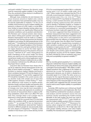
long-term implant success; and (4) diagnostic criteria for implant stability.94,95,105 EIL has been shown to significantly influence ISQ value.8,54,56,80,102,107–109 Although the stiffness of the implant is generally constant, it can sometimes vary in the presence of other contributing stiffnesses (Table 5). Classification of ISQs based upon various conditions may be a grand task. However, if these variables are ignored, the reliability of the measure- ment will be low.101,102 Therefore, only series of intra- patient RFA values over various time points may pro- vide useful information as to the stability of an implant under investigation. Furthermore, these series of values may not indicate the success or fail- ure of the implants.105 This concurs the research of Friberg et al with respect to cutting torque resistance measure- ment.28,29,83 Insertion torque was also highly associ- ated with resonance frequency of implants.30 Lower resistance and lower resonance frequency values were associated with poor bone quality. This may be related to the finding that implant success and sur- vival rates are greater in the mandible than in the maxilla.101,113,114 Prolonged healing time is required in cases with poor bone quality. Therefore, even though an implant placement in softer bone shows low stability, it seems to “catch up” to dense bone sites over time.28 The prognostic value of RFA machines such as Osstell and Implomates has, therefore, been investi- gated. The most challenging factors to overcome are the dynamic characteristics (eg, damping effect, total mass, and stiffness) of various factors surrounding the object of interest,111 bone-implant interface. Without controlling these factors, information gained from RFA is no better than guessing value. To improve its prognostic value of RFA, longitudinal studies and comparison of RFA values with histologic studies are essential. Development of simulation models on various EILs associated with various defect types may further assist in the assessment of implant stability. The shape of the transducer (an L shape) restricts its orientation, which adds a significant length to the exposed implant length, potentially masking a small amount of bone resorption.54 Osstell Mentor (Inte- gration Diagnostics) eliminates the use of an attached L-shape transducer by generating “pulse trains” from a contact-free probe. Impact signals are then picked up by a receptor called a “smart-peg.” Hence, the measurement is believed to be more accurate than the original Osstell machine. Moreover, in cases of Kennedy III partial edentulism, this con- tact-free smart-peg allows assessment of implant stability from any direction. However, due to the dif- ference in EIL and various bending forces from the different design of the transducer, data collected with the original Ostell machine and that collected with the new contact-free Osstell should be com- pared with caution. The establishment of diagnostic criteria for suc- cess, survival, and/or failure is another clinical appli- cation with RFA. However, RFA can only give informa- tion regarding success; it cannot provide information with respect to survival or failure. ISQ can be fairly reliable when an implant has achieved osseointegra- tion and the bone-implant interface is rigid.98 In cases where rigid integration is doubtful, however, the ISQ tends to fluctuate. Some doubtful implants result in failure, whereas some implants showing low ISQ later stabilize and achieve a satisfactory out- come.83 Hence, clinicians will continue to test the implant stability until they get a reasonable value. When unacceptable values are displayed, however, these values are often rejected. If the repeated mea- surements still indicate an unfavorable result, these values are unwillingly accepted. Hence, small stan- dard deviation is often reported with high ISQ. The evaluation of implant stability using RFA machines such as Osstell and Implomates still has some uncertain issues. It is clinically being used with- out much conclusive data on the bone-implant inter- face and resonance frequency values.79,91 Further research is needed to establish higher reliability of these diagnostic devices. 750 Volume 22, Number 5, 2007 Atsumi et al Table 5 Factors that Influence RFA Constants Implant length Implant diameter Implant geometry (implant system) Implant surface characteristics Placement position Abutment length Variables Bone quality Bone quantity Damping effect of marginal mucosa BIC (3-dimensional) EIL Connection of transducer Primary stability Secondary stability Atsumi.qxd 9/17/07 3:19 PM Page 750 COPYRIGHT © 2007 BY QUINTESSENCE PUBLISHING CO, INC. PRINTING OF THIS DOCUMENT IS RESTRICTED TO PERSONAL USE ONLY. NO PART OF THIS ARTICLE MAY BE REPRODUCED OR TRANSMITTED IN ANY FORM WITHOUT WRITTEN PERMISSION FROM THE PUBLISHER
9.
CONCLUSION To date, no
definite method to evaluate implant sta- bility has been established. Although the theory behind RFA is sound, the technology cannot provide a critical value that can determine the success, fail- ure, or long-term prognosis of an implant. Hence, present position from this review is that information should be assembled from many diagnostic aids to assure long-term implant stability. More research in this field is certainly needed. ACKNOWLEDGMENT This work was partially supported by the University of Michigan Periodontal Graduate Student Research Fund. REFERENCES 1. Brånemark P-I,Hansson BO,Adell R,et al.Osseointegrated implants in the treatment of the edentulous jaw.Experience from a 10-year period.Scand J Plast Reconstr Surg 1977; 16(suppl):1–132. 2. Brånemark P,Zarb G,Albrektsson T.Introduction to osseointe- gration.In:Brånemark PI,Zarb GA,Albrektsson T (eds).Tissue- Integrated Prostheses:Osseointegration in Clinical Dentistry. Chicago:Quintessence,1985:11–76. 3. Schröeder A,van der Zypen E,Stich H,Sutter F.The reactions of bone,connective tissue,and epithelium to endosteal implants with titanium-sprayed surfaces.J Maxillofac Surg 1981;9:15–25. 4. Roberts WE.Bone tissue interface.J Dent Educ 1988;52: 804–809. 5. Cochran DL,Schenk RK,Lussi A,Higginbottom FL,Buser D. Bone response to unloaded and loaded titanium implants with a sandblasted and acid-etched surface:A histometric study in the canine mandible.J Biomed Mater Res 1998;40: 1–11. 6. Berglundh T,Abrahamsson I,Lang NP,Lindhe J.De novo alve- olar bone formation adjacent to endosseous implants.Clin Oral Implants Res 2003;14:251–262. 7. Buser D,Broggini N,Wieland M,et al.Enhanced bone apposi- tion to a chemically modified SLA titanium surface.J Dent Res 2004;83:529–533. 8. Meredith N.Assessment of implant stability as a prognostic determinant.Int J Prosthodont 1998;11:491–501. 9. Brunski JB.Biomechanical factors affecting the bone-dental implant interface.Clin Mater 1992;10:153–201. 10. Sennerby L,Roos J.Surgical determinants of clinical success of osseointegrated oral implants:A review of the literature. Int J Prosthodont 1998;11:408–420. 11. Jensen O.The Carter hypothesis.In:Buser D,Dahlin C,Schenk RK (eds).Guided Bone Regeneration in Implant Dentistry. Hong Kong:Quintessence,1994:238–239. 12. Davies JE.Mechanisms of endosseous integration.Int J Prosthodont 1998;11:391–401. 13. Raghavendra S,Wood MC,Taylor TD.Early wound healing around endosseous implants:A review of the literature.Int J Oral Maxillofac Implants 2005;20:425–431. 14. Attard NJ,Zarb GA.Long-term treatment outcomes in eden- tulous patients with implant overdentures:The Toronto study.Int J Prosthodont 2004;17:425–433. 15. Attard NJ,Zarb GA.Long-term treatment outcomes in eden- tulous patients with implant-fixed prostheses:The Toronto study.Int J Prosthodont 2004;17:417–424. 16. Hermann JS,Schoolfield JD,Nummikoski PV,Buser D,Schenk RK,Cochran DL.Crestal bone changes around titanium implants:A methodologic study comparing linear radi- ographic with histometric measurements.Int J Oral Maxillo- fac Implants 2001;16:475–485. 17. van Steenberghe D,Lekholm U,Bolender C,et al.Applicability of osseointegrated oral implants in the rehabilitation of par- tial edentulism:A prospective multicenter study on 558 fix- tures.Int J Oral Maxillofac Implants 1990;5:272–281. 18. Adell R,Lekholm U,Rockler B,Brånemark P-I.A 15-year study of osseointegrated implants in the treatment of the edentu- lous jaw.Int J Oral Surg 1981;10:387–416. 19. Albrektsson T,Zarb G,Worthington P,Eriksson AR.The long- term efficacy of currently used dental implants:A review and proposed criteria of success.Int J Oral Maxillofac Implants 1986;1:11–25. 20. Smith DE,Zarb GA.Criteria for success of osseointegrated endosseous implants.J Prosthet Dent 1989;62:567–572. 21. Misch C.An implant is not a tooth:A comparison of periodon- tal indexes.In:Misch C (ed).Dental Implant Prosthetics.St Louis:Elsevier Mosby,2005:18–31. 22. Goodson JM,Haffajee AD,Socransky SS.The relationship between attachment level loss and alveolar bone loss.J Clin Periodontol 1984;11:348–359. 23. Wyatt CC,Pharoah MJ.Imaging techniques and image inter- pretation for dental implant treatment.Int J Prosthodont 1998;11:442–452. 24. Kircos L,Misch C.Diagnostic imaging and techniques.In: Misch C (ed).Dental Implant Prosthetics.St Louis:Elsevier Mosby,2005:53–70. 25. Johansson P,Strid K.Assessment of bone quality from cutting resistance during implant surgery.Int J Oral Maxillofac Implants 1994;9:279–288. 26. Friberg B,Sennerby L,Roos J,Lekholm U.Identification of bone quality in conjunction with insertion of titanium implants.A pilot study in jaw autopsy specimens.Clin Oral Implants Res 1995;6:213–219. 27. Friberg B,Sennerby L,Roos J,Johansson P,Strid CG,Lekholm U.Evaluation of bone density using cutting resistance mea- surements and microradiography:An in vitro study in pig ribs.Clin Oral Implants Res 1995;6:164–171. 28. Friberg B,Sennerby L,Meredith N,Lekholm U.A comparison between cutting torque and resonance frequency measure- ments of maxillary implants.A 20-month clinical study.Int J Oral Maxillofac Surg 1999;28:297–303. 29. Friberg B,Sennerby L,Grondahl K,Bergstrom C,Back T, Lekholm U.On cutting torque measurements during implant placement:A 3-year clinical prospective study.Clin Implant Dent Relat Res 1999;1:75–83. 30. O’Sullivan D,Sennerby L,Jagger D,Meredith N.A comparison of two methods of enhancing implant primary stability.Clin Implant Dent Relat Res 2004;6:48–57. 31. Lekholm U,Zarb G.Patient selection and preparation.In: Brånemark P-I,Zarb GA,Albrektsson T (eds).Tissue-Integrated Prostheses:Osseointegration in Clinical Dentistry.Chicago: Quintessence,1985:199–209. 32. Engquist B,Bergendal T,Kallus T,Linden U.A retrospective multicenter evaluation of osseointegrated implants support- ing overdentures.Int J Oral Maxillofac Implants 1988;3: 129–134. The International Journal of Oral & Maxillofacial Implants 751 Atsumi et al Atsumi.qxd 9/17/07 3:19 PM Page 751 COPYRIGHT © 2007 BY QUINTESSENCE PUBLISHING CO, INC. PRINTING OF THIS DOCUMENT IS RESTRICTED TO PERSONAL USE ONLY. NO PART OF THIS ARTICLE MAY BE REPRODUCED OR TRANSMITTED IN ANY FORM WITHOUT WRITTEN PERMISSION FROM THE PUBLISHER
10.
33. Friberg B,Jemt
T,Lekholm U.Early failures in 4,641 consecu- tively placed Brånemark dental implants:A study from stage 1 surgery to the connection of completed prostheses.Int J Oral Maxillofac Implants 1991;6:142–146. 34. Jemt T,Lekholm U,Adell R.Osseointegrated implants in the treatment of partially edentulous patients:A preliminary study on 876 consecutively placed fixtures.Int J Oral Maxillo- fac Implants 1989;4:211–217. 35. Misch C,Meffert RM.Implant quality of health scale:A clinical assessment of the health disease continuum.In:Misch C (ed). Dental Implant Prosthetics.St Louis:Elsevier Mosby,2005: 596–603. 36. Buser D,Mericske-Stern R,Bernard JP,et al.Long-term evalua- tion of non-submerged ITI implants.Part 1:8-year life table analysis of a prospective multi-center study with 2359 implants.Clin Oral Implants Res 1997;8:161–172. 37. Lindh T,Gunne J,Tillberg A,Molin M.A meta-analysis of implants in partial edentulism.Clin Oral Implants Res 1998; 9:80–90. 38. Roberts WE,Smith RK,Zilberman Y,Mozsary PG,Smith RS. Osseous adaptation to continuous loading of rigid endosseous implants.Am J Orthod 1984;86:95–111. 39. Johansson C,Albrektsson T.Integration of screw implants in the rabbit:A 1-year follow-up of removal torque of titanium implants.Int J Oral Maxillofac Implants 1987;2:69–75. 40. Johansson CB,Albrektsson T.A removal torque and histomor- phometric study of commercially pure niobium and titanium implants in rabbit bone.Clin Oral Implants Res 1991;2:24–29. 41. Johansson CB,Sennerby L,Albrektsson T.A removal torque and histomorphometric study of bone tissue reactions to commercially pure titanium and Vitallium implants.Int J Oral Maxillofac Implants 1991;6:437–441. 42. Roberts WE,Helm FR,Marshall KJ,Gongloff RK.Rigid endosseous implants for orthodontic and orthopedic anchorage.Angle Orthod 1989;59:247–256. 43. Tjellstrom A,Jacobsson M,Albrektsson T.Removal torque of osseointegrated craniofacial implants:A clinical study.Int J Oral Maxillofac Implants 1988;3:287–289. 44. Buser D,Nydegger T,Hirt HP,Cochran DL,Nolte LP.Removal torque values of titanium implants in the maxilla of miniature pigs.Int J Oral Maxillofac Implants 1998;13:611–619. 45. Sullivan DY,Sherwood RL,Collins TA,Krogh PH.The reverse- torque test:A clinical report.Int J Oral Maxillofac Implants 1996;11:179–185. 46. Cunningham JL,Kenwright J,Kershaw CJ.Biomechanical measurement of fracture healing.J Med Eng Technol 1990; 14:92–101. 47. Nakatsuchi Y,Tsuchikane A,Nomura A.The vibrational mode of the tibia and assessment of bone union in experimental fracture healing using the impulse response method.Med Eng Phys 1996;18:575–583. 48. Natali AN,Pavan PG,Scarpa C.Numerical analysis of tooth mobility:Formulation of a non-linear constitutive law for the periodontal ligament.Dent Mater 2004;20:623–629. 49. Olsen S,Ferguson SJ,Sigrist C,et al.A novel computational method for real-time preoperative assessment of primary dental implant stability.Clin Oral Implants Res 2005;16:53–59. 50. Simmons CA,Meguid SA,Pilliar RM.Mechanical regulation of localized and appositional bone formation around bone- interfacing implants.J Biomed Mater Res 2001;55:63–71. 51. Van Oosterwyck H,Duyck J,Vander Sloten J,Van Der Perre G, Naert I.Peri-implant bone tissue strains in cases of dehis- cence:A finite element study.Clin Oral Implants Res 2002; 13:327–333. 52. Lee SY,Huang HM,Lin CY,Shih YH.In vivo and in vitro natural frequency analysis of periodontal conditions:An innovative method.J Periodontol 2000;71:632–640. 53. Nagamatsu A.Introduction to Modal Analysis,ed 4.Tokyo: Corona,1993. 54. Meredith N,Alleyne D,Cawley P.Quantitative determination of the stability of the implant-tissue interface using reso- nance frequency analysis.Clin Oral Implants Res 1996;7: 261–267. 55. Meredith N,Friberg B,Sennerby L,Aparicio C.Relationship between contact time measurements and PTV values when using the Periotest to measure implant stability.Int J Prostho- dont 1998;11:269–275. 56. Rasmusson L,Meredith N,Kahnberg KE,Sennerby L.Stability assessments and histology of titanium implants placed simultaneously with autogenous onlay bone in the rabbit tibia.Int J Oral Maxillofac Surg 1998;27:229–235. 57. Sekiguhi J.An attempt to measure viscoelasticity of human facial skin by impact hammer method [in Japanese].Kana- gawa Shigaku 1992;26:387–411. 58. Kaneko T.Pulsed oscillation technique for assessing the mechanical state of the dental implant-bone interface.Bio- materials 1991;12:555–560. 59. Kaneko T,Nagai Y,Ogino M,Futami T,Ichimura T.Acoustoelec- tric technique for assessing the mechanical state of the den- tal implant-bone interface.J Biomed Mater Res 1986;20: 169–176. 60. Aoki H.The mobility of healthy teeth as measured with the impact hammer method [in Japanese].Kanagawa Shigaku 1987;22:13–31. 61. Hirakawa W.An attempt to measure tooth mobility in terms of time domain wave forms [in Japanese].Kanagawa Shigaku 1986;21:529–543. 62. Matsuo E,Hirakawa K,Hamada S.Tooth mobility measure- ment techniques using ECM impact hammer method.Bull Kanagawa Dent Coll 1989;17:9–19. 63. Elias JJ,Brunski JB,Scarton HA.A dynamic modal testing tech- nique for noninvasive assessment of bone-dental implant interfaces.Int J Oral Maxillofac Implants 1996;11:728–734. 64. Olive J,Aparicio C.Periotest method as a measure of osseoin- tegrated oral implant stability.Int J Oral Maxillofac Implants 1990;5:390–400. 65. Naert IE,Rosenberg D,van Steenberghe D,Tricio JA,Nys M. The influence of splinting procedures on the periodontal and peri-implant tissue damping characteristics.A longitudinal study with the Periotest device.J Clin Periodontol 1995; 22:703–708. 66. Tricio J,Laohapand P,van Steenberghe D,Quirynen M,Naert I. Mechanical state assessment of the implant-bone contin- uum:A better understanding of the Periotest method.Int J Oral Maxillofac Implants 1995;10:43–49. 67. Tricio J,van Steenberghe D,Rosenberg D,Duchateau L. Implant stability related to insertion torque force and bone density:An in vitro study.J Prosthet Dent 1995;74:608–612. 68. van Steenberghe D,Tricio J,Naert I,Nys M.Damping charac- teristics of bone-to-implant interfaces.A clinical study with the Periotest device.Clin Oral Implants Res 1995;6:31–39. 69. Aparicio C.The use of the Periotest value as the initial success criteria of an implant:8-year report.Int J Periodontics Restorative Dent 1997;17:150–161. 70. Walker L,Morris HF,Ochi S.Periotest values of dental implants in the first 2 years after second-stage surgery:DICRG interim report no.8.Dental Implant Clinical Research Group.Implant Dent 1997;6:207–212. 752 Volume 22, Number 5, 2007 Atsumi et al Atsumi.qxd 9/17/07 3:19 PM Page 752 COPYRIGHT © 2007 BY QUINTESSENCE PUBLISHING CO, INC. PRINTING OF THIS DOCUMENT IS RESTRICTED TO PERSONAL USE ONLY. NO PART OF THIS ARTICLE MAY BE REPRODUCED OR TRANSMITTED IN ANY FORM WITHOUT WRITTEN PERMISSION FROM THE PUBLISHER
11.
71. Truhlar RS,Morris
HF,Ochi S.Stability of the bone-implant complex.Results of longitudinal testing to 60 months with the Periotest device on endosseous dental implants.Ann Periodontol 2000;5:42–55. 72. Schulte W,Lukas D.The Periotest method.Int Dent J 1992;42: 433–440. 73. Zarb GA,Albrektsson T.Osseointegration:A requiem for the periodontal ligament? [editorial].Int J Periodontics Restora- tive Dent 1991;11:88–91. 74. Iijima T,Takeda T.An observation of chronological change of mobility of ITI implants with Periotest.J Jpn Soc Oral Implan- tol 1990;3:191–199. 75. Derhami K,Wolfaardt JF,Faulkner G,Grace M.Assessment of the Periotest device in baseline mobility measurements of craniofacial implants.Int J Oral Maxillofac Implants 1995;10: 221–229. 76. Morris HE,Ochi S,Crum P,Orenstein I,Plezia R.Bone density: Its influence on implant stability after uncovering.J Oral Implantol 2003;29:263–269. 77. Hurzeler MB,Quinones CR,Schupbach P,Vlassis JM,Strub JR, Caffesse RG.Influence of the suprastructure on the peri- implant tissues in beagle dogs.Clin Oral Implants Res 1995; 6:139–148. 78. Teerlinck J,Quirynen M,Darius P,van Steenberghe D.Peri- otest:An objective clinical diagnosis of bone apposition toward implants.Int J Oral Maxillofac Implants 1991;6:55–61. 79. Salvi GE,Lang NP.Diagnostic parameters for monitoring peri- implant conditions.Int J Oral Maxillofac Implants 2004; 19(suppl):116–127. 80. Meredith N,Book K,Friberg B,Jemt T,Sennerby L.Resonance frequency measurements of implant stability in vivo.A cross- sectional and longitudinal study of resonance frequency measurements on implants in the edentulous and partially dentate maxilla.Clin Oral Implants Res 1997;8:226–233. 81. Meredith N,Shagaldi F,Alleyne D,Sennerby L,Cawley P.The application of resonance frequency measurements to study the stability of titanium implants during healing in the rabbit tibia.Clin Oral Implants Res 1997;8:234–243. 82. Rasmusson L,Meredith N,Sennerby L.Measurements of sta- bility changes of titanium implants with exposed threads subjected to barrier membrane induced bone augmentation. An experimental study in the rabbit tibia.Clin Oral Implants Res 1997;8:316–322. 83. Friberg B,Sennerby L,Linden B,Grondahl K,Lekholm U.Sta- bility measurements of one-stage Branemark implants dur- ing healing in mandibles.A clinical resonance frequency analysis study.Int J Oral Maxillofac Surg 1999;28:266–272. 84. Rasmusson L,Meredith N,Cho IH,Sennerby L.The influence of simultaneous versus delayed placement on the stability of titanium implants in onlay bone grafts.A histologic and bio- mechanic study in the rabbit.Int J Oral Maxillofac Surg 1999; 28:224–231. 85. Rasmusson L,Meredith N,Kahnberg KE,Sennerby L.Effects of barrier membranes on bone resorption and implant stability in onlay bone grafts.An experimental study.Clin Oral Implants Res 1999;10:267–277. 86. Friberg B,Jisander S,Widmark G,et al.One-year prospective three-center study comparing the outcome of a“soft bone implant”(prototype Mk IV) and the standard Brånemark implant.Clin Implant Dent Relat Res 2003;5:71–77. 87. Monov G,Fuerst G,Tepper G,Watzak G,Zechner W,Watzek G. The effect of platelet-rich plasma upon implant stability mea- sured by resonance frequency analysis in the lower anterior mandibles.Clin Oral Implants Res 2005;16:461–465. 88. O’Sullivan D,Sennerby L,Meredith N.Measurements compar- ing the initial stability of five designs of dental implants:A human cadaver study.Clin Implant Dent Relat Res 2000;2: 85–92. 89. Sul YT,Johansson CB,Jeong Y,Wennerberg A,Albrektsson T. Resonance frequency and removal torque analysis of implants with turned and anodized surface oxides.Clin Oral Implants Res 2002;13:252–259. 90. Balshi SF,Allen FD,Wolfinger GJ,Balshi TJ.A resonance fre- quency analysis assessment of maxillary and mandibular immediately loaded implants.Int J Oral Maxillofac Implants 2005;20:584–594. 91. Barewal RM,Oates TW,Meredith N,Cochran DL.Resonance frequency measurement of implant stability in vivo on implants with a sandblasted and acid-etched surface.Int J Oral Maxillofac Implants 2003;18:641–651. 92. Bischof M,Nedir R,Szmukler-Moncler S,Bernard JP,Samson J. Implant stability measurement of delayed and immediately loaded implants during healing.Clin Oral Implants Res 2004; 15:529–539. 93. da Cunha HA,Francischone CE,Filho HN,de Oliveira RC.A comparison between cutting torque and resonance fre- quency in the assessment of primary stability and final torque capacity of standard and TiUnite single-tooth implants under immediate loading.Int J Oral Maxillofac Implants 2004;19:578–585. 94. Glauser R,Sennerby L,Meredith N,et al.Resonance frequency analysis of implants subjected to immediate or early func- tional occlusal loading.Successful vs failing implants.Clin Oral Implants Res 2004;15:428–434. 95. Becker W,Sennerby L,Bedrossian E,Becker BE,Lucchini JP. Implant stability measurements for implants placed at the time of extraction:A cohort,prospective clinical trial.J Peri- odontol 2005;76:391–397. 96. Gedrange T,Hietschold V,Mai R,Wolf P,Nicklisch M,Harzer W. An evaluation of resonance frequency analysis for the deter- mination of the primary stability of orthodontic palatal implants.A study in human cadavers.Clin Oral Implants Res 2005;16:425–431. 97. Hallman M,Sennerby L,Zetterqvist L,Lundgren S.A 3-year prospective follow-up study of implant-supported fixed pros- theses in patients subjected to maxillary sinus floor augmen- tation with a 80:20 mixture of deproteinized bovine bone and autogenous bone.Clinical,radiographic and resonance frequency analysis.Int J Oral Maxillofac Surg 2005;34: 273–280. 98. Nedir R,Bischof M,Szmukler-Moncler S,Bernard JP,Samson J. Predicting osseointegration by means of implant primary sta- bility.Clin Oral Implants Res 2004;15:520–528. 99. Sjostrom M,Lundgren S,Nilson H,Sennerby L.Monitoring of implant stability in grafted bone using resonance frequency analysis.A clinical study from implant placement to 6 months of loading.Int J Oral Maxillofac Surg 2005;34:45–51. 100. Cehreli MC,Akkocaoglu M,Comert A,Tekdemir I,Akca K. Human ex vivo bone tissue strains around natural teeth vs. immediate oral implants.Clin Oral Implants Res 2005;16: 540–548. 101. Lachmann S,Jager B,Axmann D,Gomez-Roman G,Groten M, Weber H.Resonance frequency analysis and damping capac- ity assessment.Part I:An in vitro study on measurement relia- bility and a method of comparison in the determination of primary dental implant stability.Clin Oral Implants Res 2006; 17:75–79. The International Journal of Oral & Maxillofacial Implants 753 Atsumi et al Atsumi.qxd 9/17/07 3:19 PM Page 753 COPYRIGHT © 2007 BY QUINTESSENCE PUBLISHING CO, INC. PRINTING OF THIS DOCUMENT IS RESTRICTED TO PERSONAL USE ONLY. NO PART OF THIS ARTICLE MAY BE REPRODUCED OR TRANSMITTED IN ANY FORM WITHOUT WRITTEN PERMISSION FROM THE PUBLISHER
12.
102. Lachmann S,Laval
JY,Jager B,et al.Resonance frequency analysis and damping capacity assessment.Part 2:Peri- implant bone loss follow-up.An in vitro study with the Peri- otest and Osstell instruments.Clin Oral Implants Res 2006;17: 80–84. 103. Zix J,Kessler-Liechti G,Mericske-Stern R.Stability measure- ments of 1-stage implants in the maxilla by means of reso- nance frequency analysis:A pilot study.Int J Oral Maxillofac Implants 2005;20:747–752. 104. Gahleitner A,Monov G.Assessment of bone quality:Tech- niques,procedures,and limitations.In:Watzek G (ed). Implants in Qualitatively Compromised Bone.Chicago:Quin- tessence,2004:55–66. 105. Ersanli S,Karabuda C,Beck F,Leblebicioglu B.Resonance fre- quency analysis of one-stage dental implant stability during the osseointegration period.J Periodontol 2005;76(7): 1066–1071. 106. Meredith N.A review of nondestructive test methods and their application to measure the stability and osseointegra- tion of bone anchored endosseous implants.Crit Rev Biomed Eng 1998;26:275–291. 107. Huang HM,Chiu CL,Yeh CY,Lee SY.Factors influencing the resonance frequency of dental implants.J Oral Maxillofac Surg 2003;61:1184–1188. 108. Huang HM,Lee SY,Yeh CY,Lin CT.Resonance frequency assessment of dental implant stability with various bone qualities:A numerical approach.Clin Oral Implants Res 2002; 13:65–74. 109. Huang HM,Pan LC,Lee SY,Chiu CL,Fan KH,Ho KN.Assessing the implant/bone interface by using natural frequency analy- sis.Oral Surg Oral Med Oral Pathol Oral Radiol Endod 2000; 90(3):285–291. 110. Huang HM,Chiu CL,Yeh CY,Lin CT,Lin LH,Lee SY.Early detec- tion of implant healing process using resonance frequency analysis.Clin Oral Implants Res 2003;14:437–443. 111. Pattijn V,Jaecques SV,De Smet E,Muraru L,Van Lierde C,Van der Perre G,et al.Resonance frequency analysis of implants in the guinea pig model:Influence of boundary conditions and orientation of the transducer.Med Eng Phys 2007;29:182–190. 112. Garg AK.Bone physiology for dental implantology.In:Garg AK (ed).Bone Biology,Harvesting and Grafting for Dental Implants:Rationale and Clinical Applications.Chicago:Quin- tessence,2004:3–19. 113. Adell R,Eriksson B,Lekholm U,Brånemark P-I,Jemt T.Long- term follow-up study of osseointegrated implants in the treatment of totally edentulous jaws.Int J Oral Maxillofac Implants 1990;5(4):347–359. 114. Lindquist LW,Carlsson GE,Jemt T.A prospective 15-year fol- low-up study of mandibular fixed prostheses supported by osseointegrated implants.Clinical results and marginal bone loss.Clin Oral Implants Res 1996;7:329–336. 754 Volume 22, Number 5, 2007 Atsumi et al Atsumi.qxd 9/17/07 3:19 PM Page 754 COPYRIGHT © 2007 BY QUINTESSENCE PUBLISHING CO, INC. PRINTING OF THIS DOCUMENT IS RESTRICTED TO PERSONAL USE ONLY. NO PART OF THIS ARTICLE MAY BE REPRODUCED OR TRANSMITTED IN ANY FORM WITHOUT WRITTEN PERMISSION FROM THE PUBLISHER
Download
![746 Volume 22, Number 5, 2007
Atsumi et al
However, this method has been criticized as being
destructive.8 Brånemark et al2 cautioned about the
risk of irreversible plastic deformation within peri-
implant bone and of implant failure if unnecessary
load was applied to an implant that was still under-
going osseointegration. Furthermore, a 20-Ncm
threshold RTV for successful osseointegration has
not yet been supported by scientific data. The
threshold limit varies among patients depending on
the implant material and the bone quality and quan-
tity. A threshold RTV may be lower in type 4 bone
than in denser bone, for instance. Hence, subjecting
implants placed in this bone type to RTV may result
in a shearing of BIC interface and cause implant fail-
ure. Furthermore, RTV can only provide information
as to “all or none” outcome (osseointegrated or
failed); it cannot quantify degree of osseointegration.
Hence,RTT is mainly used in experiments.
MODAL ANALYSIS
Modal analysis measures the natural frequency or
displacement signal of a system in resonance, which
is initiated by external steady-state waves or a tran-
sient impulse force (Table 4). Modal analysis, in other
words, is a vibration analysis. It is widely used as an
effective test method for structural analysis in engi-
neering and the health-care field.46,47 Dental applica-
tions include the quantification of osseointegra-
tion.48–51 Modal analysis can be performed in 2
models:theoretical and experimental.52
Two or 3-dimensional finite element modeling
(FEM) is an example of computer-simulated theoreti-
cal modal analysis, which is mathematically con-
structed using known biomechanical properties (eg,
Young’s modulus [Pa], Poisson ratio, and density in
g/cm3) of structures of interest. Theoretical modal
analysis such as FEM may be useful in investigation
of the vibrational characteristics of objects that may
be difficult to excite because of a damping effect
from boundary conditions such as the periodontal
ligament (PDL) in an in vivo model.49 By altering
boundary conditions such as the bone level, FEM can
theoretically be used to calculate the anticipated
stress and strain in various simulated peri-implant
bone levels.50,51
Experimental or dynamic modal analysis, on the
other hand, measures structural changes and
dynamic characteristics (eg, natural characteristic fre-
quency, characteristic mode, and attenuation) of a
system that is excited in an in vitro model via vibra-
tion testing (eg, impactor or hammer). This in vitro
approach provides a more reliable assessment of an
object than a theoretical model. This analysis has
been applied in dentistry to quantify the degree of
osseointegration and implant stability.49 Frequency
analysis and mechanical impedance analysis can be
used for detecting response waves in modal
analysis.52 By combining the vibration and response
detecting methods, various kinds of vibration analy-
ses can be performed.53 Some techniques derived
from these theoretical concepts are being tested for
use in evaluating implant mobility.
Percussion Test
A percussion test is one of the simplest methods that
can be used to estimate the level of osseointe-
gration.8,54–56 This test is based upon vibrational-
acoustic science and impact-response theory. A clini-
cal judgment on osseointegration is made based on
the sound heard upon percussion with a metallic
instrument. A clearly ringing “crystal” sound indicates
successful osseointegration, whereas a “dull” sound
may indicate no osseointegration. However, this
method heavily relies on the clinician’s experience
level and subjective belief.Therefore,it cannot be used
experimentally as a standardized testing method.
Impact Hammer Method
Impact hammer method is another example of tran-
sient impact as a source of excitement force during
experimental modal analysis.53,57 It is an improved
version of the percussion test except that sound gen-
erated from a contact between a hammer and an
object is processed through fast Fourier transform
(FFT) for analysis of transfer characteristics. By
enhancing the response detection using various
devices, such as a microphone, an accelerometer, or a
strain gauge, and by processing the detected
response with FFT, it becomes possible to quantify
and qualify the response wave in the form of disloca-
tion, speed, acceleration, stress, distortion, sound, and
other physical properties. Periotest (Siemens, Ben-
sheim, Germany) and Dental Mobility Checker (DMC;
Table 4 Implant Stability Measurement Based on
Modal or Vibration Analysis
Theoretical Modal Analysis
1. Finite element method
Experimental Modal Analysis
1. Percussion test
2. Impact hammer method (Periotest, Siemens, Bensheim,
Germany; Dental Mobility Checker, J. Morita, Suita, Japan)
3. RFA (Osstell, Integration Diagnostics, Göteborg, Sweden;
Implomate, Bio Tech One, Taipei, Taiwan)
4. Others (pulsed oscillation waveform by Kaneko)
Atsumi.qxd 9/17/07 3:19 PM Page 746
COPYRIGHT © 2007 BY QUINTESSENCE PUBLISHING CO, INC. PRINTING OF THIS DOCUMENT IS RESTRICTED TO PERSONAL USE ONLY. NO PART OF THIS
ARTICLE MAY BE REPRODUCED OR TRANSMITTED IN ANY FORM WITHOUT WRITTEN PERMISSION FROM THE PUBLISHER](https://image.slidesharecdn.com/methodsusedtoassessimplantstability-180717064058-d319c2/85/Methods-used-to_assess_implant_stability-unlocked-4-320.jpg)
![33. Friberg B,Jemt T,Lekholm U.Early failures in 4,641 consecu-
tively placed Brånemark dental implants:A study from stage
1 surgery to the connection of completed prostheses.Int J
Oral Maxillofac Implants 1991;6:142–146.
34. Jemt T,Lekholm U,Adell R.Osseointegrated implants in the
treatment of partially edentulous patients:A preliminary
study on 876 consecutively placed fixtures.Int J Oral Maxillo-
fac Implants 1989;4:211–217.
35. Misch C,Meffert RM.Implant quality of health scale:A clinical
assessment of the health disease continuum.In:Misch C (ed).
Dental Implant Prosthetics.St Louis:Elsevier Mosby,2005:
596–603.
36. Buser D,Mericske-Stern R,Bernard JP,et al.Long-term evalua-
tion of non-submerged ITI implants.Part 1:8-year life table
analysis of a prospective multi-center study with 2359
implants.Clin Oral Implants Res 1997;8:161–172.
37. Lindh T,Gunne J,Tillberg A,Molin M.A meta-analysis of
implants in partial edentulism.Clin Oral Implants Res 1998;
9:80–90.
38. Roberts WE,Smith RK,Zilberman Y,Mozsary PG,Smith RS.
Osseous adaptation to continuous loading of rigid
endosseous implants.Am J Orthod 1984;86:95–111.
39. Johansson C,Albrektsson T.Integration of screw implants in
the rabbit:A 1-year follow-up of removal torque of titanium
implants.Int J Oral Maxillofac Implants 1987;2:69–75.
40. Johansson CB,Albrektsson T.A removal torque and histomor-
phometric study of commercially pure niobium and titanium
implants in rabbit bone.Clin Oral Implants Res 1991;2:24–29.
41. Johansson CB,Sennerby L,Albrektsson T.A removal torque
and histomorphometric study of bone tissue reactions to
commercially pure titanium and Vitallium implants.Int J Oral
Maxillofac Implants 1991;6:437–441.
42. Roberts WE,Helm FR,Marshall KJ,Gongloff RK.Rigid
endosseous implants for orthodontic and orthopedic
anchorage.Angle Orthod 1989;59:247–256.
43. Tjellstrom A,Jacobsson M,Albrektsson T.Removal torque of
osseointegrated craniofacial implants:A clinical study.Int J
Oral Maxillofac Implants 1988;3:287–289.
44. Buser D,Nydegger T,Hirt HP,Cochran DL,Nolte LP.Removal
torque values of titanium implants in the maxilla of miniature
pigs.Int J Oral Maxillofac Implants 1998;13:611–619.
45. Sullivan DY,Sherwood RL,Collins TA,Krogh PH.The reverse-
torque test:A clinical report.Int J Oral Maxillofac Implants
1996;11:179–185.
46. Cunningham JL,Kenwright J,Kershaw CJ.Biomechanical
measurement of fracture healing.J Med Eng Technol 1990;
14:92–101.
47. Nakatsuchi Y,Tsuchikane A,Nomura A.The vibrational mode
of the tibia and assessment of bone union in experimental
fracture healing using the impulse response method.Med
Eng Phys 1996;18:575–583.
48. Natali AN,Pavan PG,Scarpa C.Numerical analysis of tooth
mobility:Formulation of a non-linear constitutive law for the
periodontal ligament.Dent Mater 2004;20:623–629.
49. Olsen S,Ferguson SJ,Sigrist C,et al.A novel computational
method for real-time preoperative assessment of primary
dental implant stability.Clin Oral Implants Res 2005;16:53–59.
50. Simmons CA,Meguid SA,Pilliar RM.Mechanical regulation of
localized and appositional bone formation around bone-
interfacing implants.J Biomed Mater Res 2001;55:63–71.
51. Van Oosterwyck H,Duyck J,Vander Sloten J,Van Der Perre G,
Naert I.Peri-implant bone tissue strains in cases of dehis-
cence:A finite element study.Clin Oral Implants Res 2002;
13:327–333.
52. Lee SY,Huang HM,Lin CY,Shih YH.In vivo and in vitro natural
frequency analysis of periodontal conditions:An innovative
method.J Periodontol 2000;71:632–640.
53. Nagamatsu A.Introduction to Modal Analysis,ed 4.Tokyo:
Corona,1993.
54. Meredith N,Alleyne D,Cawley P.Quantitative determination
of the stability of the implant-tissue interface using reso-
nance frequency analysis.Clin Oral Implants Res 1996;7:
261–267.
55. Meredith N,Friberg B,Sennerby L,Aparicio C.Relationship
between contact time measurements and PTV values when
using the Periotest to measure implant stability.Int J Prostho-
dont 1998;11:269–275.
56. Rasmusson L,Meredith N,Kahnberg KE,Sennerby L.Stability
assessments and histology of titanium implants placed
simultaneously with autogenous onlay bone in the rabbit
tibia.Int J Oral Maxillofac Surg 1998;27:229–235.
57. Sekiguhi J.An attempt to measure viscoelasticity of human
facial skin by impact hammer method [in Japanese].Kana-
gawa Shigaku 1992;26:387–411.
58. Kaneko T.Pulsed oscillation technique for assessing the
mechanical state of the dental implant-bone interface.Bio-
materials 1991;12:555–560.
59. Kaneko T,Nagai Y,Ogino M,Futami T,Ichimura T.Acoustoelec-
tric technique for assessing the mechanical state of the den-
tal implant-bone interface.J Biomed Mater Res 1986;20:
169–176.
60. Aoki H.The mobility of healthy teeth as measured with the
impact hammer method [in Japanese].Kanagawa Shigaku
1987;22:13–31.
61. Hirakawa W.An attempt to measure tooth mobility in terms
of time domain wave forms [in Japanese].Kanagawa Shigaku
1986;21:529–543.
62. Matsuo E,Hirakawa K,Hamada S.Tooth mobility measure-
ment techniques using ECM impact hammer method.Bull
Kanagawa Dent Coll 1989;17:9–19.
63. Elias JJ,Brunski JB,Scarton HA.A dynamic modal testing tech-
nique for noninvasive assessment of bone-dental implant
interfaces.Int J Oral Maxillofac Implants 1996;11:728–734.
64. Olive J,Aparicio C.Periotest method as a measure of osseoin-
tegrated oral implant stability.Int J Oral Maxillofac Implants
1990;5:390–400.
65. Naert IE,Rosenberg D,van Steenberghe D,Tricio JA,Nys M.
The influence of splinting procedures on the periodontal and
peri-implant tissue damping characteristics.A longitudinal
study with the Periotest device.J Clin Periodontol 1995;
22:703–708.
66. Tricio J,Laohapand P,van Steenberghe D,Quirynen M,Naert I.
Mechanical state assessment of the implant-bone contin-
uum:A better understanding of the Periotest method.Int J
Oral Maxillofac Implants 1995;10:43–49.
67. Tricio J,van Steenberghe D,Rosenberg D,Duchateau L.
Implant stability related to insertion torque force and bone
density:An in vitro study.J Prosthet Dent 1995;74:608–612.
68. van Steenberghe D,Tricio J,Naert I,Nys M.Damping charac-
teristics of bone-to-implant interfaces.A clinical study with
the Periotest device.Clin Oral Implants Res 1995;6:31–39.
69. Aparicio C.The use of the Periotest value as the initial success
criteria of an implant:8-year report.Int J Periodontics
Restorative Dent 1997;17:150–161.
70. Walker L,Morris HF,Ochi S.Periotest values of dental implants
in the first 2 years after second-stage surgery:DICRG interim
report no.8.Dental Implant Clinical Research Group.Implant
Dent 1997;6:207–212.
752 Volume 22, Number 5, 2007
Atsumi et al
Atsumi.qxd 9/17/07 3:19 PM Page 752
COPYRIGHT © 2007 BY QUINTESSENCE PUBLISHING CO, INC. PRINTING OF THIS DOCUMENT IS RESTRICTED TO PERSONAL USE ONLY. NO PART OF THIS
ARTICLE MAY BE REPRODUCED OR TRANSMITTED IN ANY FORM WITHOUT WRITTEN PERMISSION FROM THE PUBLISHER](https://image.slidesharecdn.com/methodsusedtoassessimplantstability-180717064058-d319c2/85/Methods-used-to_assess_implant_stability-unlocked-10-320.jpg)
![71. Truhlar RS,Morris HF,Ochi S.Stability of the bone-implant
complex.Results of longitudinal testing to 60 months with
the Periotest device on endosseous dental implants.Ann
Periodontol 2000;5:42–55.
72. Schulte W,Lukas D.The Periotest method.Int Dent J 1992;42:
433–440.
73. Zarb GA,Albrektsson T.Osseointegration:A requiem for the
periodontal ligament? [editorial].Int J Periodontics Restora-
tive Dent 1991;11:88–91.
74. Iijima T,Takeda T.An observation of chronological change of
mobility of ITI implants with Periotest.J Jpn Soc Oral Implan-
tol 1990;3:191–199.
75. Derhami K,Wolfaardt JF,Faulkner G,Grace M.Assessment of
the Periotest device in baseline mobility measurements of
craniofacial implants.Int J Oral Maxillofac Implants 1995;10:
221–229.
76. Morris HE,Ochi S,Crum P,Orenstein I,Plezia R.Bone density:
Its influence on implant stability after uncovering.J Oral
Implantol 2003;29:263–269.
77. Hurzeler MB,Quinones CR,Schupbach P,Vlassis JM,Strub JR,
Caffesse RG.Influence of the suprastructure on the peri-
implant tissues in beagle dogs.Clin Oral Implants Res 1995;
6:139–148.
78. Teerlinck J,Quirynen M,Darius P,van Steenberghe D.Peri-
otest:An objective clinical diagnosis of bone apposition
toward implants.Int J Oral Maxillofac Implants 1991;6:55–61.
79. Salvi GE,Lang NP.Diagnostic parameters for monitoring peri-
implant conditions.Int J Oral Maxillofac Implants 2004;
19(suppl):116–127.
80. Meredith N,Book K,Friberg B,Jemt T,Sennerby L.Resonance
frequency measurements of implant stability in vivo.A cross-
sectional and longitudinal study of resonance frequency
measurements on implants in the edentulous and partially
dentate maxilla.Clin Oral Implants Res 1997;8:226–233.
81. Meredith N,Shagaldi F,Alleyne D,Sennerby L,Cawley P.The
application of resonance frequency measurements to study
the stability of titanium implants during healing in the rabbit
tibia.Clin Oral Implants Res 1997;8:234–243.
82. Rasmusson L,Meredith N,Sennerby L.Measurements of sta-
bility changes of titanium implants with exposed threads
subjected to barrier membrane induced bone augmentation.
An experimental study in the rabbit tibia.Clin Oral Implants
Res 1997;8:316–322.
83. Friberg B,Sennerby L,Linden B,Grondahl K,Lekholm U.Sta-
bility measurements of one-stage Branemark implants dur-
ing healing in mandibles.A clinical resonance frequency
analysis study.Int J Oral Maxillofac Surg 1999;28:266–272.
84. Rasmusson L,Meredith N,Cho IH,Sennerby L.The influence
of simultaneous versus delayed placement on the stability of
titanium implants in onlay bone grafts.A histologic and bio-
mechanic study in the rabbit.Int J Oral Maxillofac Surg 1999;
28:224–231.
85. Rasmusson L,Meredith N,Kahnberg KE,Sennerby L.Effects of
barrier membranes on bone resorption and implant stability
in onlay bone grafts.An experimental study.Clin Oral
Implants Res 1999;10:267–277.
86. Friberg B,Jisander S,Widmark G,et al.One-year prospective
three-center study comparing the outcome of a“soft bone
implant”(prototype Mk IV) and the standard Brånemark
implant.Clin Implant Dent Relat Res 2003;5:71–77.
87. Monov G,Fuerst G,Tepper G,Watzak G,Zechner W,Watzek G.
The effect of platelet-rich plasma upon implant stability mea-
sured by resonance frequency analysis in the lower anterior
mandibles.Clin Oral Implants Res 2005;16:461–465.
88. O’Sullivan D,Sennerby L,Meredith N.Measurements compar-
ing the initial stability of five designs of dental implants:A
human cadaver study.Clin Implant Dent Relat Res 2000;2:
85–92.
89. Sul YT,Johansson CB,Jeong Y,Wennerberg A,Albrektsson T.
Resonance frequency and removal torque analysis of
implants with turned and anodized surface oxides.Clin Oral
Implants Res 2002;13:252–259.
90. Balshi SF,Allen FD,Wolfinger GJ,Balshi TJ.A resonance fre-
quency analysis assessment of maxillary and mandibular
immediately loaded implants.Int J Oral Maxillofac Implants
2005;20:584–594.
91. Barewal RM,Oates TW,Meredith N,Cochran DL.Resonance
frequency measurement of implant stability in vivo on
implants with a sandblasted and acid-etched surface.Int J
Oral Maxillofac Implants 2003;18:641–651.
92. Bischof M,Nedir R,Szmukler-Moncler S,Bernard JP,Samson J.
Implant stability measurement of delayed and immediately
loaded implants during healing.Clin Oral Implants Res 2004;
15:529–539.
93. da Cunha HA,Francischone CE,Filho HN,de Oliveira RC.A
comparison between cutting torque and resonance fre-
quency in the assessment of primary stability and final
torque capacity of standard and TiUnite single-tooth
implants under immediate loading.Int J Oral Maxillofac
Implants 2004;19:578–585.
94. Glauser R,Sennerby L,Meredith N,et al.Resonance frequency
analysis of implants subjected to immediate or early func-
tional occlusal loading.Successful vs failing implants.Clin
Oral Implants Res 2004;15:428–434.
95. Becker W,Sennerby L,Bedrossian E,Becker BE,Lucchini JP.
Implant stability measurements for implants placed at the
time of extraction:A cohort,prospective clinical trial.J Peri-
odontol 2005;76:391–397.
96. Gedrange T,Hietschold V,Mai R,Wolf P,Nicklisch M,Harzer W.
An evaluation of resonance frequency analysis for the deter-
mination of the primary stability of orthodontic palatal
implants.A study in human cadavers.Clin Oral Implants Res
2005;16:425–431.
97. Hallman M,Sennerby L,Zetterqvist L,Lundgren S.A 3-year
prospective follow-up study of implant-supported fixed pros-
theses in patients subjected to maxillary sinus floor augmen-
tation with a 80:20 mixture of deproteinized bovine bone
and autogenous bone.Clinical,radiographic and resonance
frequency analysis.Int J Oral Maxillofac Surg 2005;34:
273–280.
98. Nedir R,Bischof M,Szmukler-Moncler S,Bernard JP,Samson J.
Predicting osseointegration by means of implant primary sta-
bility.Clin Oral Implants Res 2004;15:520–528.
99. Sjostrom M,Lundgren S,Nilson H,Sennerby L.Monitoring of
implant stability in grafted bone using resonance frequency
analysis.A clinical study from implant placement to 6 months
of loading.Int J Oral Maxillofac Surg 2005;34:45–51.
100. Cehreli MC,Akkocaoglu M,Comert A,Tekdemir I,Akca K.
Human ex vivo bone tissue strains around natural teeth vs.
immediate oral implants.Clin Oral Implants Res 2005;16:
540–548.
101. Lachmann S,Jager B,Axmann D,Gomez-Roman G,Groten M,
Weber H.Resonance frequency analysis and damping capac-
ity assessment.Part I:An in vitro study on measurement relia-
bility and a method of comparison in the determination of
primary dental implant stability.Clin Oral Implants Res 2006;
17:75–79.
The International Journal of Oral & Maxillofacial Implants 753
Atsumi et al
Atsumi.qxd 9/17/07 3:19 PM Page 753
COPYRIGHT © 2007 BY QUINTESSENCE PUBLISHING CO, INC. PRINTING OF THIS DOCUMENT IS RESTRICTED TO PERSONAL USE ONLY. NO PART OF THIS
ARTICLE MAY BE REPRODUCED OR TRANSMITTED IN ANY FORM WITHOUT WRITTEN PERMISSION FROM THE PUBLISHER](https://image.slidesharecdn.com/methodsusedtoassessimplantstability-180717064058-d319c2/85/Methods-used-to_assess_implant_stability-unlocked-11-320.jpg)