This study evaluated the stability and rate of osseointegration of 33 immediately loaded dental implants in 7 patients that were treated with ultraviolet light photofunctionalization. Photofunctionalization increased the hydrophilicity, cleanliness, and positive charge of the implant surfaces. The average stability of the photofunctionalized implants at 6 weeks was higher than typically reported for untreated implants after 2-6 months of healing. No stability dips were observed for the photofunctionalized implants. Photofunctionalization accelerated osseointegration as shown by higher stability increases per month compared to literature reports on untreated implants. The results suggest photofunctionalization provides benefits for immediate loading by enhancing


![Implant Stability Measurement and
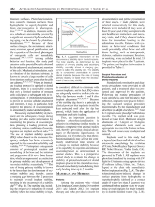
Osseointegration Speed Index
Implant stability was evaluated by
measuring the ISQ at implant
placement (ISQi) and during the heal-
ing period with a 1-week interval up
to 11 weeks using Osstell ISQ
(Osstell AB, Gothenburg, Sweden).
Furthermore, the rate of establishing
implant stability was evaluated by the
osseointegration speed index (OSI)
defined as an ISQ increase per month,
that is, ([ISQ at week 6] − [ISQi])/1.5.
Statistical Analysis
The effect of healing time on ISQ
values was evaluated by ANOVA; P ,
0.05 indicated statistical significance.
When the effect was significant, further
post hoc analysis of Bonferroni was
performed to compare the ISQi with
the ISQ at each of the subsequent time
points. The ISQ values were compared
among implants with different lengths
using ANOVA. Furthermore, the effect
of different bone types where implants
were placed was evaluated.
RESULTS
Implant Dimensions and Bone Type
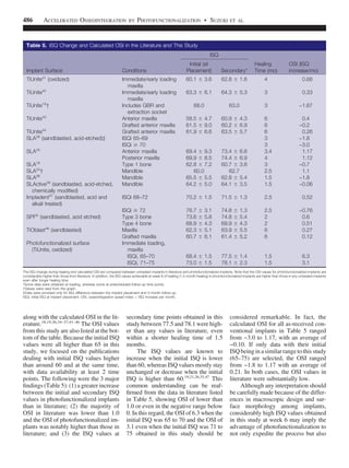
The diameter of all implants used in
this study was 4.3 mm, whereas their
length varied; 13 mm implants were
used most often (Table 1). A majority of
implants (57.6%) were placed in the
type 2 bone, whereas 24.2% and
18.2% implants were placed in the type
1 and type 3 bones, respectively. There
was no type 4 bone because the cases
included in the study were selected for
immediate loading.
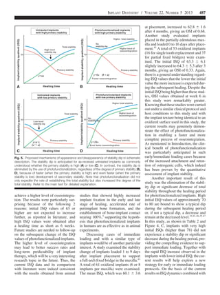
Change in Implant Stability
To visualize the overall trend of
change in implant stability, ISQi values
and the ISQ values at week 6 were
individually plotted (Fig. 3). The ISQi
varied widely from 65 to 85, whereas
the ISQ values at week 6 were con-
verged to the higher level. There was
a variation in ISQ fluctuation between
the time of implant placement and week
6, an increase, no change, or a decrease,
for implants with ISQi that were 77 or
higher. In contrast, all implants with IS-
Qi 75 or lower showed an increase at
week 6. There was a clear trend that
lower the ISQi, the greater the subse-
quent ISQ increase. As a result, the
ISQ values at week 6 were all 75 or
higher.
Next, the implants were divided
into 3 groups depending on the range of
their ISQi (ISQi 65–70, ISQi 71–75,
and ISQi $ 76), and the ISQ values
Fig. 4. Change in implant stability for photofunctionalized implants, evaluated by ISQ values at
implant placement and subsequent healing time. Line graphs are drawn in 3 different groups
depending on the initial ISQ values at implant placement (ISQi). A, ISQi 65 -70; B, ISQi 71 –75;
and C, ISQi $ 76. *P , 0.05, **P , 0.01, statistically significant difference from the ISQi.
484 ACCELERATED OSSEOINTEGRATION BY PHOTOFUNCTIONALIZATION SUZUKI ET AL](https://image.slidesharecdn.com/implantstability2013-180717064045/85/Implant-stability-2013-4-320.jpg)
![indirectly, in the products or informa-
tion listed in the article.
REFERENCES
1. Ogawa T. UV photofunctionalization
of titanium implants. J Craniofac Tissue
Eng. 2012;2:151–158.
2. Att W, Ogawa T. Biological aging of
implant surfaces and their restoration with
ultraviolet light treatment: A novel under-
standing of osseointegration. Int J Oral
Maxillofac Implants. 2012;27:753–761.
3. Lee JH, Ogawa T. The biological
aging of titanium implants. Implant Dent.
2012;21:415–421.
4. Ogawa T. Photofunctionalization of
TiO2 for optimal integration of titanium with
bone. In: Kamat P, Anpo M, eds. Benign
Photocatalysts. Applications of Titanium
Oxide-based Materials. New York:
Springer; 2010:699–713.
5. Aita H, Hori N, Takeuchi M, et al.
The effect of ultraviolet functionalization of
titanium on integration with bone. Bioma-
terials. 2009;30:1015–1025.
6. Tsukimura N, Yamada M, Iwasa F,
et al. Synergistic effects of UV photofunc-
tionalization and micro-nano hybrid topog-
raphy on the biological properties of
titanium. Biomaterials. 2011;32:4358–4368.
7. Ueno T, Yamada M, Hori N, et al.
Effect of ultraviolet photoactivation of tita-
nium on osseointegration in a rat model.
Int J Oral Maxillofac Implants. 2010;25:
287–294.
8. Ueno T, Yamada M, Suzuki T, et al.
Enhancement of bone-titanium integration
profile with UV-photofunctionalized tita-
nium in a gap healing model. Biomaterials.
2010;31:1546–1557.
9. Suzuki T, Hori N, Att W, et al. Ultra-
violet treatment overcomes time-related
degrading bioactivity of titanium. Tissue
Eng Part A. 2009;15:3679–3688.
10. Iwasa F, Tsukimura N, Sugita Y, et al.
TiO2 micro-nano-hybrid surface to alleviate
biological aging of UV-photofunctionalized
titanium. Int J Nanomedicine. 2011;6:
1327–1341.
11. Iwasa F, Hori N, Ueno T, et al.
Enhancement of osteoblast adhesion to
UV-photofunctionalized titanium via an
electrostatic mechanism. Biomaterials.
2010;31:2717–2727.
12. Hori N, Ueno T, Minamikawa H,
et al. Electrostatic control of protein
adsorption on UV-photofunctionalized tita-
nium. Acta Biomater. 2010;6:4175–4180.
13. Hori N, Att W, Ueno T, et al. Age-
dependent degradation of the protein
adsorption capacity of titanium. J Dent
Res. 2009;88:663–667.
14. Att W, Hori N, Takeuchi M, et al.
Time-dependent degradation of titanium
osteoconductivity: An implication of bio-
logical aging of implant materials. Bioma-
terials. 2009;30:5352–5363.
15. Yamada M, Miyauchi T, Yamamoto
A, et al. Enhancement of adhesion
strength and cellular stiffness of osteo-
blasts on mirror-polished titanium surface
by UV-photofunctionalization. Acta Bio-
mater. 2010;6:4578–4588.
16. Miyauchi T, Yamada M, Yamamoto
A, et al. The enhanced characteristics of
osteoblast adhesion to photofunctional-
ized nanoscale TiO2 layers on biomaterials
surfaces. Biomaterials. 2010;31:3827–3839.
17. Ueno T, Tsukimura N, Yamada M,
et al. Enhanced bone-integration capability
of alkali- and heat-treated nanopolymor-
phic titanium in micro-to-nanoscale hierar-
chy. Biomaterials. 2011;32:7297–7308.
18. Bischof M, Nedir R, Szmukler-
Moncler S, et al. Implant stability measure-
ment of delayed and immediately loaded
implants during healing. Clin Oral Implants
Res. 2004;15:529–539.
19. Glauser R, Sennerby L, Meredith
N, et al. Resonance frequency analysis of
implants subjected to immediate or early
functional occlusal loading. Successful vs.
failing implants. Clin Oral Implants Res.
2004;15:428–434.
20. Han J, Lulic M, Lang NP. Factors
influencing resonance frequency analysis
assessed by Osstell mentor during implant
tissue integration: II. Implant surface mod-
ifications and implant diameter. Clin Oral
Implants Res. 2010;21:605–611.
21. Makary C, Rebaudi A, Sammartino
G, et al. Implant primary stability deter-
mined by resonance frequency analysis:
Correlation with insertion torque, histologic
bone volume, and torsional stability at 6
weeks. Implant Dent. 2012;21:474–480.
22. Javed F, Almas K, Crespi R, et al.
Implant surface morphology and primary
stability: Is there a connection? Implant
Dent. 2011;20:40–46.
23. Lee HJ, Aparecida de Mattias
Sartori I, Alcântara PR, et al. Implant sta-
bility measurements of two immediate
loading protocols for the edentulous man-
dible: Rigid and semi-rigid splinting of the
implants. Implant Dent. 2012;21:486–490.
24. Chan HL, El-Kholy K, Fu JH, et al.
Implant primary stability determined by
resonance frequency analysis in surgically
created defects: A pilot cadaver study.
Implant Dent. 2010;19:509–519.
25. Gupta RK, Padmanabhan TV. An
evaluation of the resonance frequency anal-
ysis device: Examiner reliability and repeat-
ability of readings [published online ahead of
print October 4, 2011]. J Oral Implantol. doi:
doi.org/10.1563/AAID-JOI-D-11-00099.
26. Nedir R, Bischof M, Szmukler-
Moncler S, et al. Predicting osseointegra-
tion by means of implant primary stability.
Clin Oral Implants Res. 2004;15:520–528.
27. Meredith N, Alleyne D, Cawley P.
Quantitative determination of the stability
of the implant-tissue interface using reso-
nance frequency analysis. Clin Oral Im-
plants Res. 1996;7:261–267.
28. Huang HL, Tsai MT, Su KC, et al.
Relation between initial implant stability
quotient and bone-implant contact per-
centage: An in vitro model study [pub-
lished online ahead of print August 23,
2012]. Oral Surg Oral Med Oral Pathol Oral
Radiol. doi.10.1016/j.oooo.2012.01.037.
29. Park KJ, Kwon JY, Kim SK, et al.
The relationship between implant stability
quotient values and implant insertion vari-
ables: A clinical study. J Oral Rehabil.
2012;39:151–159.
30. Sennerby L, Meredith N. Implant
stability measurements using resonance
frequency analysis: Biological and biome-
chanical aspects and clinical implications.
Periodontol 2000. 2008;47:51–66.
31. Ogawa T, Nishimura I. Different
bone integration profiles of turned and
acid-etched implants associated with
modulated expression of extracellular
matrix genes. Int J Oral Maxillofac Im-
plants. 2003;18:200–210.
32. Aparicio C, Lang NP, Rangert B.
Validity and clinical significance of biome-
chanical testing of implant/bone interface.
Clin Oral Implants Res. 2006;17(suppl 2):2–7.
33. Atsumi M, Park SH, Wang HL.
Methods used to assess implant stability:
Current status. Int J Oral Maxillofac Im-
plants. 2007;22:743–754.
34. Barewal RM, Oates TW, Meredith
N, et al. Resonance frequency measure-
ment of implant stability in vivo on implants
with a sandblasted and acid-etched sur-
face. Int J Oral Maxillofac Implants. 2003;
18:641–651.
35. Sençimen M, Gülses A, Ozen J, et al.
Early detection of alterations in the reso-
nance frequency assessment of oral implant
stability on various bone types: A clinical
study. J Oral Implantol. 2011;37:411–419.
36. Oates TW, Valderrama P, Bischof
M, et al. Enhanced implant stability with
a chemically modified SLA surface: A ran-
domized pilot study. Int J Oral Maxillofac
Implants. 2007;22:755–760.
37. Simunek A, Kopecka D, Brazda T,
et al. Development of implant stability dur-
ing early healing of immediately loaded im-
plants. Int J Oral Maxillofac Implants.
2012;27:619–627.
38. Maló P, de Araújo Nobre M, Lopes
A, et al. “All-on-4” immediate-function con-
cept for completely edentulous maxillae: A
clinical report on the medium (3 years) and
long-term (5 years) outcomes. Clin Implant
Dent Relat Res. 2012;14(suppl 1):e139–
e150.
39. Malo P, de Araújo Nobre M, Lopes
A, et al. A longitudinal study of the survival
IMPLANT DENTISTRY / VOLUME 22, NUMBER 5 2013 489](https://image.slidesharecdn.com/implantstability2013-180717064045/85/Implant-stability-2013-9-320.jpg)