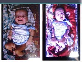
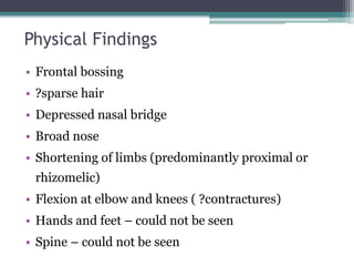
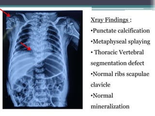
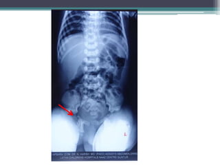
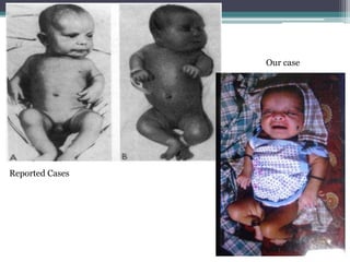
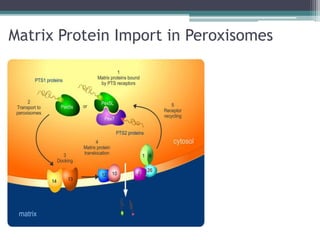
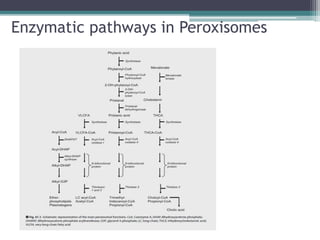
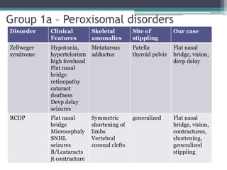
CASE PRESENTATION
By Kanika Singh
DNB Student Medical Genetics
SGRH
15 months 3 months
Presenting Complaints :
First child
• Antenatal history – uneventful, no h/o
oligo/polyhydramnios, fetal movements ?
• Birth history – Term/NVD/Cried after birth/
Bwt – 3kg. No immediate perinatal
complications
• After birth noted to have short limbs and
contractures. B/L knees and elbows in flexion
and restriction of movement at shoulder joints.
• H/o global developmental delay present – no
neck holding, social smile, did not grasp objects
• Vision impairment - Unable to fix gaze on an
object or parents noticed at 3 months of age
• Acyanotic heart disease detected at 6 months of
age
• Not gaining weight
• Expired at 15 months of age
• Terminal event – fever ?sepsis
• Weight at death 2.5 kg (Failure to thrive)
Second Child
• Antenatal history - ? Polyhydramnios
• Term born/3.5kgs/Respiratory distress at birth
Nicu stay x 20 days. Echo – small ASD
• Short limbs and contractures
• Feeding – Normal
• Vision - ? Parents unsure
• Expired at 3 months of age. Cause ?
Physical Findings
• Frontal bossing
• ?sparse hair
• Depressed nasal bridge
• Broad nose
• Shortening of limbs (predominantly proximal or
rhizomelic)
• Flexion at elbow and knees ( ?contractures)
• Hands and feet – could not be seen
• Spine – could not be seen
Summary
• Consanguinous family
• Two siblings affected, both boy and girl
• Flat nasal bridge + rhizomelic shortening +
contractures + failure to thrive + developmental
delay + congenital heart disease + Vision
involvement
Differential Diagnosis
Rhizomelic Skeletal dysplasias:
• Achondroplasia
• Hypochondroplasia
• Kyphomelic dysplasia
• Spondylo epiphyseal dysplasia congenita
• Rhizomelic Chondro dysplasia Punctata
• Osteogenesis imperfecta type IV
Xray Findings :
•Punctate calcification
•Metaphyseal splaying
• Thoracic Vertebral
segmentation defect
•Normal ribs scapulae
clavicle
•Normal
mineralization
Revised Differential Diagnosis :
Rhizomelic shortening + flat nasal bridge +
punctate epiphysis
• Genetic defects in
1. Peroxisomal metabolism
2. Cholesterol metabolism
3. Vitamin K metabolism
• Acquired embryopathies
1. Maternal malabsorption of Vit K
2. Drugs –Warfarin
3. Maternal SLE
Rhizomelic Chondrodysplasia Punctata
• Disorder of peroxismal import
• Clinical Features :
Flat nasal bridge
Rhizomelic shortening (humerus > femur)
Postnatal growth deficiency
Cataracts
Severe intellectual disability
Seizures
Punctate calcification usually disappearing after
1-3 yr of age
Epiphyseal and metaphyseal abnormalities
Vertebral coronal clefts
Others – cleft of the soft palate
Congenital heart disease
UPJ obstruction
Brain MRI – cerebral and cerebellar atrophy
with enlargement of ventricles
Brain MRS – delayed myelination, decreased
choline to creatinine ratios
Death in infancy
Reported Cases
Our case
Xray from published
literature
Our Case
Vertebral Coronal Clefts
•Failure of fusion of
anterior and posterior
ossification centers
•Best seen on lateral
Xray films
•Radiolucent band
running through the
vertebral bodies
Matrix Protein Import in Peroxisomes
Enzymatic pathways in Peroxisomes
Classification
• Multiple enzyme deficiencies:Peroxisomal
Biogenesis Disorders (PBD)
▫ Zellweger spectrum disorder
▫ Rhizomelic chondrodysplasia punctata spectrum
(RCDP)
▫ Neonatal ALD
▫ Infantile Refsum disease
• Single enzyme deficiencies
▫ X-linked adrenoleukodystrophy (X-ALD)
▫ Acyl-CoA oxidase deficiency
▫ Adult Refsum disease
▫ Hyperoxaluria Type I
Diagnosis & Testing of RCDP
Biochemical tests:
• Deficiency of plasmalogens in RBC’s
• Elevated plasma concentration of phytanic acid
• Normal plasma VLCFA
Done in limited no. Of laboratories worldwide
Assays in cultured skin fibroblasts
• Possible to demonstrate defective plasmalogen biosynthesis,
defective phytanic acid oxidation and normal VLCFA oxidation
Genetic Testing
• Autosomal Recessive
• Three types of RCDP – same clinical phenotype
• Type 1 – Mutations in PEX7 gene encodes for
peroxisomal type 2 targeting signal receptor. Located at
6q22-q24
• Type 2 – deficiency of the peroxisomal enzyme
dihydroxyacetone phosphate acyltransferase, encoded
by GNPAT
• Type 3 - deficiency of the alkyl-dihydroxyacetone
phosphate synthase, encoded by AGPS
• Diagnosis is by sequencing of the genes
• Correlations between the predicted severity
of PEX7 pathogenic variants and phenotype
• Homozygosity for the p.Leu292Ter pathogenic variant
have classic RCDP1.
• Phenotype of Compound heterozygotes
for p.Leu292Ter and another pathogenic
variant,depends on the other allele. Several PEX7 alleles
that are associated with a milder RCDP phenotype, adult
Refsum disease, or isolated congenital cataracts have
been identified
Group 1a – Peroxisomal disorders
Disorder Clinical
Features
Skeletal
anomalies
Site of
stippling
Our case
Zellweger
syndrome
Hypotonia,
hypertelorism
high forehead
Flat nasal
bridge
retinopathy
cataract
deafness
Devp delay
seizures
Metatarsus
adductus
Patella
thyroid pelvis
Flat nasal
bridge, vision,
devp delay
RCDP Flat nasal
bridge
Microcephaly
SNHL
seizures
B/Lcataracts
jt contracture
Symmetric
shortening of
limbs
Vertebral
coronal clefts
generalized Flat nasal
bridge, vision,
contractures,
shortening,
generalized
stippling
Group 1b – Cholesterol biosynthesis defects
Disorder Clinical
Features
Skeletal
anomalies
Site of
stippling
Our case
Greenberg
Dysplasia
Polyamnios
Hydrops
Omphalocele
Cystic
hygroma
Postaxial
polydactyl
Lethal
Moth eaten
appearance
scapulae
pelvis, short
thorax
Trachea ribs
sternum
pelvis
platyspondyly
nil
CDP X-linked
Dominant
SNHL
icthyosis
sparse hair
cataract (U/L)
dandy walker
Assymetric
shortening
vertebral
body defects
generalized Shortening
vision sparse
hair
CHILD MMC
ichthyosis
Hypoplastic
/absent limbs
,ribs,scapulae
scoliosis
epiphysis Epiphyseal
stippling
Group 2 - Disruption of Vit K metabolism
Disorder Clinical
features
Skeletal
anomalies
Site of stippling
Fetal warfarin
syndrome
flat nasal
bridge,cataract
cardiac defects,
upper airway obst
Shortening of
limbs, short broad
phalanges &
metacarpals
Generalized
Fetal phenytoin Hypoplastic nails,
distal phalanges,
cleft lip/palate
cardiac
Short terminal
phalanges
Craniosynostosis
Vertebral saggital
clefts
Thyroid,
sacrococcyx,
periarticular
Combined def of
vit K dep clotting
factors
Nasal hypoplasia,
bleeding manif
Short terminal
phalanges
generalized
CDP X linked
Recessive
Hypotonia Short terminal
phalanges &
metacarpal
Carpal,
metacarpal
Others
• Chromosomal - Trisomy 21 & 18 – stippling in
sacrococcyx / calcaneo talus
• Maternal SLE – hypoplastic nails, hypoplasia of facial
bones, short distal phalanges, vertebral clefts Stippling
in trachea/vertebra/periarticular
• Dappled diaphysial dysplasia – hydrops, shortening of
limbs, platyspondyly, stippling pelvis,tracha,ribs
• Pacman dysplasia – cystic hygroma, cloudy cornea,
shortening of limbs, vertebral clefts, generalized
stippling
THANK YOU

Case - Rhizomelic chondrodysplasia punctata

  • 1.
    CASE PRESENTATION By KanikaSingh DNB Student Medical Genetics SGRH
  • 2.
    15 months 3months Presenting Complaints :
  • 3.
    First child • Antenatalhistory – uneventful, no h/o oligo/polyhydramnios, fetal movements ? • Birth history – Term/NVD/Cried after birth/ Bwt – 3kg. No immediate perinatal complications • After birth noted to have short limbs and contractures. B/L knees and elbows in flexion and restriction of movement at shoulder joints.
  • 4.
    • H/o globaldevelopmental delay present – no neck holding, social smile, did not grasp objects • Vision impairment - Unable to fix gaze on an object or parents noticed at 3 months of age • Acyanotic heart disease detected at 6 months of age • Not gaining weight
  • 5.
    • Expired at15 months of age • Terminal event – fever ?sepsis • Weight at death 2.5 kg (Failure to thrive)
  • 6.
    Second Child • Antenatalhistory - ? Polyhydramnios • Term born/3.5kgs/Respiratory distress at birth Nicu stay x 20 days. Echo – small ASD • Short limbs and contractures • Feeding – Normal • Vision - ? Parents unsure • Expired at 3 months of age. Cause ?
  • 8.
    Physical Findings • Frontalbossing • ?sparse hair • Depressed nasal bridge • Broad nose • Shortening of limbs (predominantly proximal or rhizomelic) • Flexion at elbow and knees ( ?contractures) • Hands and feet – could not be seen • Spine – could not be seen
  • 9.
    Summary • Consanguinous family •Two siblings affected, both boy and girl • Flat nasal bridge + rhizomelic shortening + contractures + failure to thrive + developmental delay + congenital heart disease + Vision involvement
  • 10.
    Differential Diagnosis Rhizomelic Skeletaldysplasias: • Achondroplasia • Hypochondroplasia • Kyphomelic dysplasia • Spondylo epiphyseal dysplasia congenita • Rhizomelic Chondro dysplasia Punctata • Osteogenesis imperfecta type IV
  • 11.
    Xray Findings : •Punctatecalcification •Metaphyseal splaying • Thoracic Vertebral segmentation defect •Normal ribs scapulae clavicle •Normal mineralization
  • 13.
    Revised Differential Diagnosis: Rhizomelic shortening + flat nasal bridge + punctate epiphysis • Genetic defects in 1. Peroxisomal metabolism 2. Cholesterol metabolism 3. Vitamin K metabolism • Acquired embryopathies 1. Maternal malabsorption of Vit K 2. Drugs –Warfarin 3. Maternal SLE
  • 14.
    Rhizomelic Chondrodysplasia Punctata •Disorder of peroxismal import • Clinical Features : Flat nasal bridge Rhizomelic shortening (humerus > femur) Postnatal growth deficiency Cataracts Severe intellectual disability Seizures
  • 15.
    Punctate calcification usuallydisappearing after 1-3 yr of age Epiphyseal and metaphyseal abnormalities Vertebral coronal clefts Others – cleft of the soft palate Congenital heart disease UPJ obstruction Brain MRI – cerebral and cerebellar atrophy with enlargement of ventricles Brain MRS – delayed myelination, decreased choline to creatinine ratios Death in infancy
  • 16.
  • 17.
  • 18.
    Vertebral Coronal Clefts •Failureof fusion of anterior and posterior ossification centers •Best seen on lateral Xray films •Radiolucent band running through the vertebral bodies
  • 19.
    Matrix Protein Importin Peroxisomes
  • 20.
  • 21.
    Classification • Multiple enzymedeficiencies:Peroxisomal Biogenesis Disorders (PBD) ▫ Zellweger spectrum disorder ▫ Rhizomelic chondrodysplasia punctata spectrum (RCDP) ▫ Neonatal ALD ▫ Infantile Refsum disease • Single enzyme deficiencies ▫ X-linked adrenoleukodystrophy (X-ALD) ▫ Acyl-CoA oxidase deficiency ▫ Adult Refsum disease ▫ Hyperoxaluria Type I
  • 22.
    Diagnosis & Testingof RCDP Biochemical tests: • Deficiency of plasmalogens in RBC’s • Elevated plasma concentration of phytanic acid • Normal plasma VLCFA Done in limited no. Of laboratories worldwide Assays in cultured skin fibroblasts • Possible to demonstrate defective plasmalogen biosynthesis, defective phytanic acid oxidation and normal VLCFA oxidation
  • 23.
    Genetic Testing • AutosomalRecessive • Three types of RCDP – same clinical phenotype • Type 1 – Mutations in PEX7 gene encodes for peroxisomal type 2 targeting signal receptor. Located at 6q22-q24 • Type 2 – deficiency of the peroxisomal enzyme dihydroxyacetone phosphate acyltransferase, encoded by GNPAT • Type 3 - deficiency of the alkyl-dihydroxyacetone phosphate synthase, encoded by AGPS
  • 24.
    • Diagnosis isby sequencing of the genes • Correlations between the predicted severity of PEX7 pathogenic variants and phenotype • Homozygosity for the p.Leu292Ter pathogenic variant have classic RCDP1. • Phenotype of Compound heterozygotes for p.Leu292Ter and another pathogenic variant,depends on the other allele. Several PEX7 alleles that are associated with a milder RCDP phenotype, adult Refsum disease, or isolated congenital cataracts have been identified
  • 25.
    Group 1a –Peroxisomal disorders Disorder Clinical Features Skeletal anomalies Site of stippling Our case Zellweger syndrome Hypotonia, hypertelorism high forehead Flat nasal bridge retinopathy cataract deafness Devp delay seizures Metatarsus adductus Patella thyroid pelvis Flat nasal bridge, vision, devp delay RCDP Flat nasal bridge Microcephaly SNHL seizures B/Lcataracts jt contracture Symmetric shortening of limbs Vertebral coronal clefts generalized Flat nasal bridge, vision, contractures, shortening, generalized stippling
  • 26.
    Group 1b –Cholesterol biosynthesis defects Disorder Clinical Features Skeletal anomalies Site of stippling Our case Greenberg Dysplasia Polyamnios Hydrops Omphalocele Cystic hygroma Postaxial polydactyl Lethal Moth eaten appearance scapulae pelvis, short thorax Trachea ribs sternum pelvis platyspondyly nil CDP X-linked Dominant SNHL icthyosis sparse hair cataract (U/L) dandy walker Assymetric shortening vertebral body defects generalized Shortening vision sparse hair CHILD MMC ichthyosis Hypoplastic /absent limbs ,ribs,scapulae scoliosis epiphysis Epiphyseal stippling
  • 27.
    Group 2 -Disruption of Vit K metabolism Disorder Clinical features Skeletal anomalies Site of stippling Fetal warfarin syndrome flat nasal bridge,cataract cardiac defects, upper airway obst Shortening of limbs, short broad phalanges & metacarpals Generalized Fetal phenytoin Hypoplastic nails, distal phalanges, cleft lip/palate cardiac Short terminal phalanges Craniosynostosis Vertebral saggital clefts Thyroid, sacrococcyx, periarticular Combined def of vit K dep clotting factors Nasal hypoplasia, bleeding manif Short terminal phalanges generalized CDP X linked Recessive Hypotonia Short terminal phalanges & metacarpal Carpal, metacarpal
  • 28.
    Others • Chromosomal -Trisomy 21 & 18 – stippling in sacrococcyx / calcaneo talus • Maternal SLE – hypoplastic nails, hypoplasia of facial bones, short distal phalanges, vertebral clefts Stippling in trachea/vertebra/periarticular • Dappled diaphysial dysplasia – hydrops, shortening of limbs, platyspondyly, stippling pelvis,tracha,ribs • Pacman dysplasia – cystic hygroma, cloudy cornea, shortening of limbs, vertebral clefts, generalized stippling
  • 29.