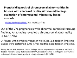
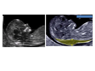
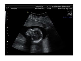
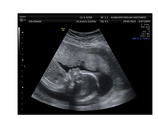
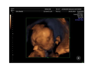
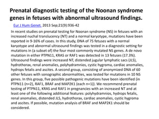
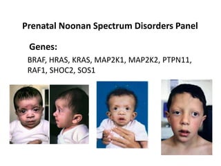
Prenatal diagnosis of frequently seen
fetal syndromes (A-Z) 
Ibrahim Bildirici,MD
Professor of OBGYN
ACIBADEM University SOM
Attending Perinatologist
ACIBADEM MASLAK Hospital
Amniotic band sequence:
Amniotic band sequence refers to a highly variable
spectrum of congenital anomalies that occur in
association with amniotic bands
The estimated incidence of ABS ranges from 1:1200
to 1:15,000 in live births, and 1:70 in stillbirths
Anomalies include:
Craniofacial abnormalities — eg, encephalocele, exencephaly,
clefts, which are often in unusual locations; anencephaly.
Body wall defects (especially if not in the midline), abdominal or
thoracic contents may herniate through a body wall defect and
into the amniotic cavity.
Limb defects — constriction rings, amputation, syndactyly,
clubfoot, hand deformities, lymphedema distal to a constriction
ring.
Visceral defects — eg, lung hypoplasia.
Other — Autotransplanted tissue on skin tags, spinal defects,
scoliosis, ambiguous genitalia, short umbilical cord due to
restricted motion of the fetus
Arthrogryposis
•Multiple congenital joint contractures/ankyloses
involving two or more body areas
•Pena Shokeir phenotype
micrognathia, multiple contractures, camptodactyly
(persistent finger flexion), polyhydramnios
*many are AR
*Lethal due to pulmonary hypoplasia
• Distal arthrogryposis
Subset of non-progressive contractures w/o associated
primary neurologic or muscle disease
Beckwith Wiedemannn Syndrome
Macrosomia
Hemihyperplasia
Macroglossia
Ventral wall defects
Predisposition to embryonal tumors
Neonatal hypoglycemia
Variable developmental delay
85% sporadic with normal karyotype
10-15% autosomal dominant inheritance
10-20% with paternal uniparental disomy
(Both copies of 11p15 derived from father)
***Imprinting related disorder
1/13 000.
Binder Phenotype
a flat profile and depressed nasal bridge.
Short nose, short columella, flat naso-labial angle and
perialar flattening
Isolated Binder Phenotype transmission would be
autosomal dominant
Binder Phenotype can also be an important sign of
chondrodysplasia punctata (CDDP)
1. Chromosomal abnormalities: As Trisomy 21/4p del
2. Metabolic congenital abnormalities: As Zellweger syndrome.
3. Disruption of vitamin K metabolism: caused by inherited or
extrinsic factors
3.1 Inherited etiology: X-linked recessive brachytelephalangic
type of chondrodysplasia punctata = CDPX1 caused by mutations
of ARSE, localized in Xp22.3. ARSE codes Aryl Sulfatase Enzyme, a
system Golgi enzyme. His activity is inhibited in vitro by
Warfarin.
3.2 Extrinsic factors:
- Prenatal exposure to Phenytoin and Alcohol
- Prenatal exposure to Coumarin derivatives: especially between
6th and 9th weeks.
- Maternal chronic disease: with important vitamin K deficiency
during first trimester.
CHARGE Association
CHARGE is a mnemonic for
coloboma,
heart defects,
choanal atresia,
retarded growth and
development,
genital abnormalities, and
ear anomalies.
TE fistula +/- esophageal atresia,
anal atresia
CHARGE syndrome, caused
by mutation of CHD7, is inherited in an autosomal
dominant manner.
Cornelia de Lange Syndrome
Abnormal facies
Growth and mental retardation
Limb defects
Gastrointestinal defects
Cardiac defects
Hypertrichosis
IUGR
Upper limb reduction defects
Micrognathia with protruding upper lip
CDH
AD
Most sporadic, rare familial cases
NIPBL mutations
Cri du Chat Syndrome (5p- syndrome)
Microcephaly
Facial dysmorphic features: hypertelorism,
micrognathia, low-set ears
Intrauterine growth restriction
Cardiac defects (ventricular, atrial septal defect, patent
ductus arteriosus, tetralogy of Fallot)
Occasional abnormalities: Cleft lip and palate, myopia, optic
atrophy, preauricular skin tag, bifid uvula, clinodactyly, absent
kidney and spleen, cryptorchidism and hemivertebra.
Absent nasal bone:
Amniocentesis: 5 p-
Cystic fibrosis
AR multisystem disorder: dysfunctional chloride ion transport
across epithelial surfaces
>1000 CFTR mutations possible
1/2000-5000
Best diagnostic clue:
Echogenic bowel in 2nd trimester progressing to bowel dilation
in 3rd trimester
Fetal echogenic bowel
DiGeorge Syndrome
Prenatal diagnosis of chromosomal abnormalities in
fetuses with abnormal cardiac ultrasound findings:
evaluation of chromosomal microarray-based
analysis.
Ultrasound Obstet Gynecol. 2013 Apr;41(4):375-82
Out of the 276 pregnancies with abnormal cardiac ultrasound
findings, karyotyping revealed a chromosomal abnormality
in 44 (15.9%).
Of fetuses with normal karyotype in which 22q11.2 deletion syndrome
studies were performed, 6.4% (5/78) had this microdeletion syndrome.
Among fetuses with abnormal cardiac findings, normal karyotype and negative or no 22q11.2
deletion syndrome study that underwent CMA, the detection rate of pathogenic copy number
variants not detected by conventional cytogenetics was 2.0% (1/51)
Down Syndrome:
Edwards syndrome (Trisomy 18)
1st trimester
Increased nuchal translucency
2nd trimester
Multiple major anomalies
Single major anomaly + Trisomy 18 marker
Choroid plexus cyst + other anomalies
Early IUGR
IUGR
Cardiac defects
Muskuloskeletal findingd
-Clenched hands + overlapping index finger
Arthrogryposis
Rocker bottom foot
Clubfoot
Radial ray malformation
Cystic hygroma
Brain anomalies
Strawvberry shaped calvarium
Meningomyelocele
Facial anomalies
GI anomalies
SUA
Fryns Syndrome
Perinatal lethal disorder
CDH with pulmonary hypoplasia
Micrognathia
Orofacial cleft
Cardiac defects
Polyhydramnios
Distal digital hypoplasia (typically not apparent on ultrasound)
***AR inheritance
Gene not known
Meckel-Gruber Syndrome
Renal cystic dysplasia 95-100%
Encephalocele 60-80%
Postaxial polydactyly 55-75%
2nd trimester oligohydramnios
Significantly increased abdominal circumference
***AR =25% recurrence risk
MKS1 17q21-24, MKS2 11q13, MKS3 8q24
Noonan Syndrome
The prenatal ultrasound findings reported in Noonan syndrome
include accumulation of nuchal fluid, femur lengths at or just
below the lower end of the normal range, pleural effusions, and
renal anomalies.
The most common prenatal features are
polyhydramnios (58%),
cystic hygroma (42%),
increased thickness of the nuchal translucency and fetal hydrops
(33%).
Cardiac anomalies are present in 60% of cases: including left
ventricular hypertrophy (25%), pulmonary stenosis (19%), atrial
septal defect (10%) or dysplastic pulmonary valve in 7%.
In recent studies on prenatal testing for Noonan syndrome (NS) in fetuses with an
increased nuchal translucency (NT) and a normal karyotype, mutations have been
reported in 9-16% of cases. In this study, DNA of 75 fetuses with a normal
karyotype and abnormal ultrasound findings was tested in a diagnostic setting for
mutations in (a subset of) the four most commonly mutated NS genes. A de novo
mutation in either PTPN11, KRAS or RAF1 was detected in 13 fetuses (17.3%).
Ultrasound findings were increased NT, distended jugular lymphatic sacs (JLS),
hydrothorax, renal anomalies, polyhydramnios, cystic hygroma, cardiac anomalies,
hydrops fetalis and ascites. A second group, consisting of anonymized DNA of 60
other fetuses with sonographic abnormalities, was tested for mutations in 10 NS
genes. In this group, five possible pathogenic mutations have been identified (in
PTPN11 (n=2), RAF1, BRAF and MAP2K1 (each n=1)). We recommend prenatal
testing of PTPN11, KRAS and RAF1 in pregnancies with an increased NT and at
least one of the following additional features: polyhydramnios, hydrops fetalis,
renal anomalies, distended JLS, hydrothorax, cardiac anomalies, cystic hygroma
and ascites. If possible, mutation analysis of BRAF and MAP2K1 should be
considered.
Prenatal diagnostic testing of the Noonan syndrome
genes in fetuses with abnormal ultrasound findings.
Eur J Hum Genet. 2013 Sep;21(9):936-42
Prenatal Noonan Spectrum Disorders Panel
BRAF, HRAS, KRAS, MAP2K1, MAP2K2, PTPN11,
RAF1, SHOC2, SOS1
Genes:
Patau Syndrome(Trisomy 13)
1st trimester
Increased NT
2nd trimester
Multiple major anomalies (>90%)
Holoprosencephaly
Midline facial anomalies
Cardiac defects
Polydactyly
Early IUGR
Smith-Lemli-Opitz Syndrome
Disorder of cholesterol biosynthesis characterized by
IUGR
Multiple congenital anomalies
Developmental delay
IUGR
Microcephaly (90%)
Cardiac defects: AV canal defect, VSD, Hypoplastic LH
Postaxial polydactyly
Genital ambiguity, cystic renal disease
Facial malformations: Hypertelorism, short up-turned
nose, low-set ears, micrognathia
Increased NT in the 1st trimester
*AR
Smith-Lemli-Opitz syndrome (SLOS) is an autosomal
recessive disorder of endogenous cholesterol
biosynthesis caused by deficiency of
7-dehydrocholesterol reductase (DHCR7).
DHCR7 mutation is a rapid and reliable method for
prenatal diagnosis of SLOS, and provides an
alternative to specialized biochemical tests for
elevated 7DHC in amniotic fluid or CVS.
***Up to 7% of stillbirths may be due to SLOS
Wolf Hirschorn Syndrome (4p-)
Severe intrauterine growth restriction
Facial dysmorphic features, midline defects
(hypertelorism, labial or labio-palatine defects, corpus
callosum agenesis)
Cardiac septal defects
Urinary tract malformations
Brain anomalies***microcephaly***
Wolf-Hirschhorn syndrome is caused by deletion of the
WHSCR of chromosome 4p16.3 by one of several genetic
mechanisms.
Severe early onset IUGR with normal doppler findings
‘Greek helmet’ facial appearance w prominent glabella, rectangular nose,
downturned mouth, hypospadias, cardiac and urinary tract defects
These children have severe developmental delays.
Other significant problems can include heart defects,
cleft lip and/or palate, hearing impairment, and eye
problems. Most children who have WHS have
seizures (approximately 90%).
array-CGH
is the way to go!
TEŞEKKÜRLER

İbrahi̇m bi̇ldi̇ri̇ci̇

  • 1.
    Prenatal diagnosis offrequently seen fetal syndromes (A-Z)  Ibrahim Bildirici,MD Professor of OBGYN ACIBADEM University SOM Attending Perinatologist ACIBADEM MASLAK Hospital
  • 2.
    Amniotic band sequence: Amnioticband sequence refers to a highly variable spectrum of congenital anomalies that occur in association with amniotic bands The estimated incidence of ABS ranges from 1:1200 to 1:15,000 in live births, and 1:70 in stillbirths
  • 3.
    Anomalies include: Craniofacial abnormalities— eg, encephalocele, exencephaly, clefts, which are often in unusual locations; anencephaly. Body wall defects (especially if not in the midline), abdominal or thoracic contents may herniate through a body wall defect and into the amniotic cavity. Limb defects — constriction rings, amputation, syndactyly, clubfoot, hand deformities, lymphedema distal to a constriction ring. Visceral defects — eg, lung hypoplasia. Other — Autotransplanted tissue on skin tags, spinal defects, scoliosis, ambiguous genitalia, short umbilical cord due to restricted motion of the fetus
  • 5.
    Arthrogryposis •Multiple congenital jointcontractures/ankyloses involving two or more body areas •Pena Shokeir phenotype micrognathia, multiple contractures, camptodactyly (persistent finger flexion), polyhydramnios *many are AR *Lethal due to pulmonary hypoplasia • Distal arthrogryposis Subset of non-progressive contractures w/o associated primary neurologic or muscle disease
  • 8.
    Beckwith Wiedemannn Syndrome Macrosomia Hemihyperplasia Macroglossia Ventralwall defects Predisposition to embryonal tumors Neonatal hypoglycemia Variable developmental delay
  • 10.
    85% sporadic withnormal karyotype 10-15% autosomal dominant inheritance 10-20% with paternal uniparental disomy (Both copies of 11p15 derived from father) ***Imprinting related disorder 1/13 000.
  • 11.
    Binder Phenotype a flatprofile and depressed nasal bridge. Short nose, short columella, flat naso-labial angle and perialar flattening Isolated Binder Phenotype transmission would be autosomal dominant Binder Phenotype can also be an important sign of chondrodysplasia punctata (CDDP)
  • 13.
    1. Chromosomal abnormalities:As Trisomy 21/4p del 2. Metabolic congenital abnormalities: As Zellweger syndrome. 3. Disruption of vitamin K metabolism: caused by inherited or extrinsic factors 3.1 Inherited etiology: X-linked recessive brachytelephalangic type of chondrodysplasia punctata = CDPX1 caused by mutations of ARSE, localized in Xp22.3. ARSE codes Aryl Sulfatase Enzyme, a system Golgi enzyme. His activity is inhibited in vitro by Warfarin. 3.2 Extrinsic factors: - Prenatal exposure to Phenytoin and Alcohol - Prenatal exposure to Coumarin derivatives: especially between 6th and 9th weeks. - Maternal chronic disease: with important vitamin K deficiency during first trimester.
  • 18.
    CHARGE Association CHARGE isa mnemonic for coloboma, heart defects, choanal atresia, retarded growth and development, genital abnormalities, and ear anomalies. TE fistula +/- esophageal atresia, anal atresia CHARGE syndrome, caused by mutation of CHD7, is inherited in an autosomal dominant manner.
  • 24.
    Cornelia de LangeSyndrome Abnormal facies Growth and mental retardation Limb defects Gastrointestinal defects Cardiac defects Hypertrichosis IUGR Upper limb reduction defects Micrognathia with protruding upper lip CDH
  • 25.
    AD Most sporadic, rarefamilial cases NIPBL mutations
  • 26.
    Cri du ChatSyndrome (5p- syndrome) Microcephaly Facial dysmorphic features: hypertelorism, micrognathia, low-set ears Intrauterine growth restriction Cardiac defects (ventricular, atrial septal defect, patent ductus arteriosus, tetralogy of Fallot) Occasional abnormalities: Cleft lip and palate, myopia, optic atrophy, preauricular skin tag, bifid uvula, clinodactyly, absent kidney and spleen, cryptorchidism and hemivertebra.
  • 28.
  • 29.
    Cystic fibrosis AR multisystemdisorder: dysfunctional chloride ion transport across epithelial surfaces >1000 CFTR mutations possible 1/2000-5000 Best diagnostic clue: Echogenic bowel in 2nd trimester progressing to bowel dilation in 3rd trimester
  • 30.
  • 32.
  • 33.
    Prenatal diagnosis ofchromosomal abnormalities in fetuses with abnormal cardiac ultrasound findings: evaluation of chromosomal microarray-based analysis. Ultrasound Obstet Gynecol. 2013 Apr;41(4):375-82 Out of the 276 pregnancies with abnormal cardiac ultrasound findings, karyotyping revealed a chromosomal abnormality in 44 (15.9%). Of fetuses with normal karyotype in which 22q11.2 deletion syndrome studies were performed, 6.4% (5/78) had this microdeletion syndrome. Among fetuses with abnormal cardiac findings, normal karyotype and negative or no 22q11.2 deletion syndrome study that underwent CMA, the detection rate of pathogenic copy number variants not detected by conventional cytogenetics was 2.0% (1/51)
  • 37.
  • 48.
    Edwards syndrome (Trisomy18) 1st trimester Increased nuchal translucency 2nd trimester Multiple major anomalies Single major anomaly + Trisomy 18 marker Choroid plexus cyst + other anomalies Early IUGR
  • 49.
    IUGR Cardiac defects Muskuloskeletal findingd -Clenchedhands + overlapping index finger Arthrogryposis Rocker bottom foot Clubfoot Radial ray malformation Cystic hygroma Brain anomalies Strawvberry shaped calvarium Meningomyelocele Facial anomalies GI anomalies SUA
  • 61.
    Fryns Syndrome Perinatal lethaldisorder CDH with pulmonary hypoplasia Micrognathia Orofacial cleft Cardiac defects Polyhydramnios Distal digital hypoplasia (typically not apparent on ultrasound) ***AR inheritance Gene not known
  • 63.
    Meckel-Gruber Syndrome Renal cysticdysplasia 95-100% Encephalocele 60-80% Postaxial polydactyly 55-75% 2nd trimester oligohydramnios Significantly increased abdominal circumference ***AR =25% recurrence risk MKS1 17q21-24, MKS2 11q13, MKS3 8q24
  • 65.
    Noonan Syndrome The prenatalultrasound findings reported in Noonan syndrome include accumulation of nuchal fluid, femur lengths at or just below the lower end of the normal range, pleural effusions, and renal anomalies. The most common prenatal features are polyhydramnios (58%), cystic hygroma (42%), increased thickness of the nuchal translucency and fetal hydrops (33%). Cardiac anomalies are present in 60% of cases: including left ventricular hypertrophy (25%), pulmonary stenosis (19%), atrial septal defect (10%) or dysplastic pulmonary valve in 7%.
  • 66.
    In recent studieson prenatal testing for Noonan syndrome (NS) in fetuses with an increased nuchal translucency (NT) and a normal karyotype, mutations have been reported in 9-16% of cases. In this study, DNA of 75 fetuses with a normal karyotype and abnormal ultrasound findings was tested in a diagnostic setting for mutations in (a subset of) the four most commonly mutated NS genes. A de novo mutation in either PTPN11, KRAS or RAF1 was detected in 13 fetuses (17.3%). Ultrasound findings were increased NT, distended jugular lymphatic sacs (JLS), hydrothorax, renal anomalies, polyhydramnios, cystic hygroma, cardiac anomalies, hydrops fetalis and ascites. A second group, consisting of anonymized DNA of 60 other fetuses with sonographic abnormalities, was tested for mutations in 10 NS genes. In this group, five possible pathogenic mutations have been identified (in PTPN11 (n=2), RAF1, BRAF and MAP2K1 (each n=1)). We recommend prenatal testing of PTPN11, KRAS and RAF1 in pregnancies with an increased NT and at least one of the following additional features: polyhydramnios, hydrops fetalis, renal anomalies, distended JLS, hydrothorax, cardiac anomalies, cystic hygroma and ascites. If possible, mutation analysis of BRAF and MAP2K1 should be considered. Prenatal diagnostic testing of the Noonan syndrome genes in fetuses with abnormal ultrasound findings. Eur J Hum Genet. 2013 Sep;21(9):936-42
  • 67.
    Prenatal Noonan SpectrumDisorders Panel BRAF, HRAS, KRAS, MAP2K1, MAP2K2, PTPN11, RAF1, SHOC2, SOS1 Genes:
  • 68.
  • 69.
    1st trimester Increased NT 2ndtrimester Multiple major anomalies (>90%) Holoprosencephaly Midline facial anomalies Cardiac defects Polydactyly Early IUGR
  • 72.
    Smith-Lemli-Opitz Syndrome Disorder ofcholesterol biosynthesis characterized by IUGR Multiple congenital anomalies Developmental delay IUGR Microcephaly (90%) Cardiac defects: AV canal defect, VSD, Hypoplastic LH Postaxial polydactyly Genital ambiguity, cystic renal disease Facial malformations: Hypertelorism, short up-turned nose, low-set ears, micrognathia Increased NT in the 1st trimester *AR
  • 73.
    Smith-Lemli-Opitz syndrome (SLOS)is an autosomal recessive disorder of endogenous cholesterol biosynthesis caused by deficiency of 7-dehydrocholesterol reductase (DHCR7). DHCR7 mutation is a rapid and reliable method for prenatal diagnosis of SLOS, and provides an alternative to specialized biochemical tests for elevated 7DHC in amniotic fluid or CVS. ***Up to 7% of stillbirths may be due to SLOS
  • 76.
    Wolf Hirschorn Syndrome(4p-) Severe intrauterine growth restriction Facial dysmorphic features, midline defects (hypertelorism, labial or labio-palatine defects, corpus callosum agenesis) Cardiac septal defects Urinary tract malformations Brain anomalies***microcephaly***
  • 77.
    Wolf-Hirschhorn syndrome iscaused by deletion of the WHSCR of chromosome 4p16.3 by one of several genetic mechanisms.
  • 78.
    Severe early onsetIUGR with normal doppler findings ‘Greek helmet’ facial appearance w prominent glabella, rectangular nose, downturned mouth, hypospadias, cardiac and urinary tract defects
  • 79.
    These children havesevere developmental delays. Other significant problems can include heart defects, cleft lip and/or palate, hearing impairment, and eye problems. Most children who have WHS have seizures (approximately 90%). array-CGH is the way to go!
  • 80.