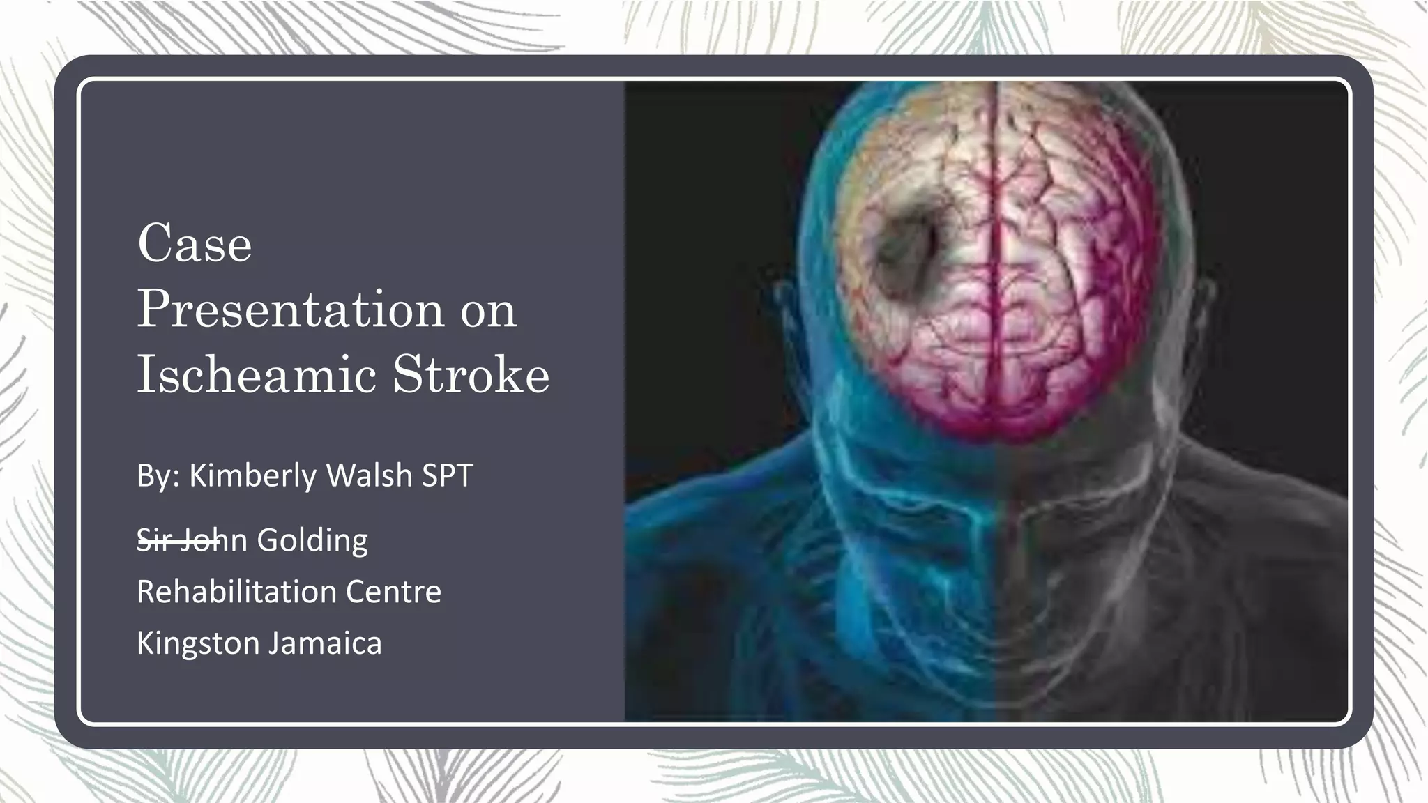
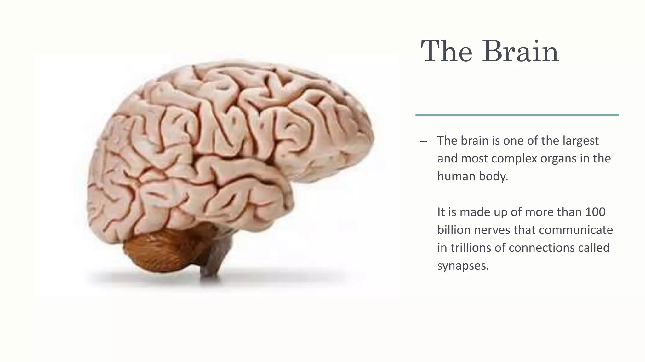
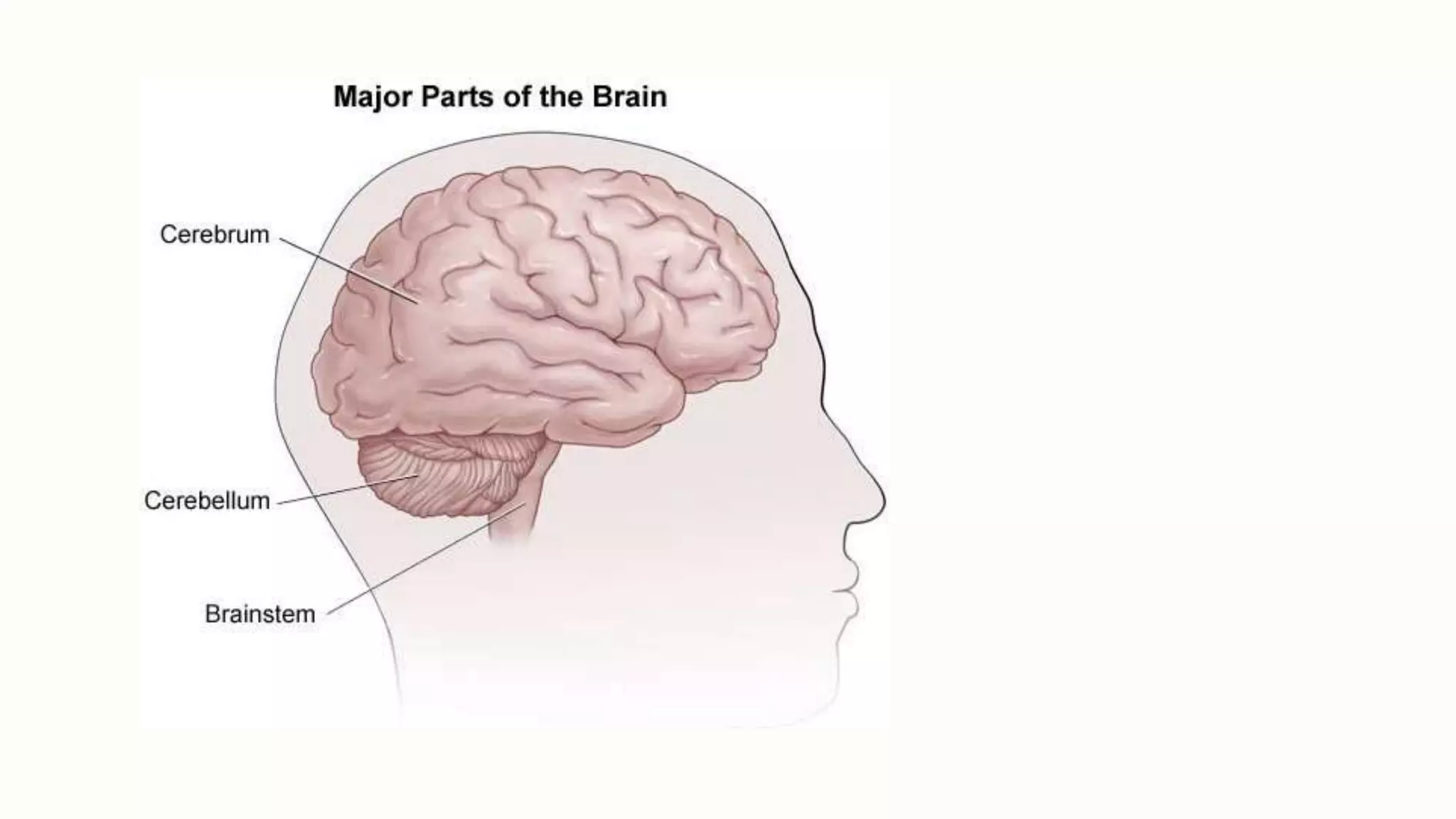
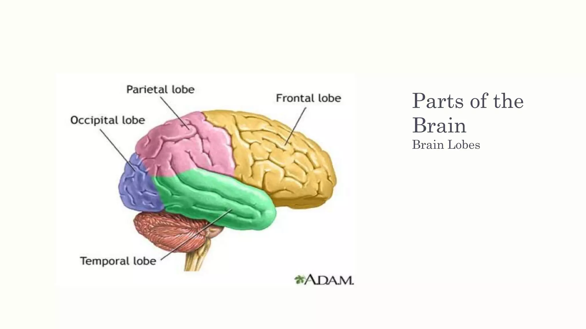
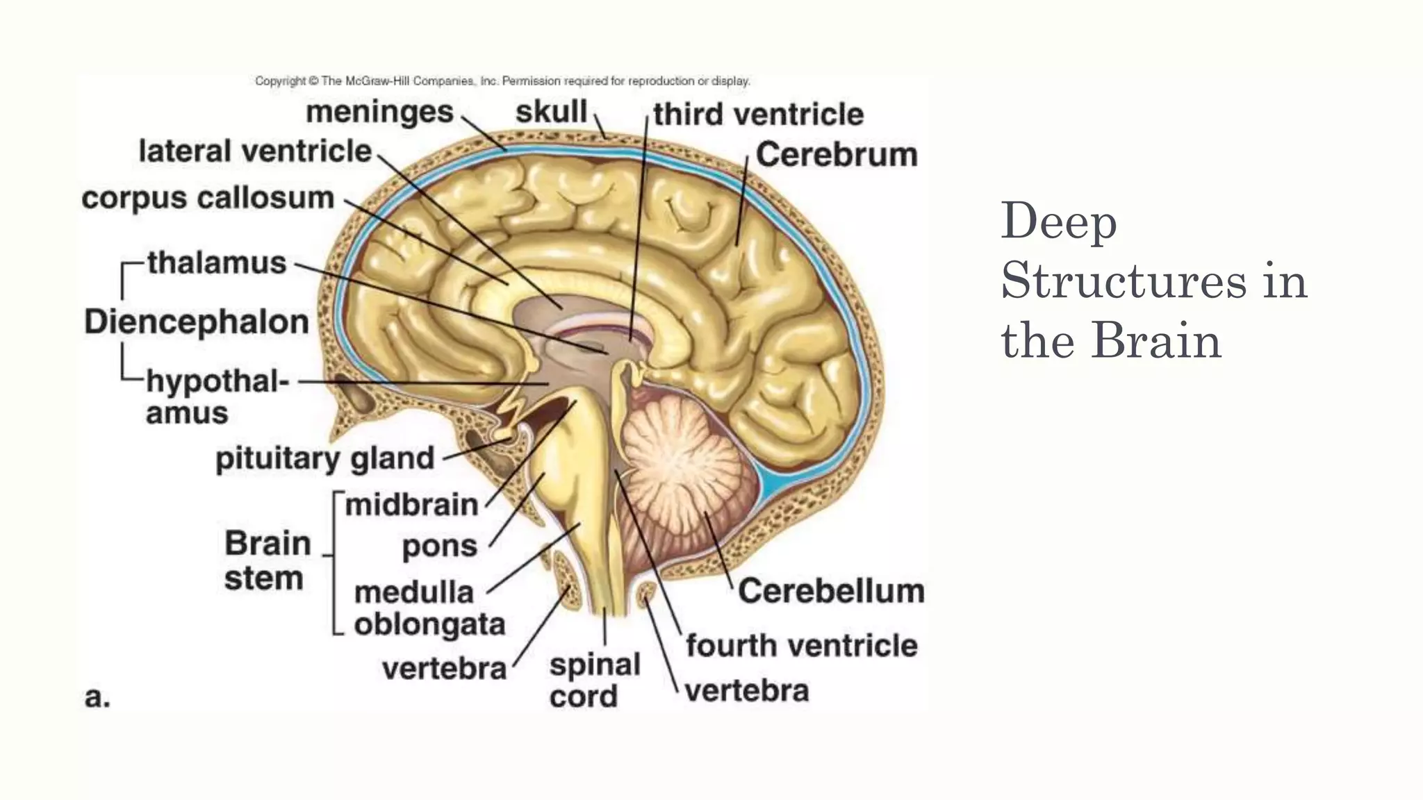
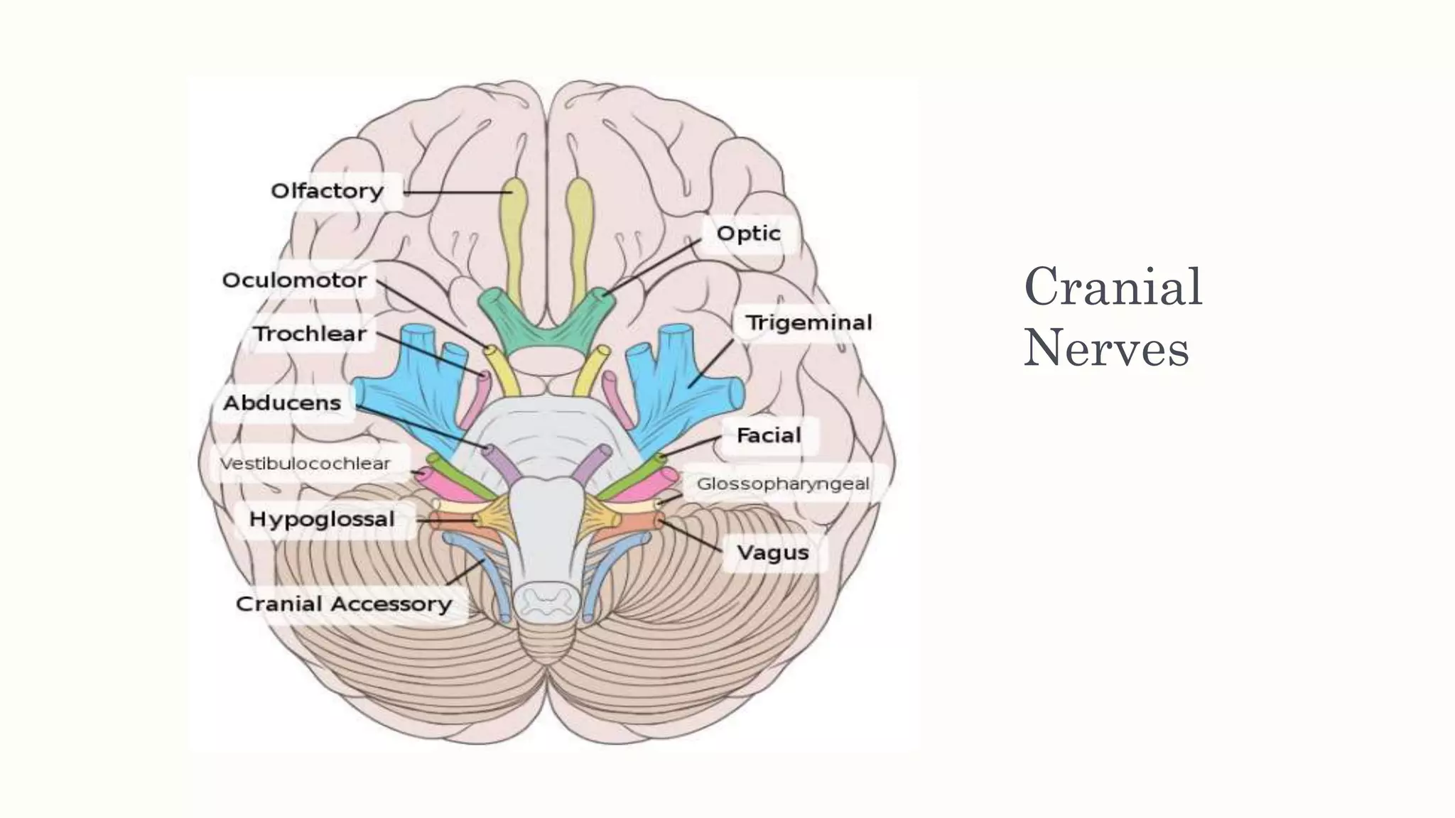
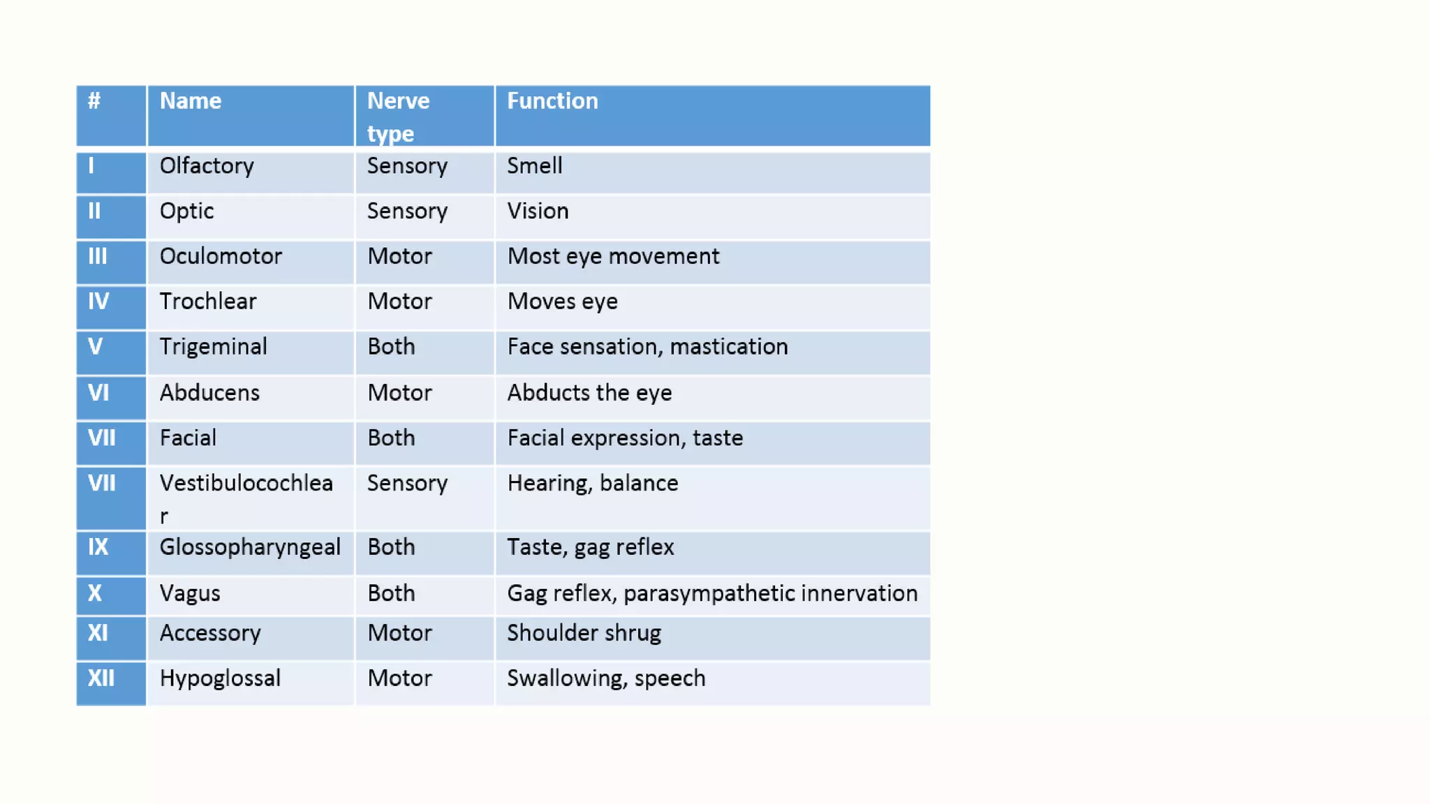
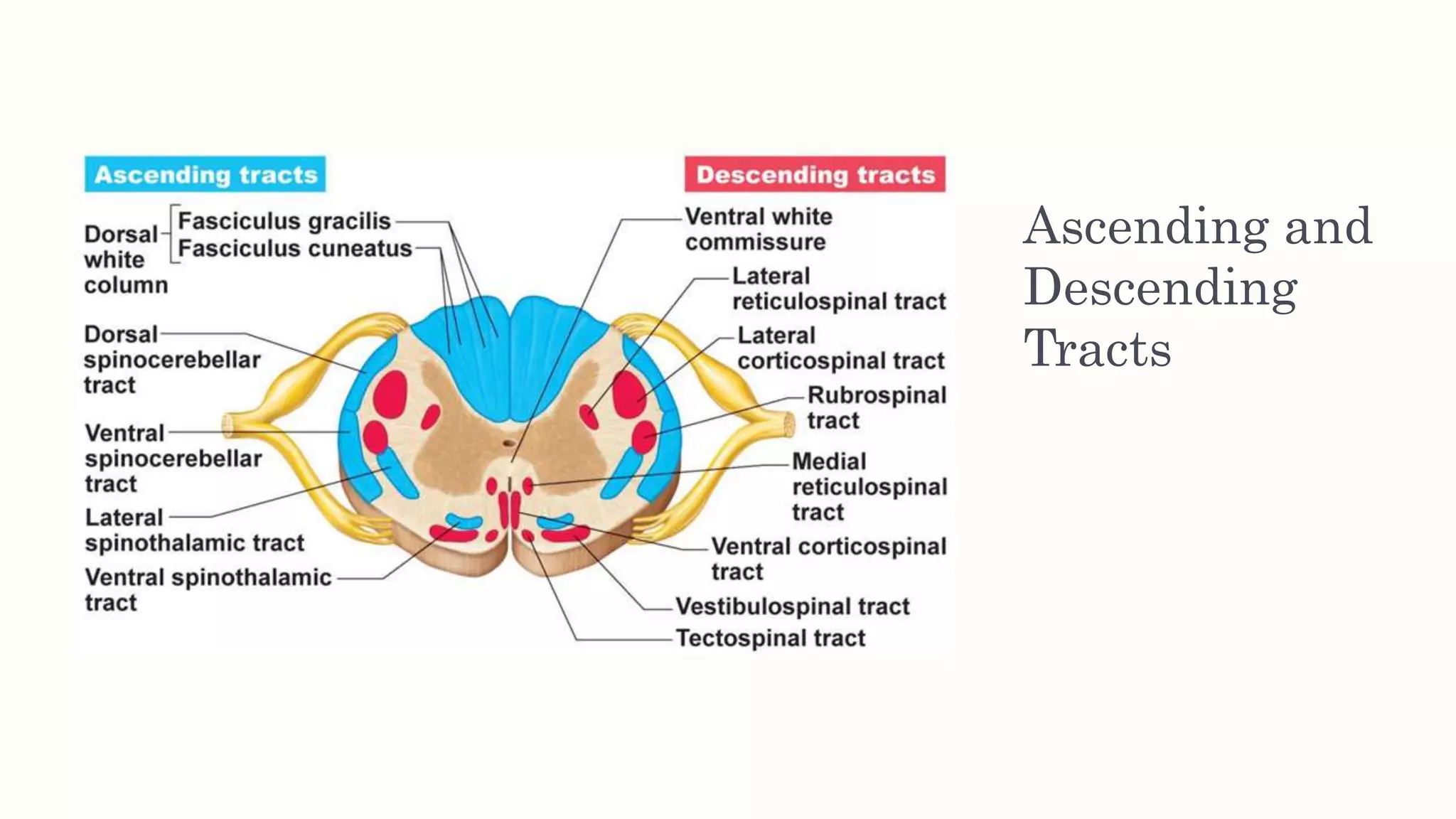
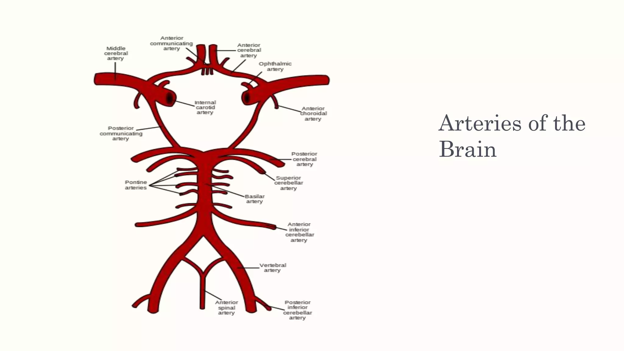
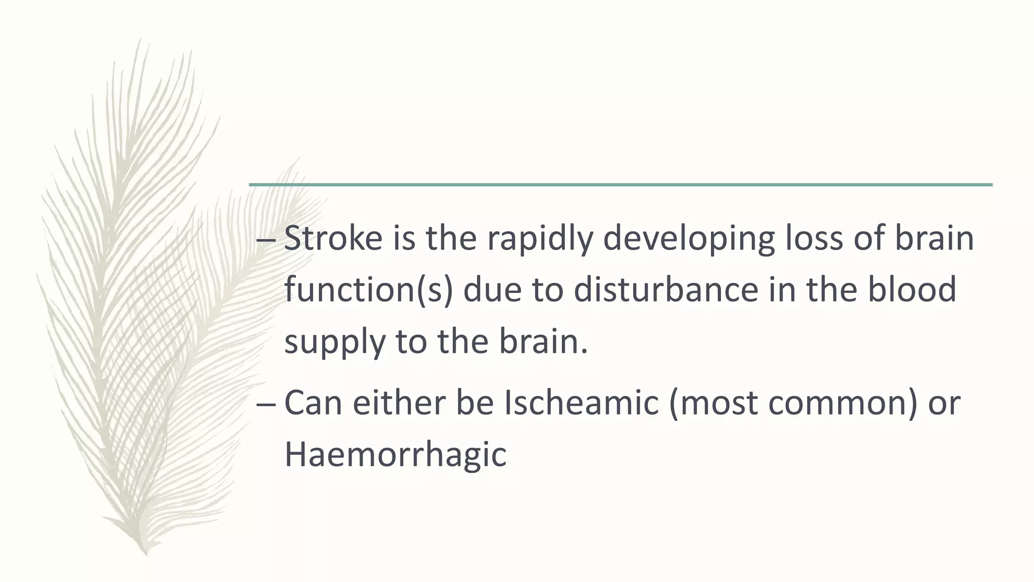
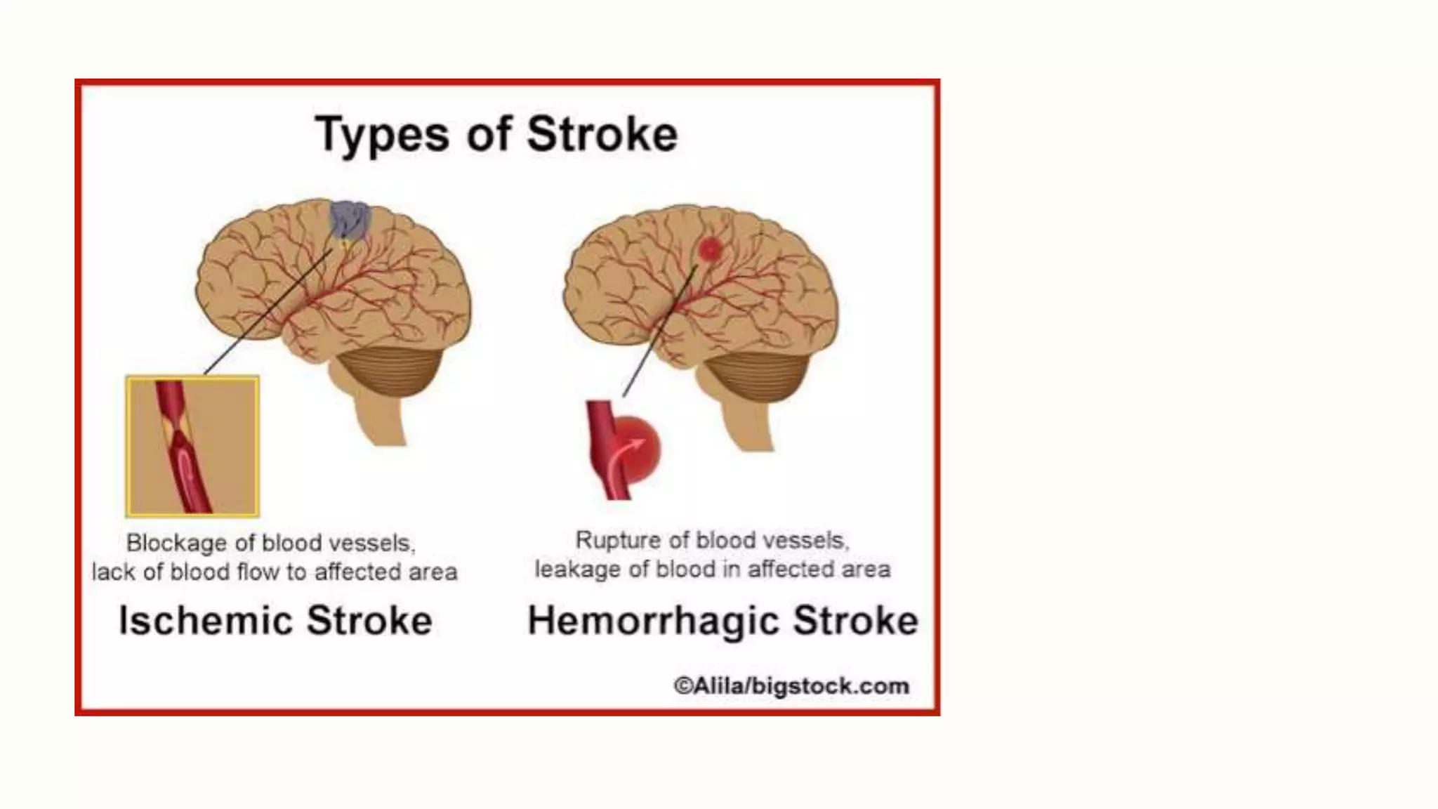
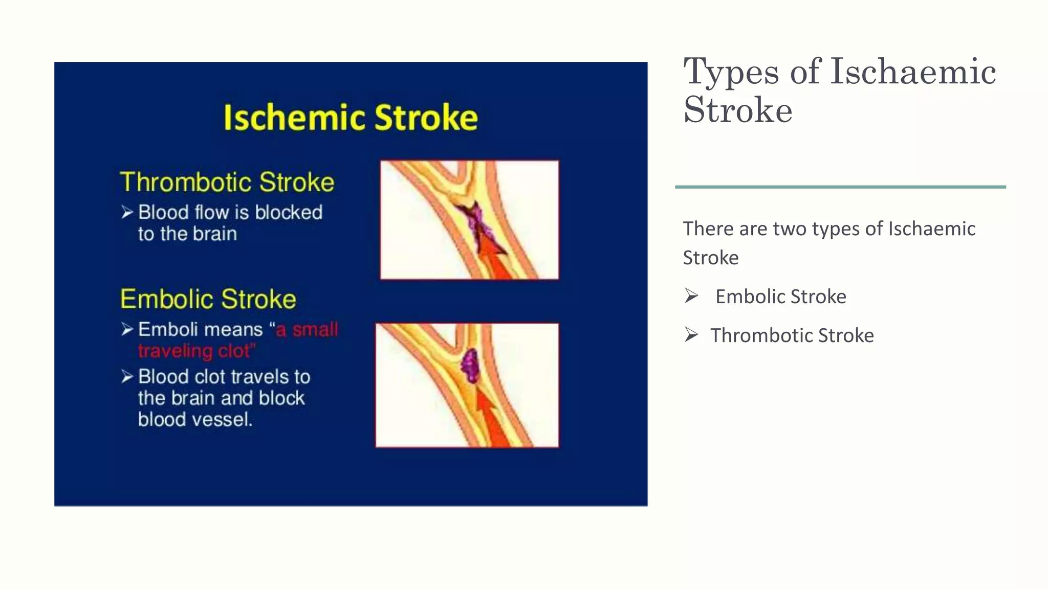
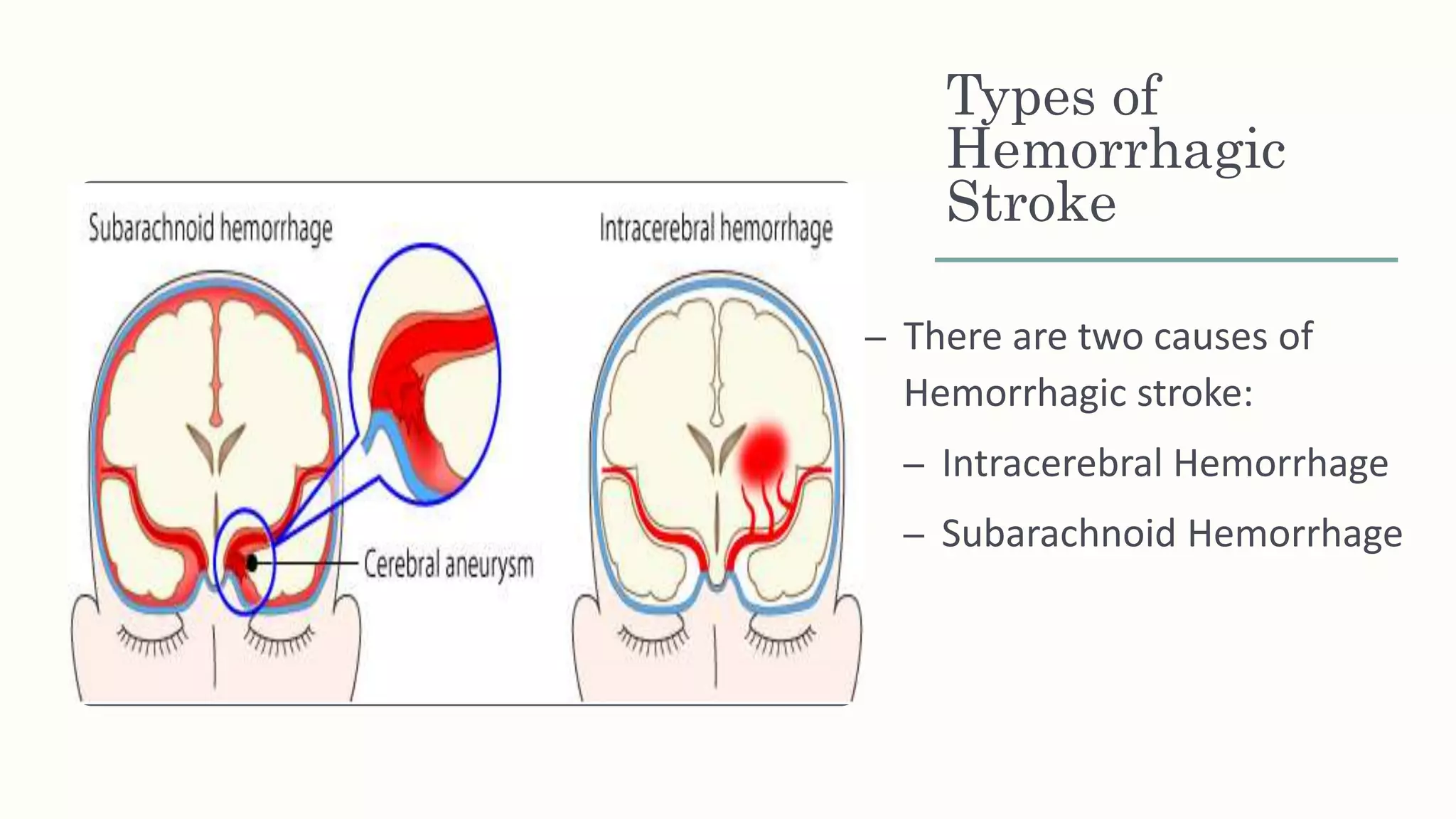
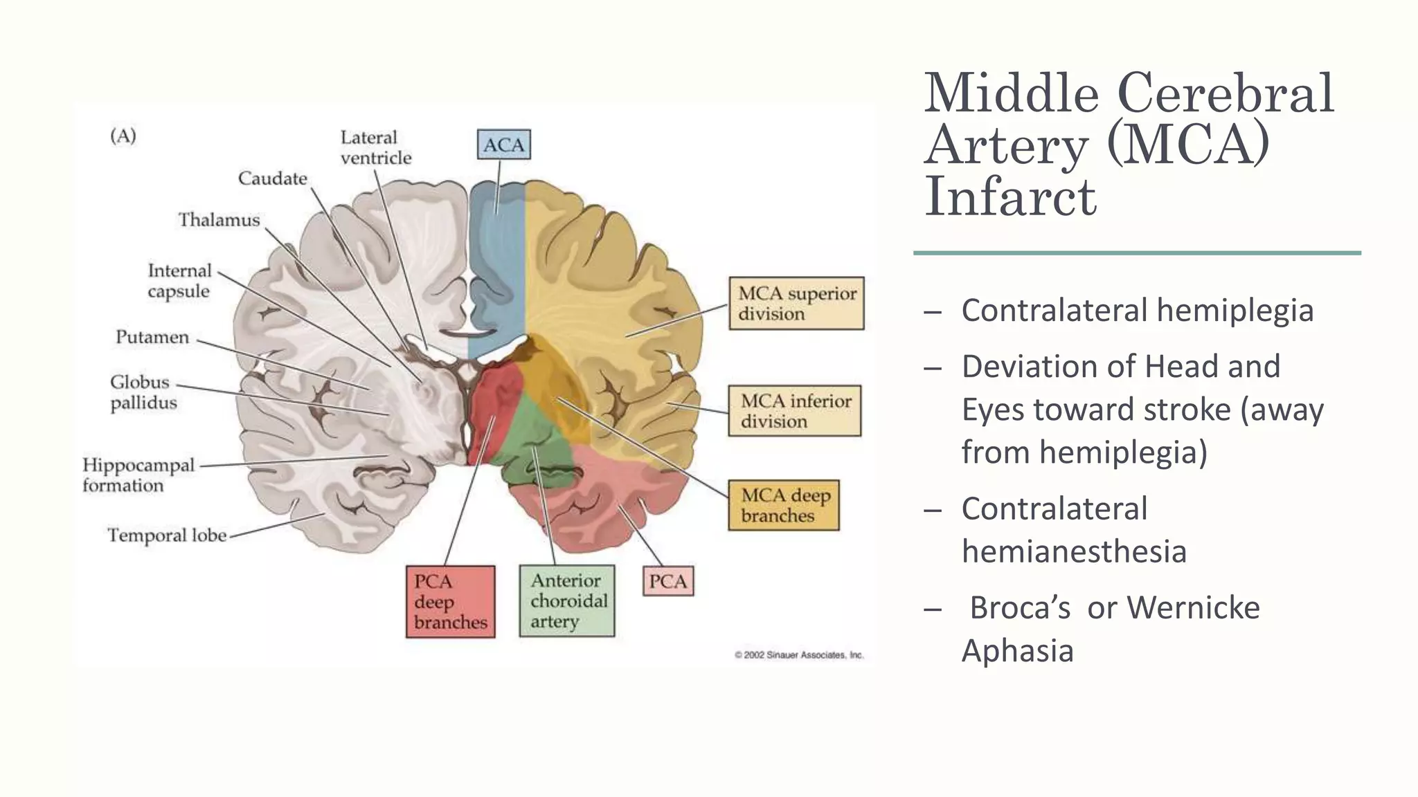
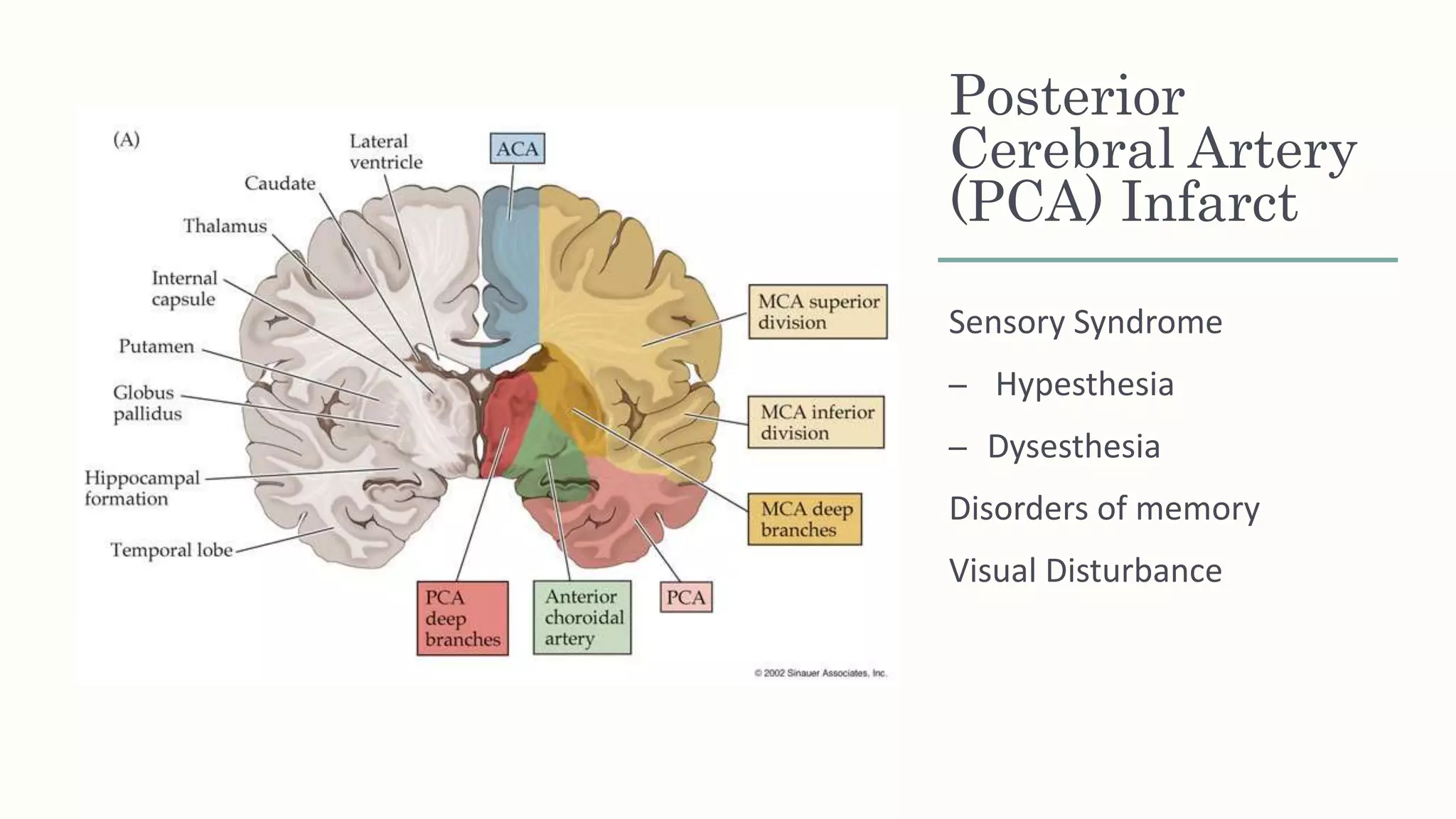
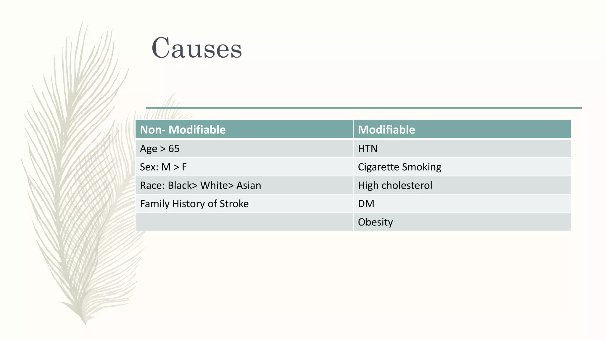
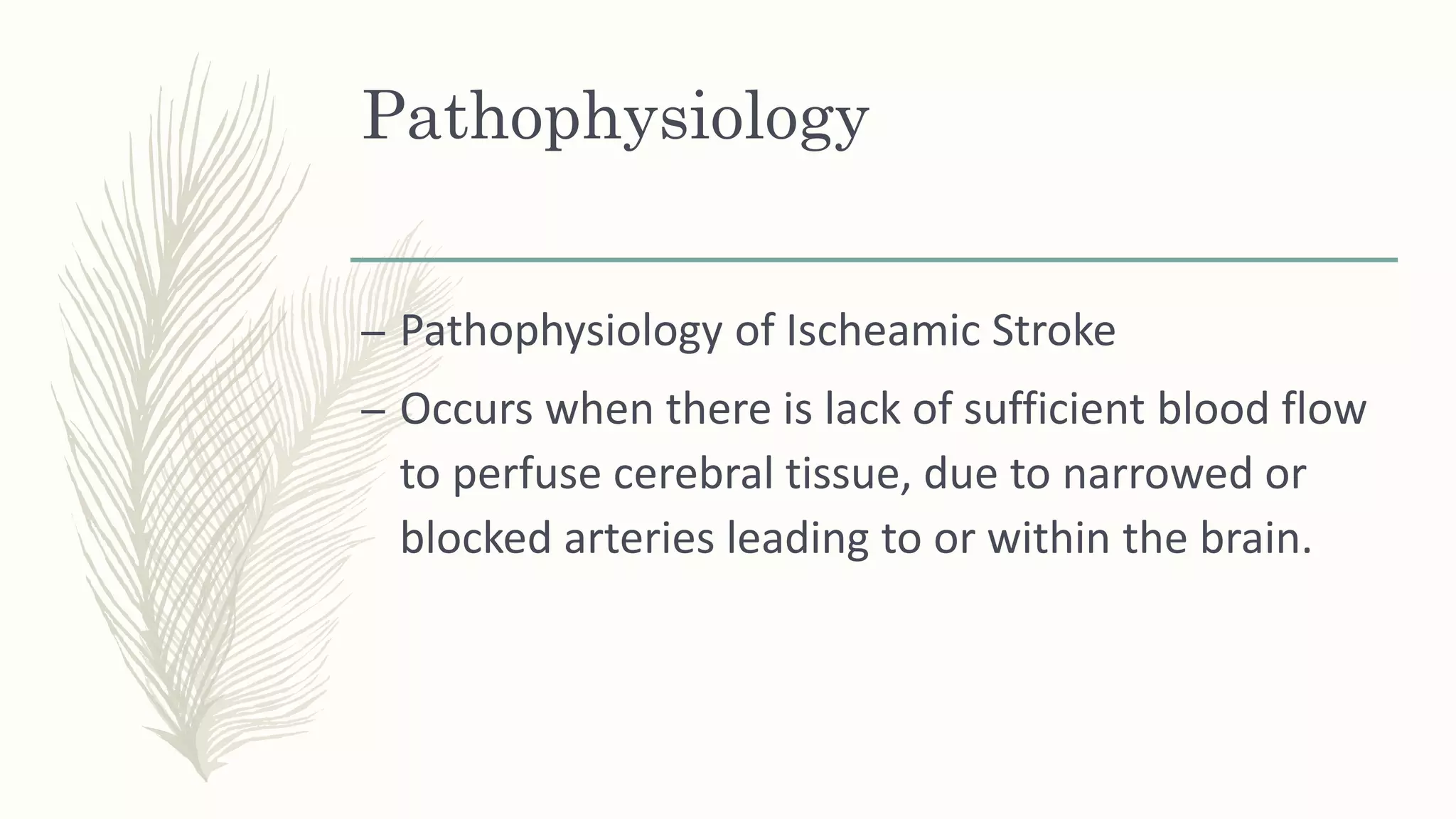
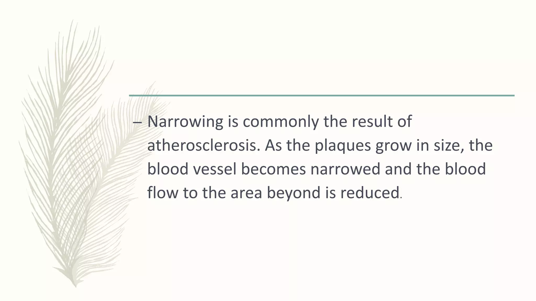
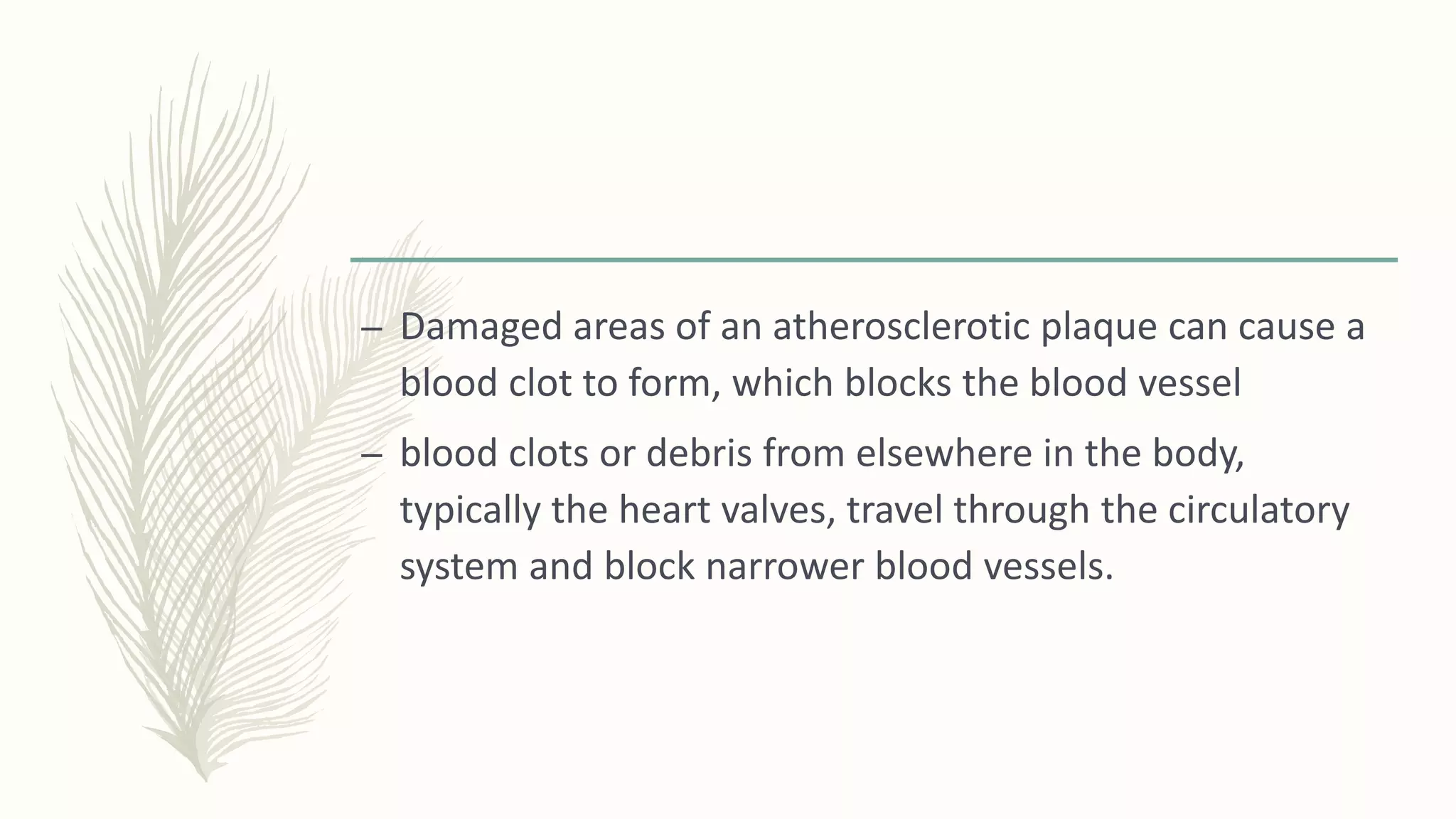
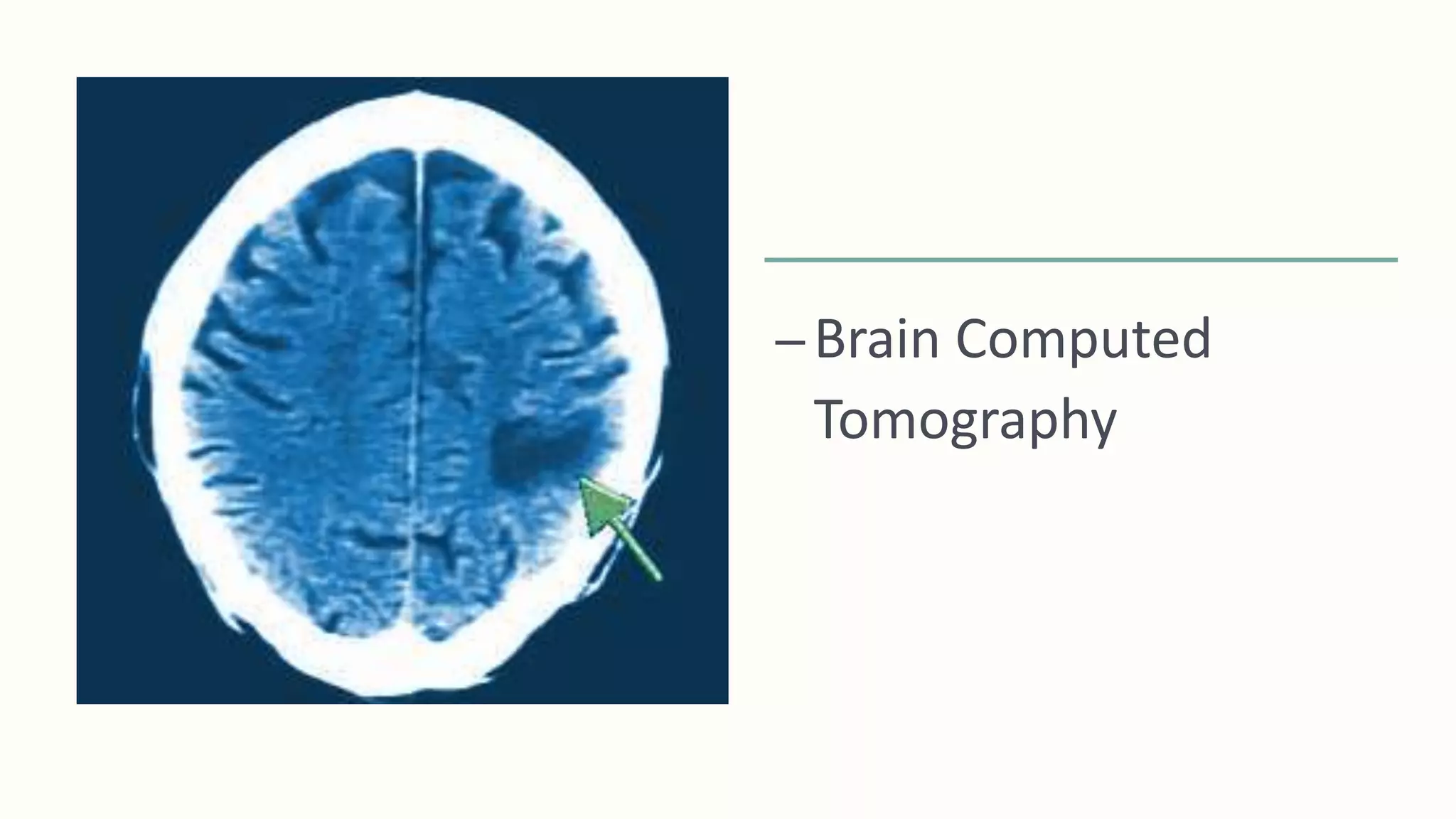
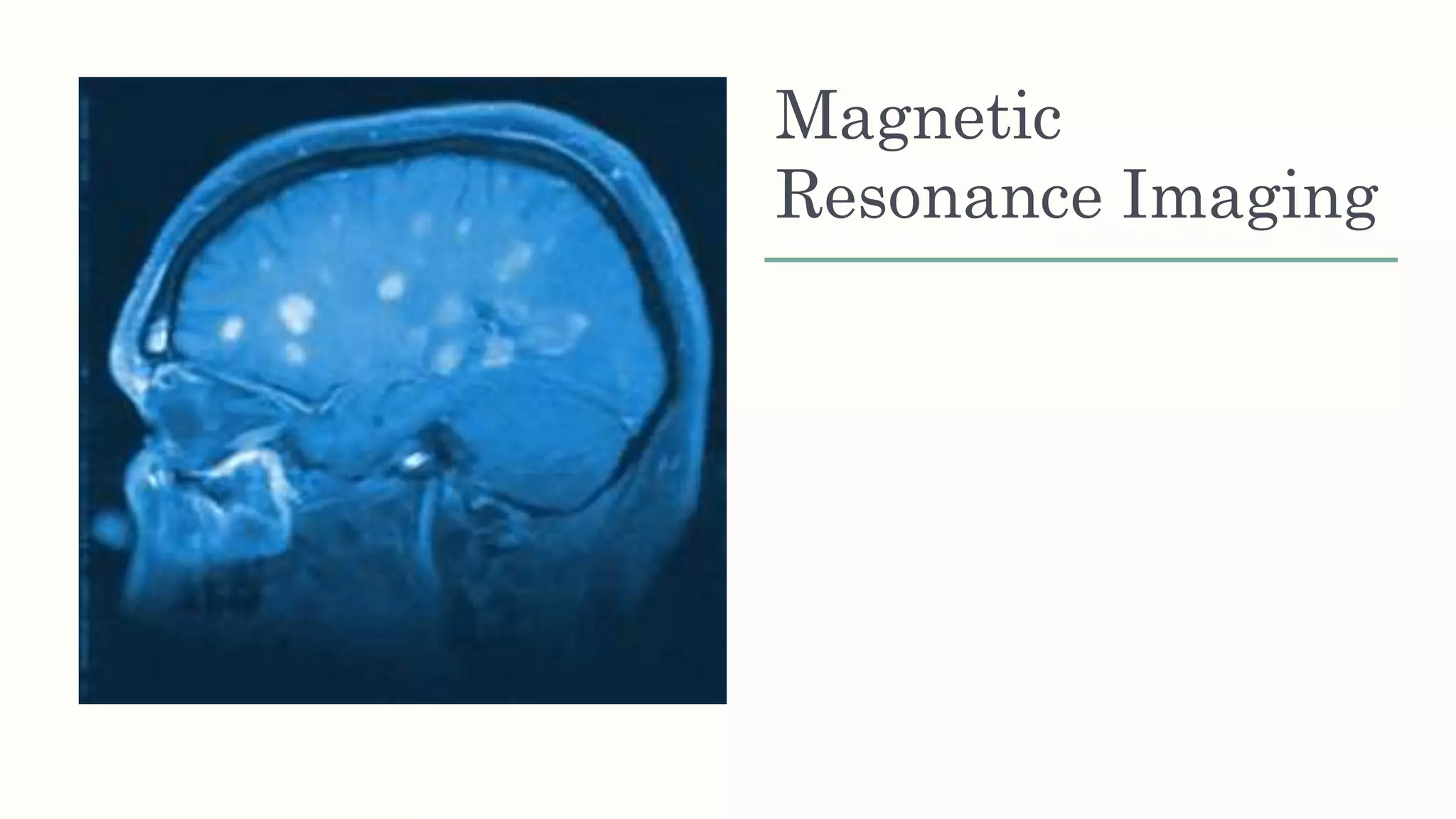
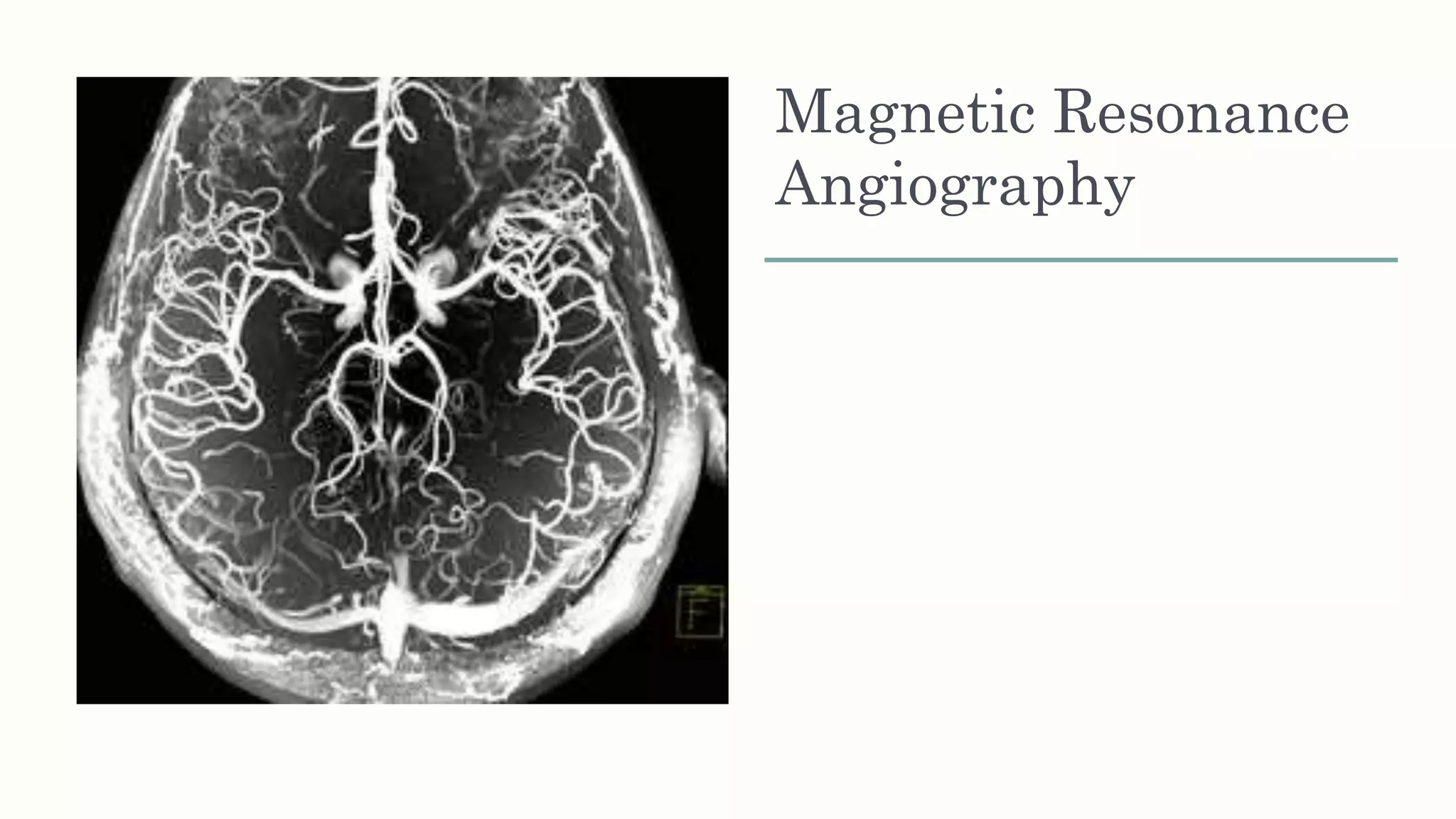
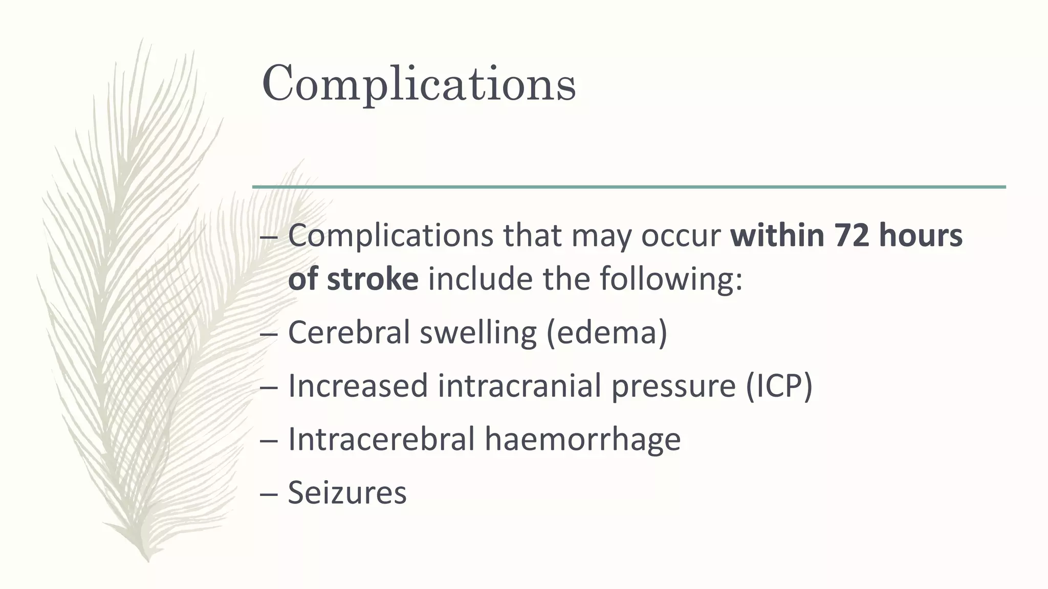
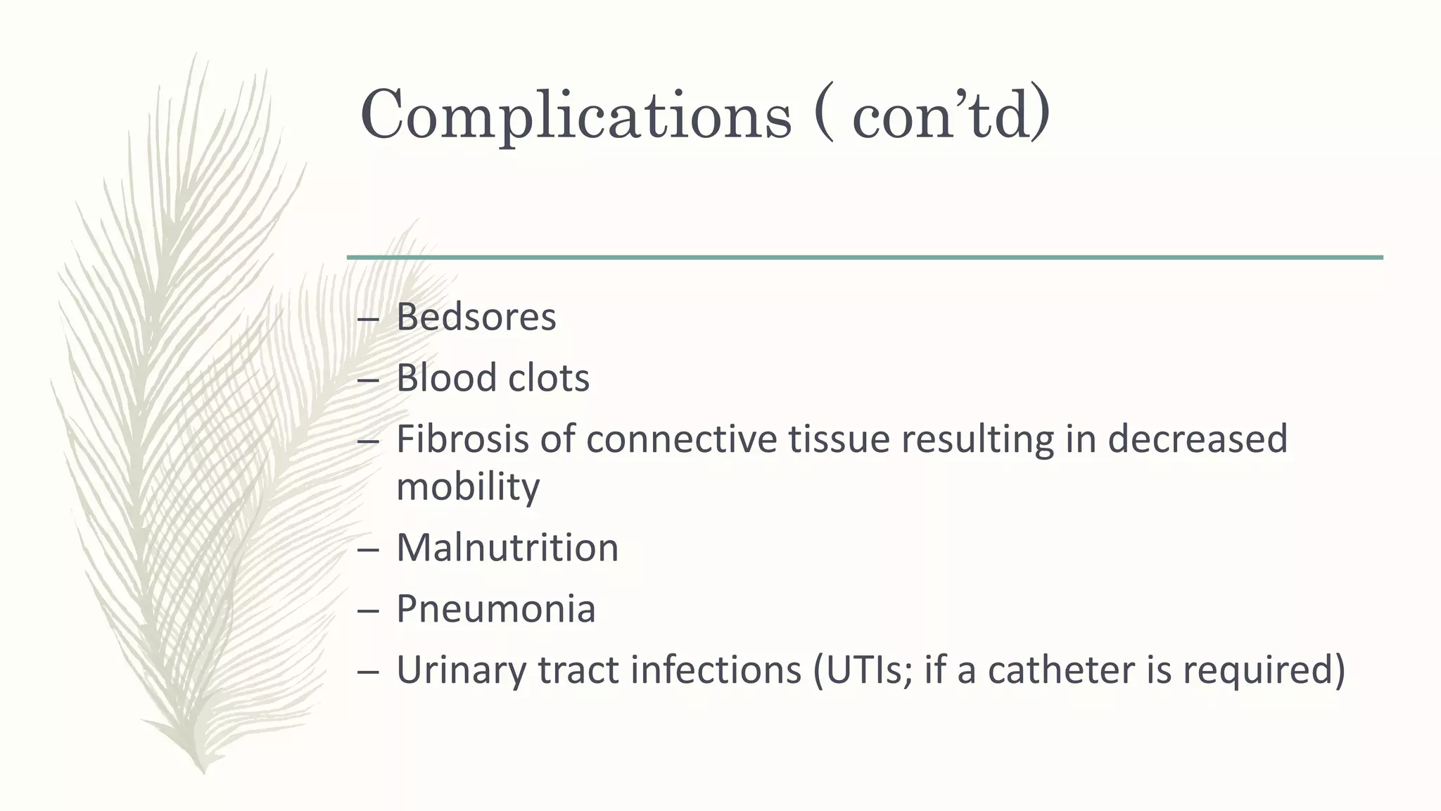
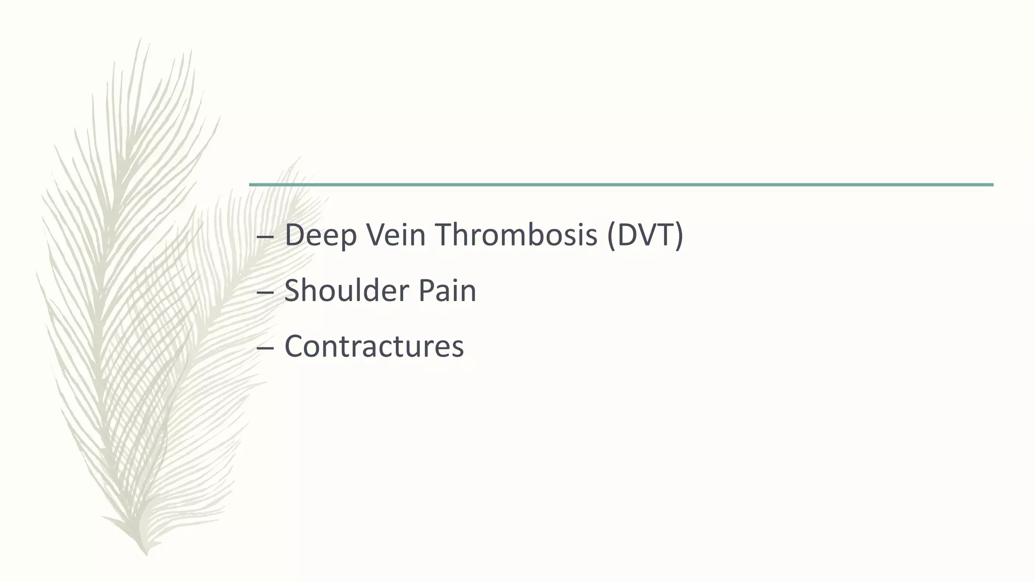
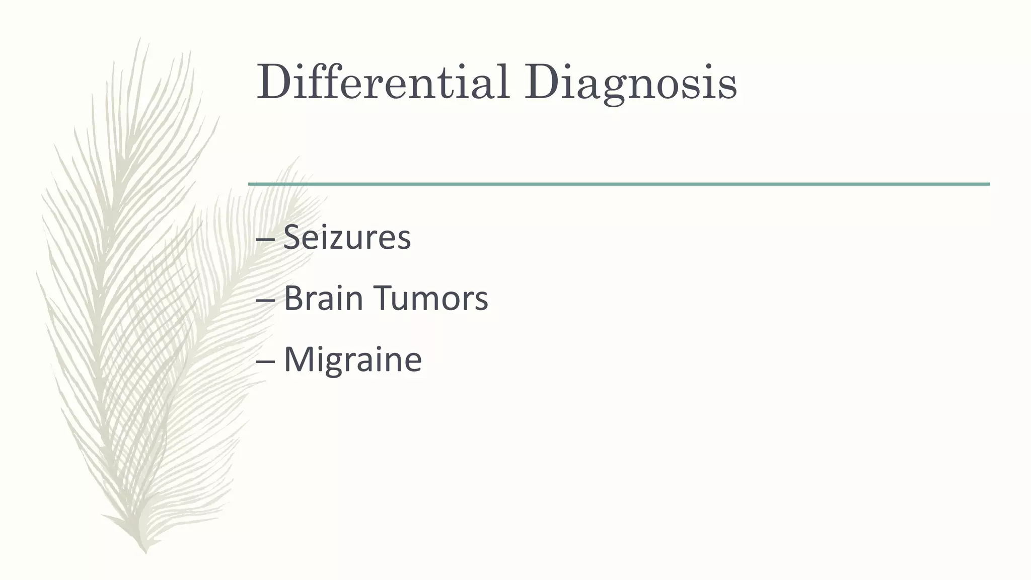
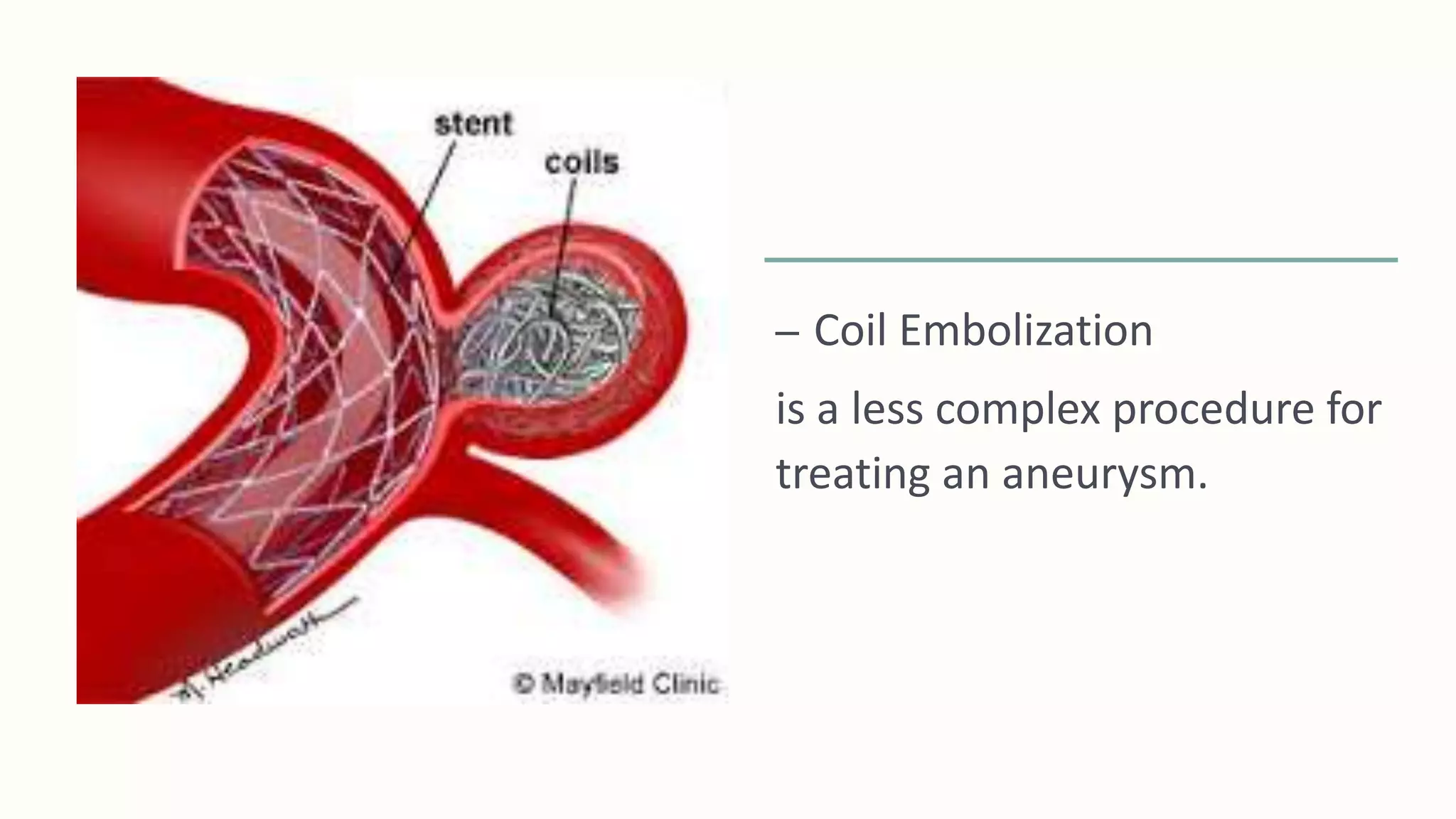
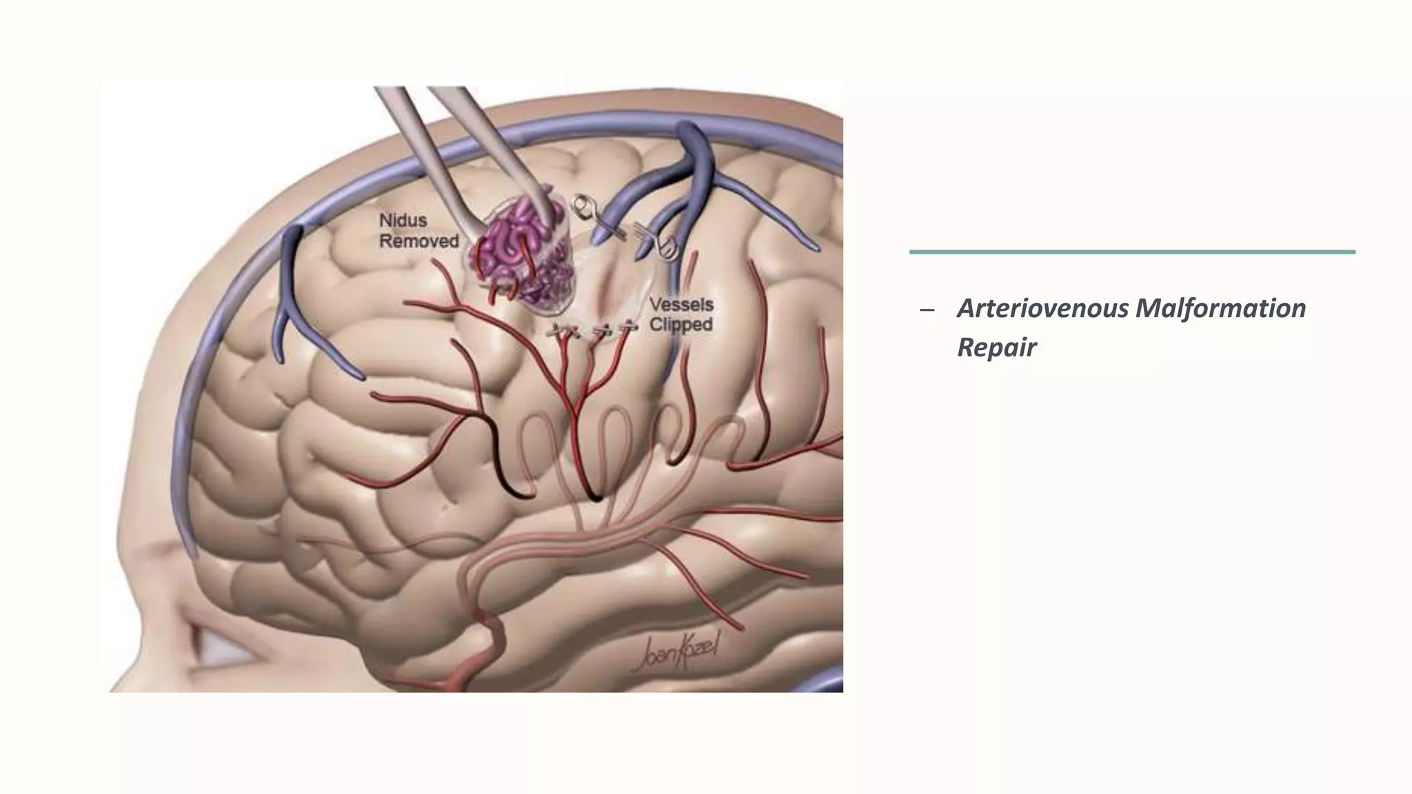
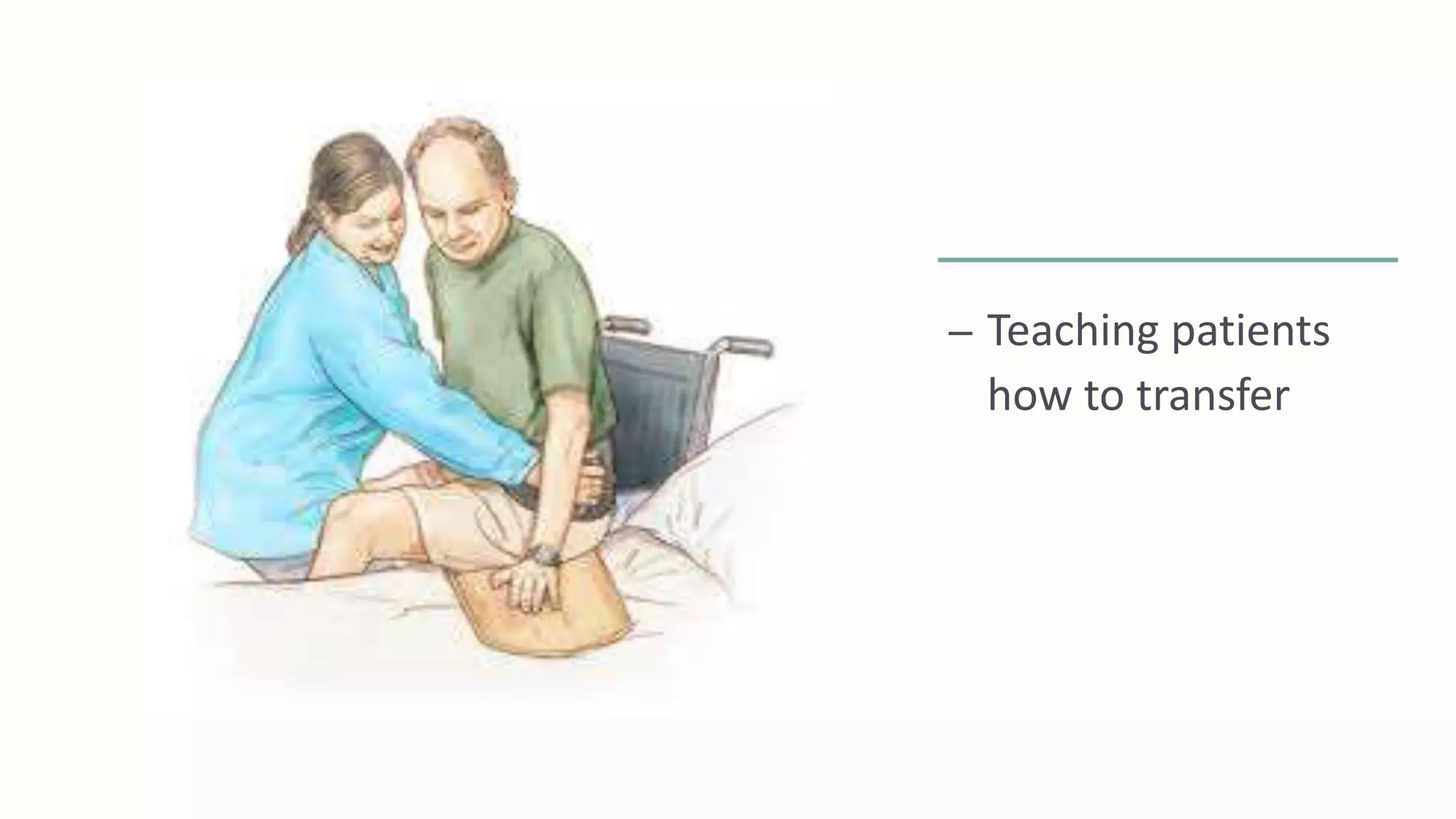
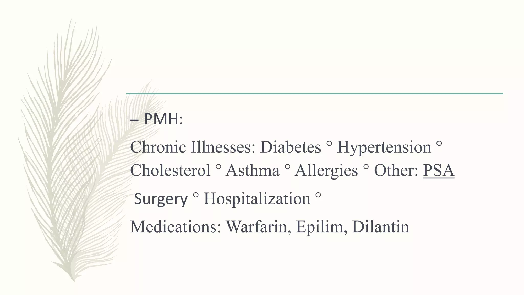
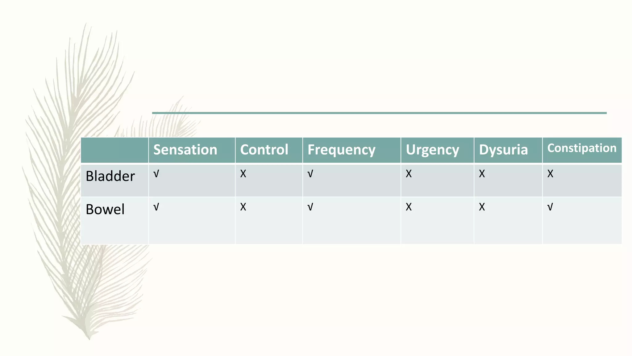
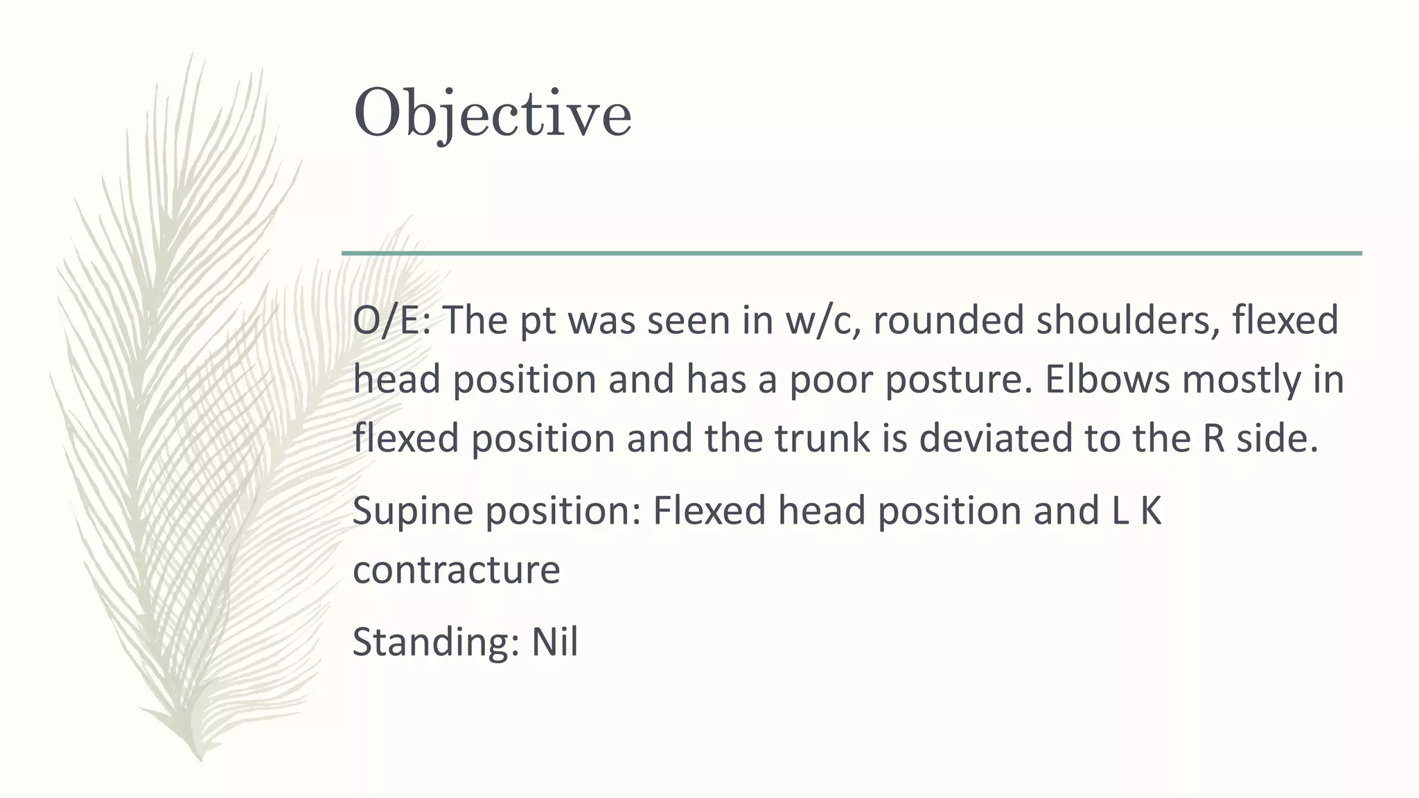
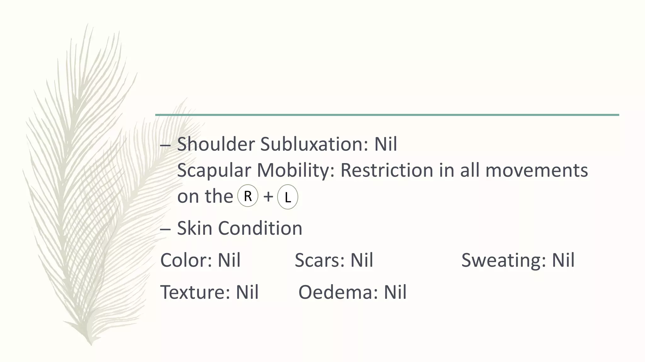
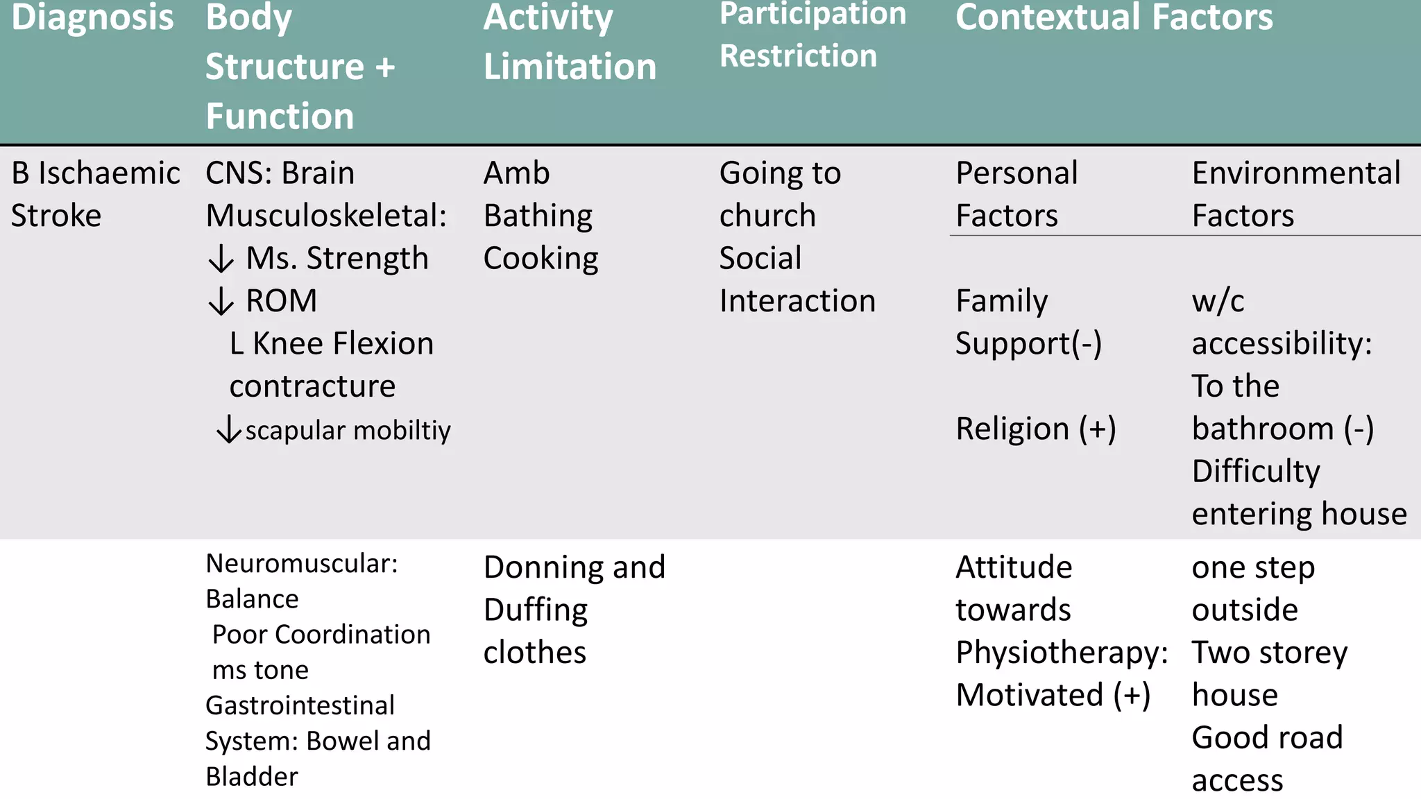
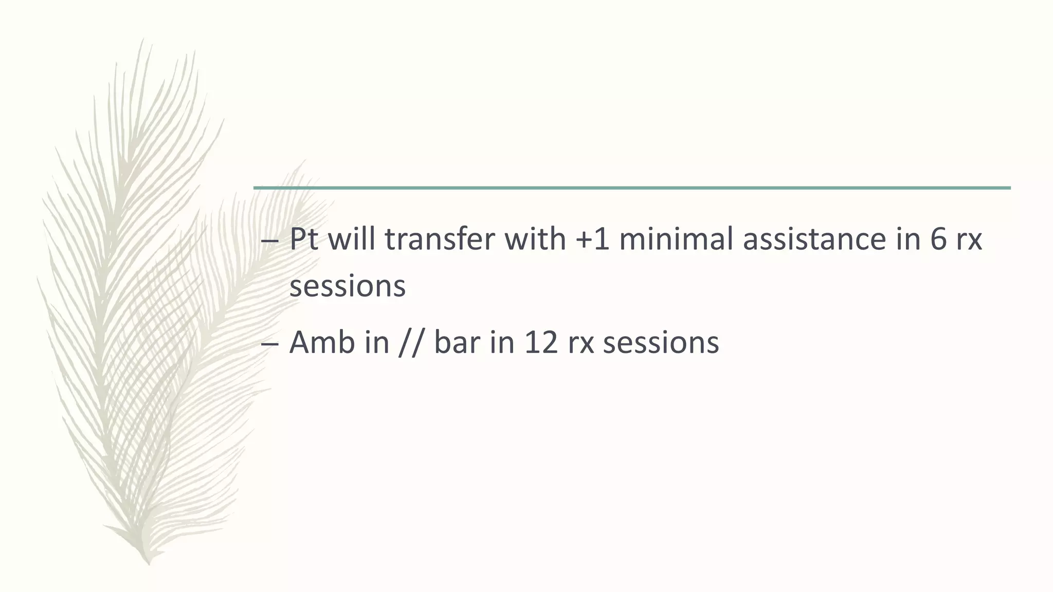
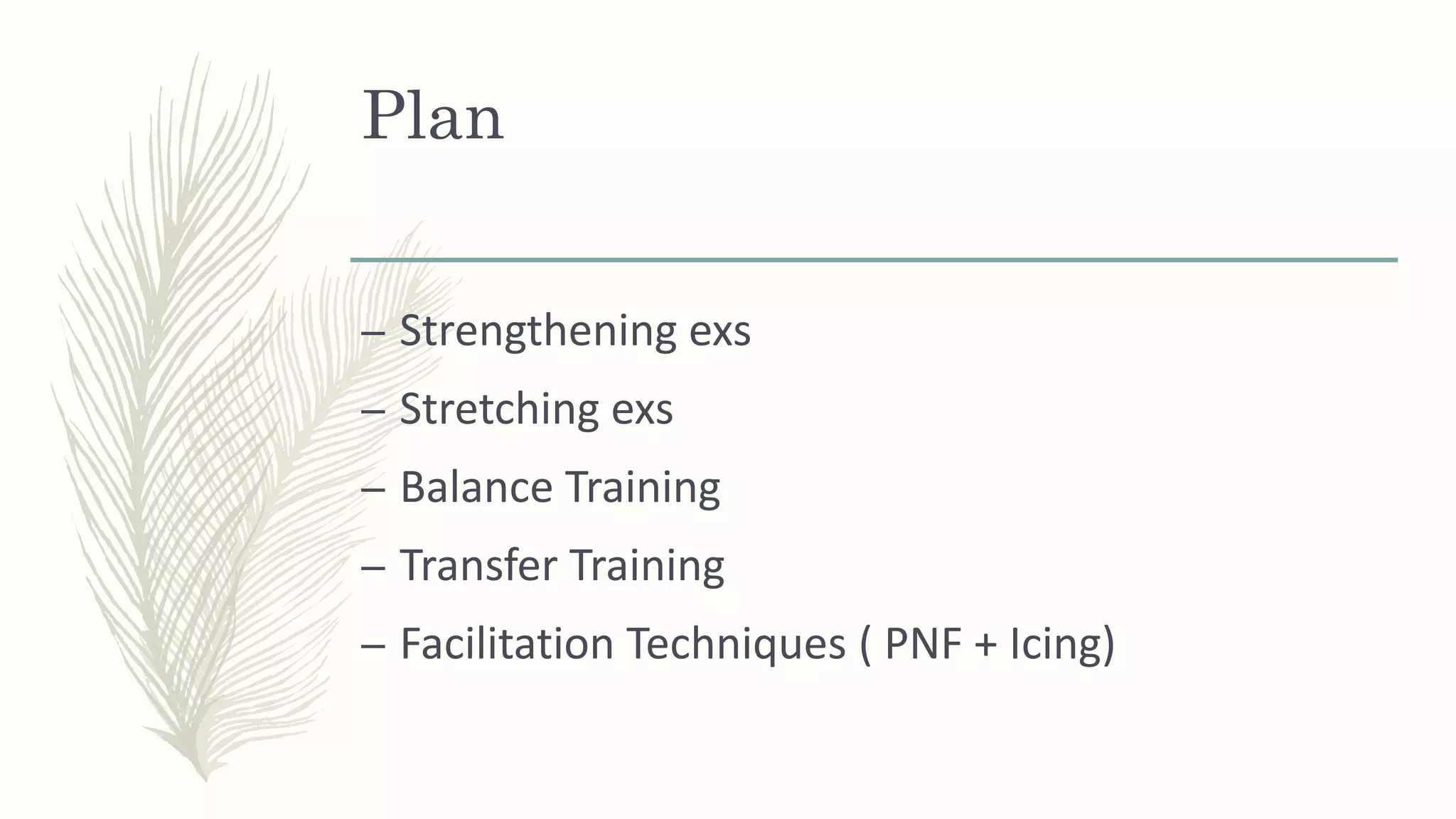
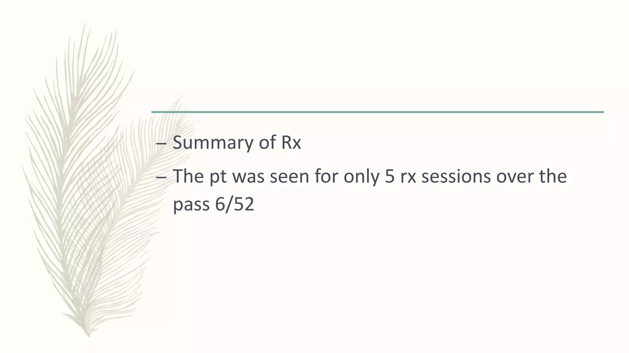
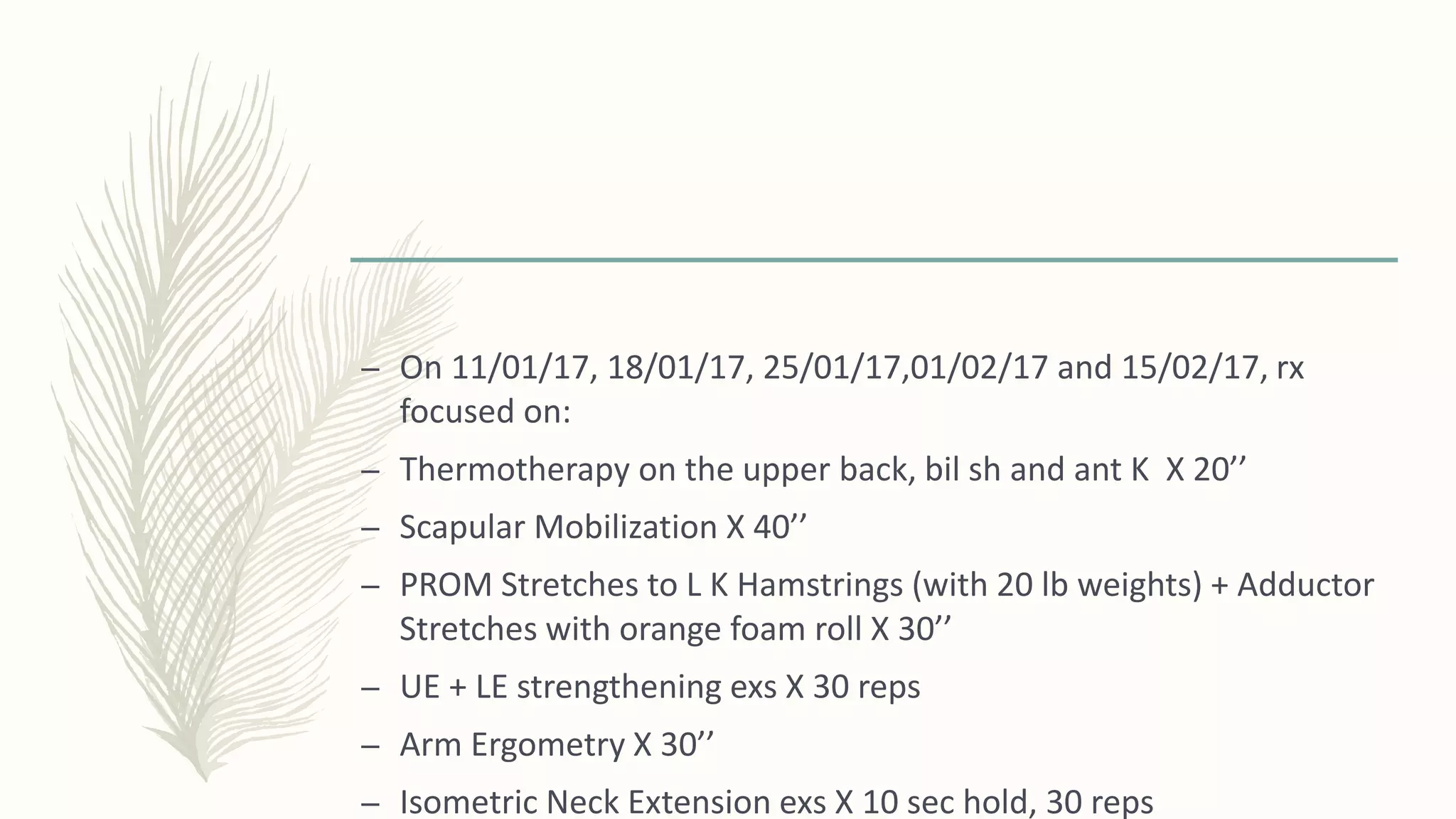
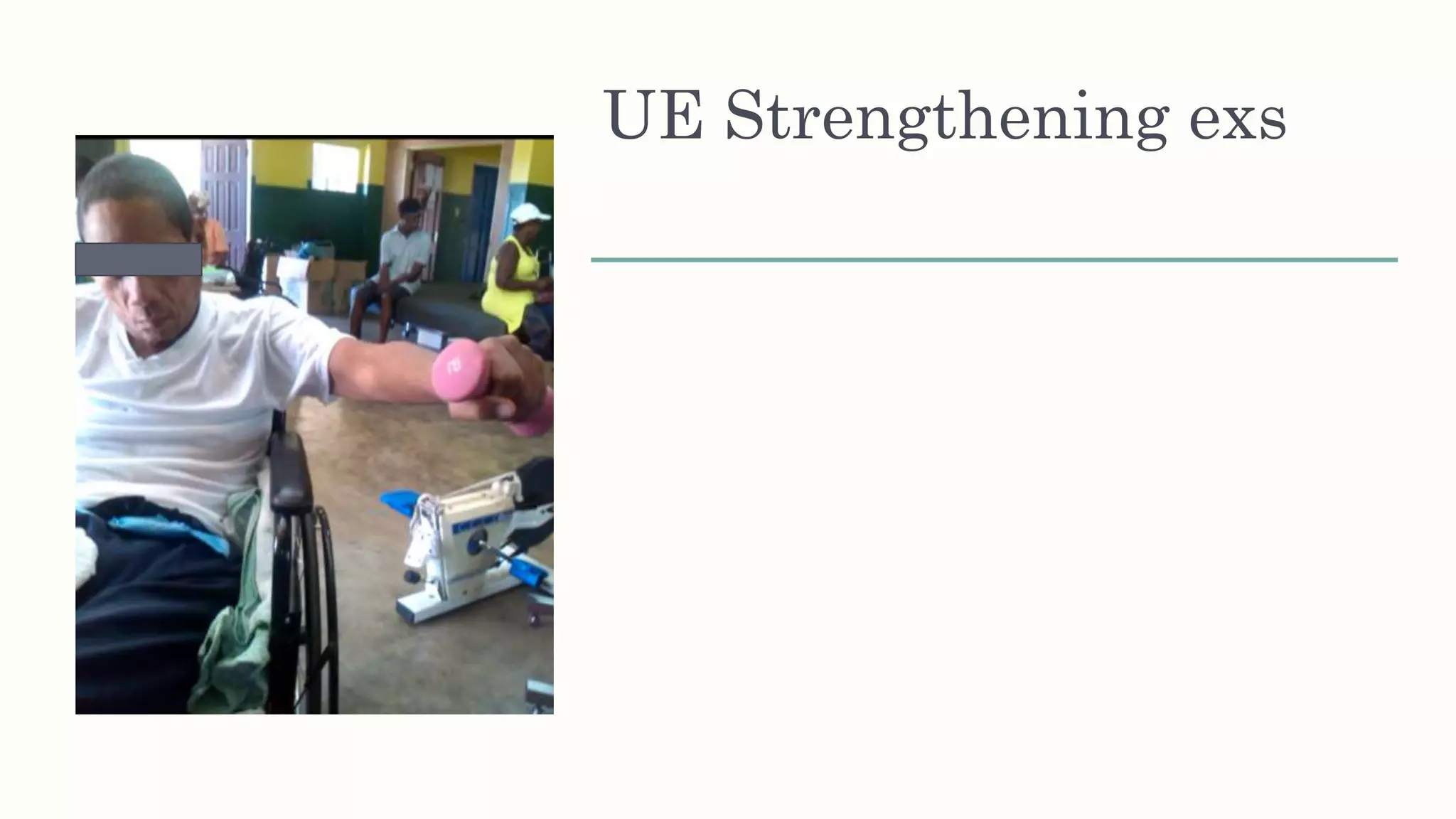
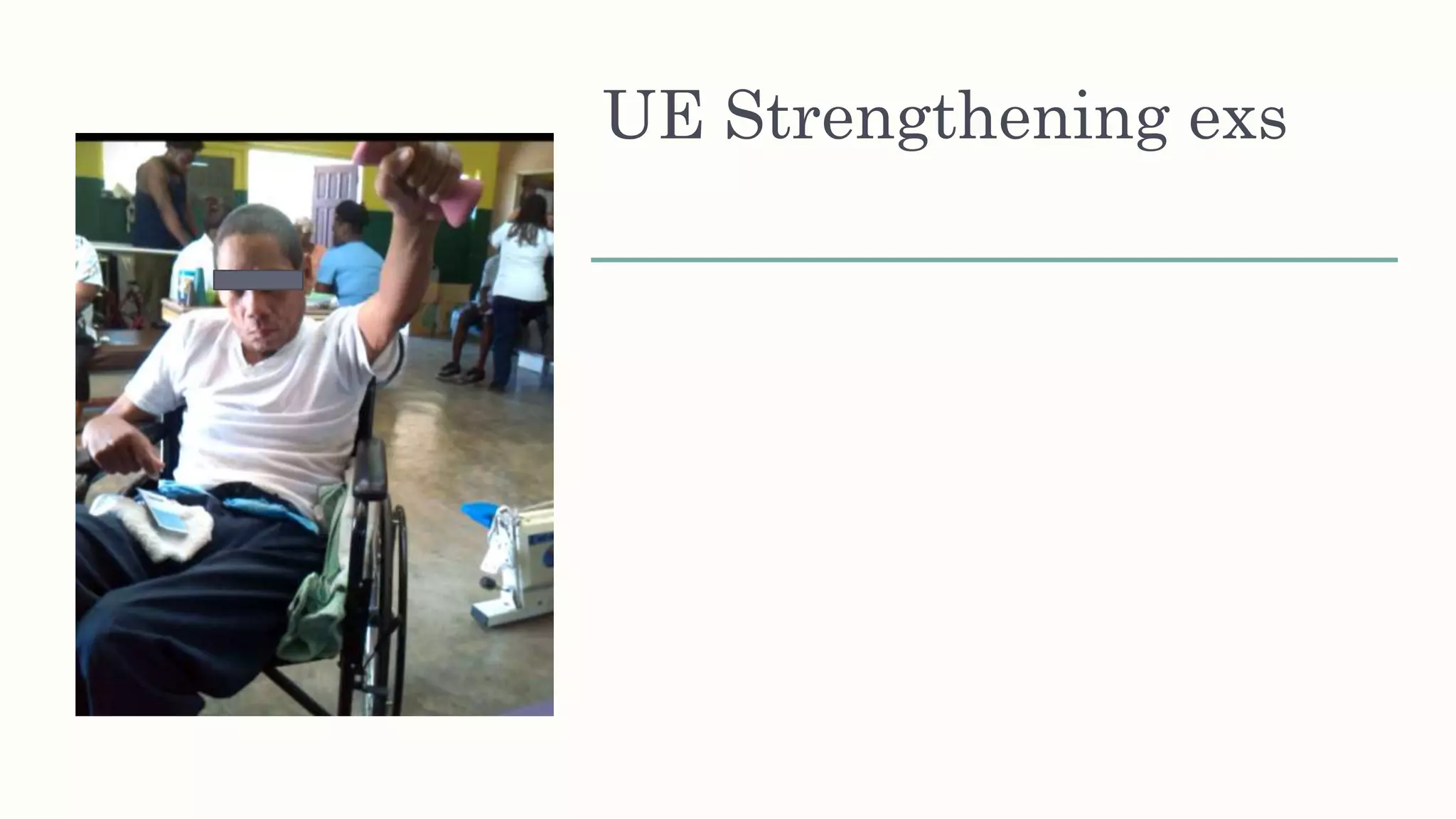
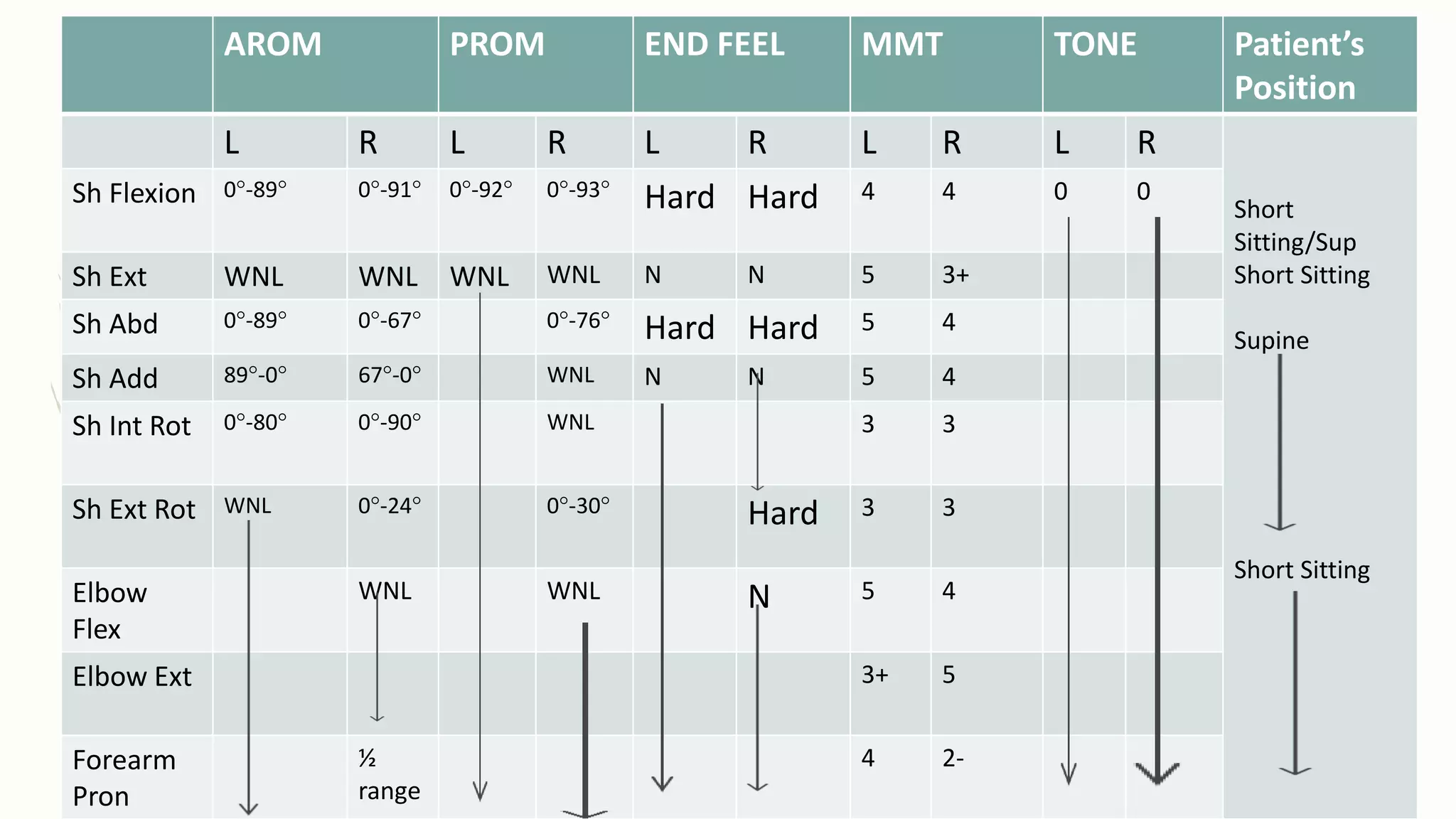
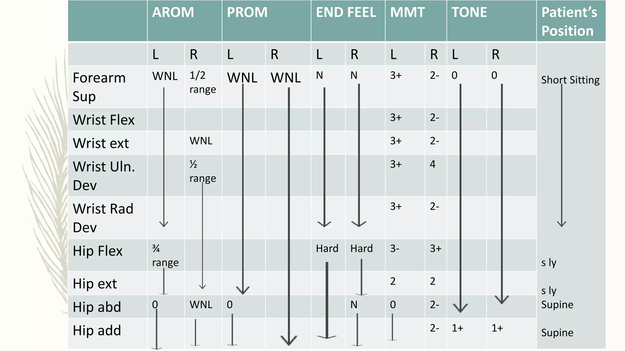
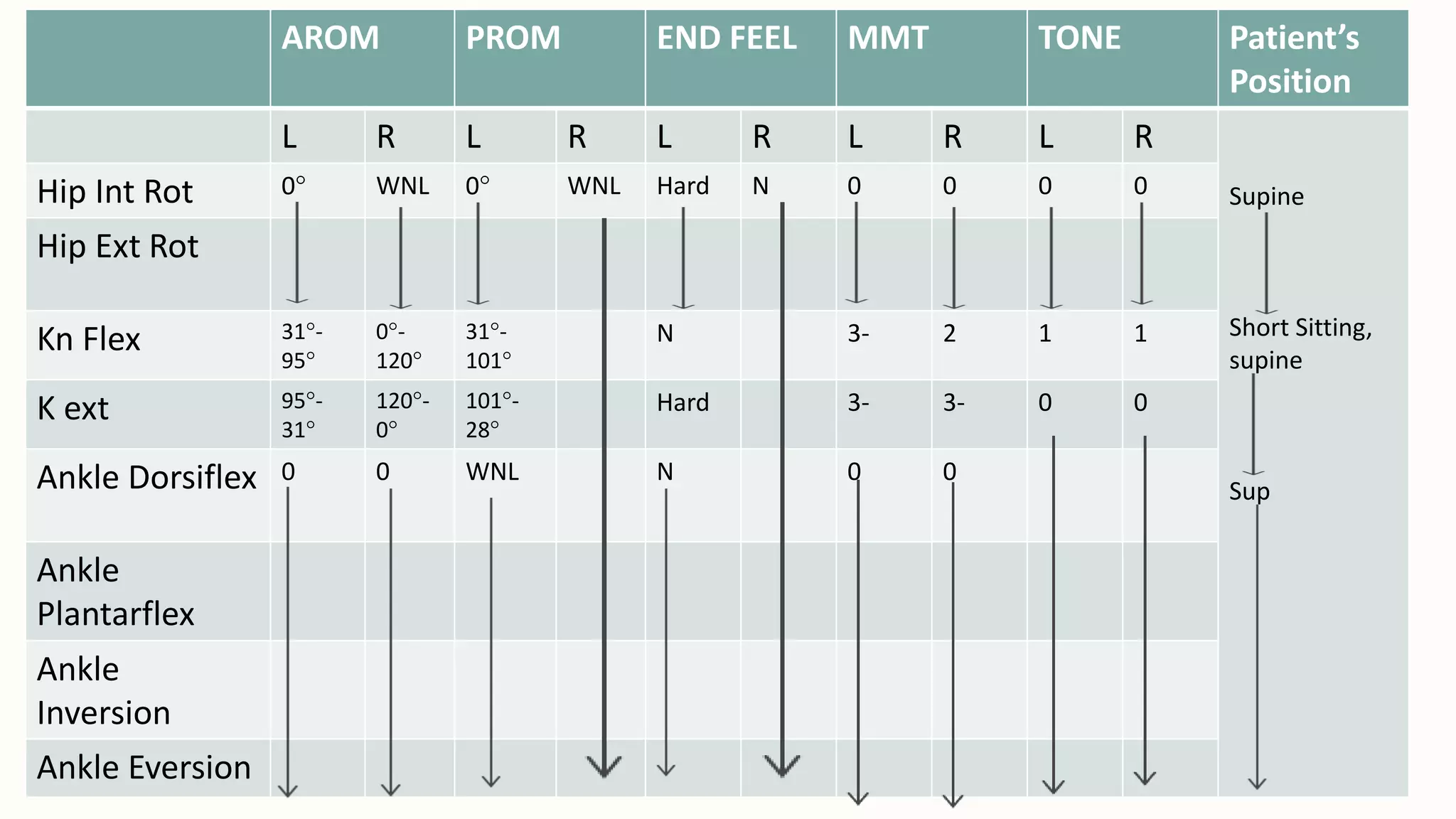
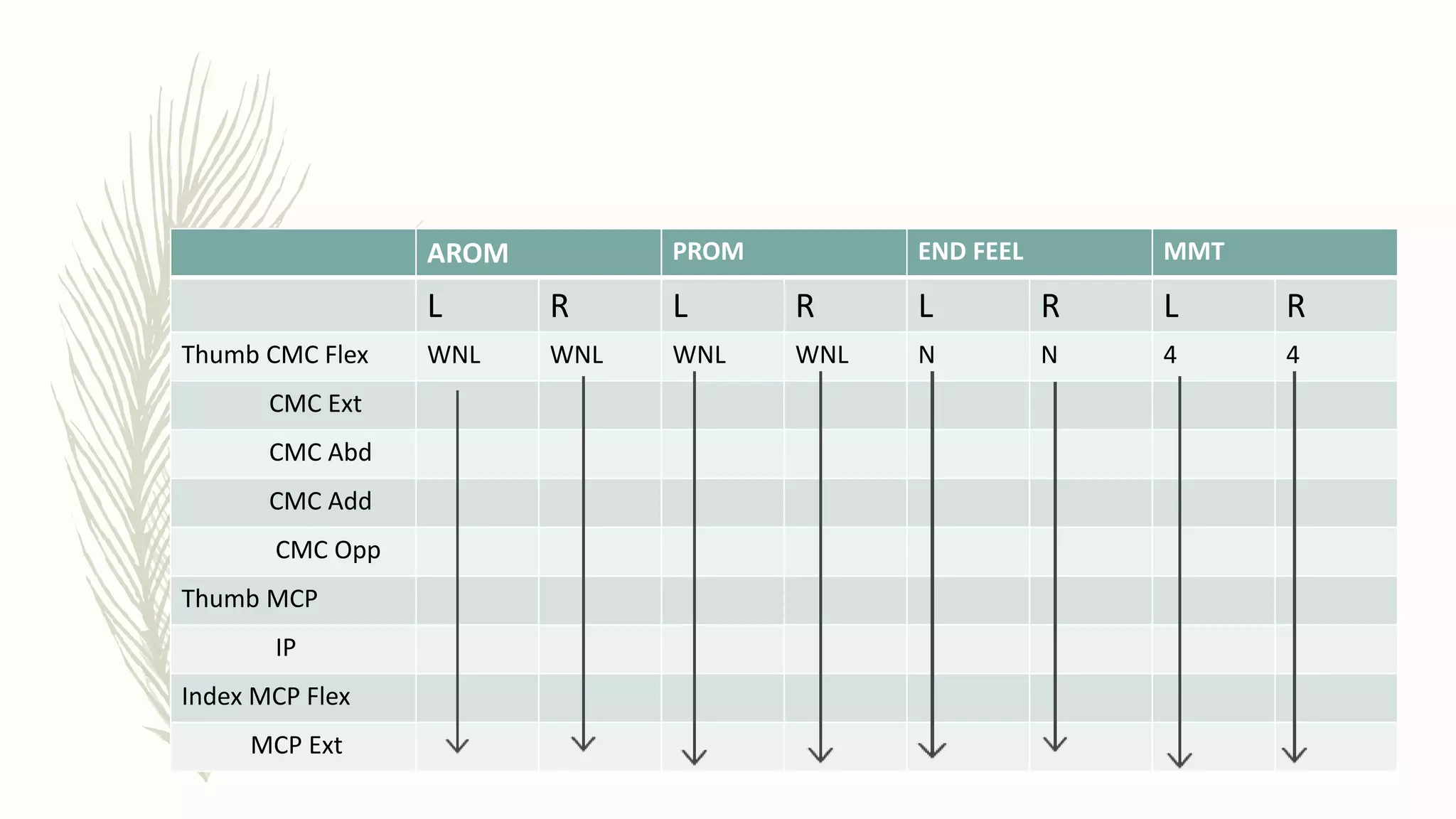
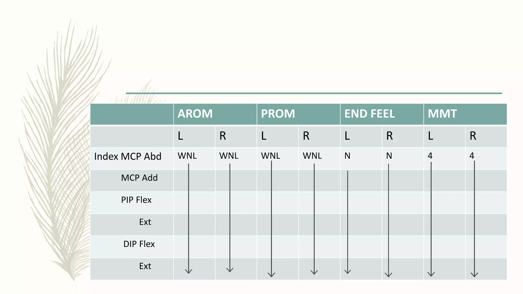
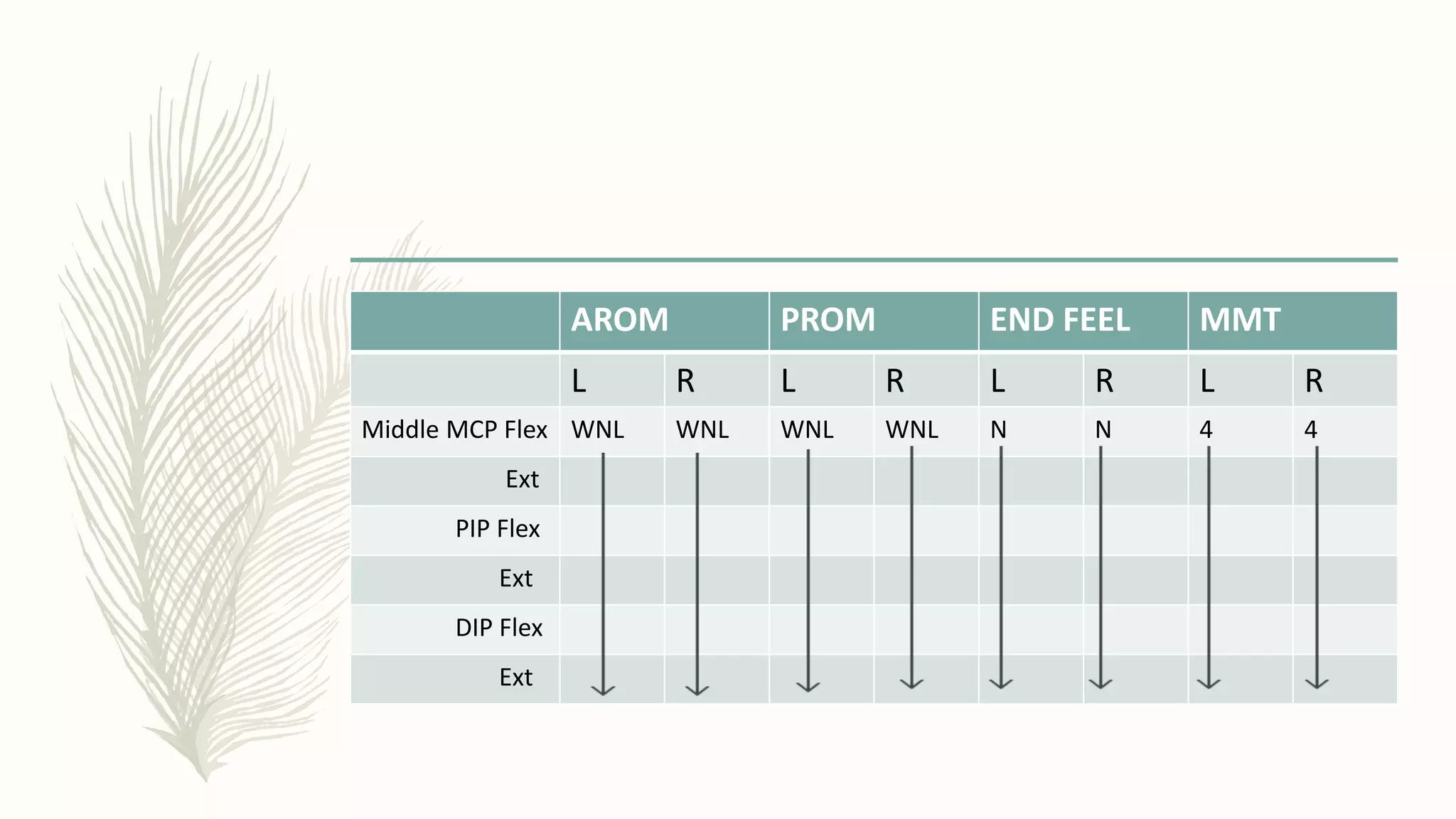
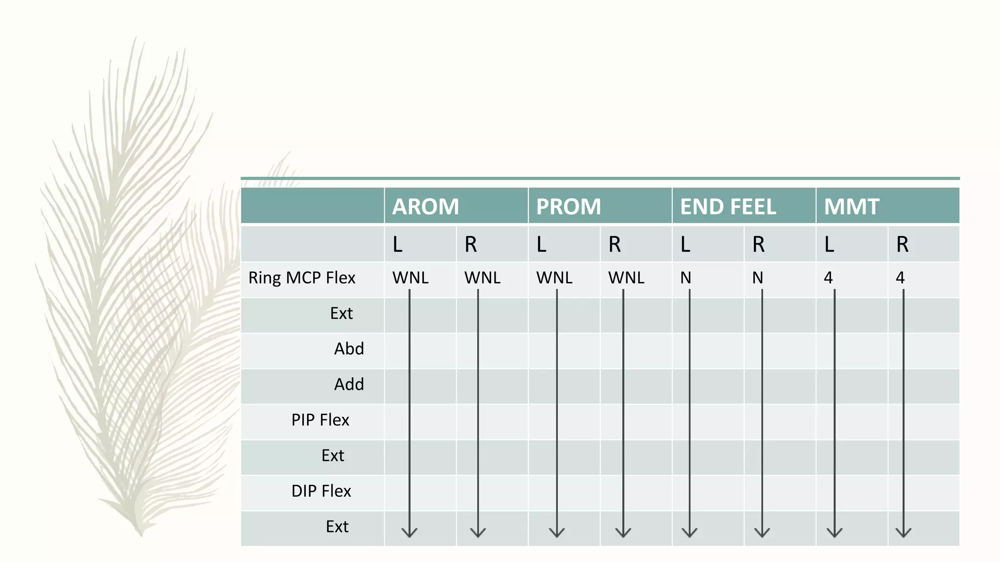
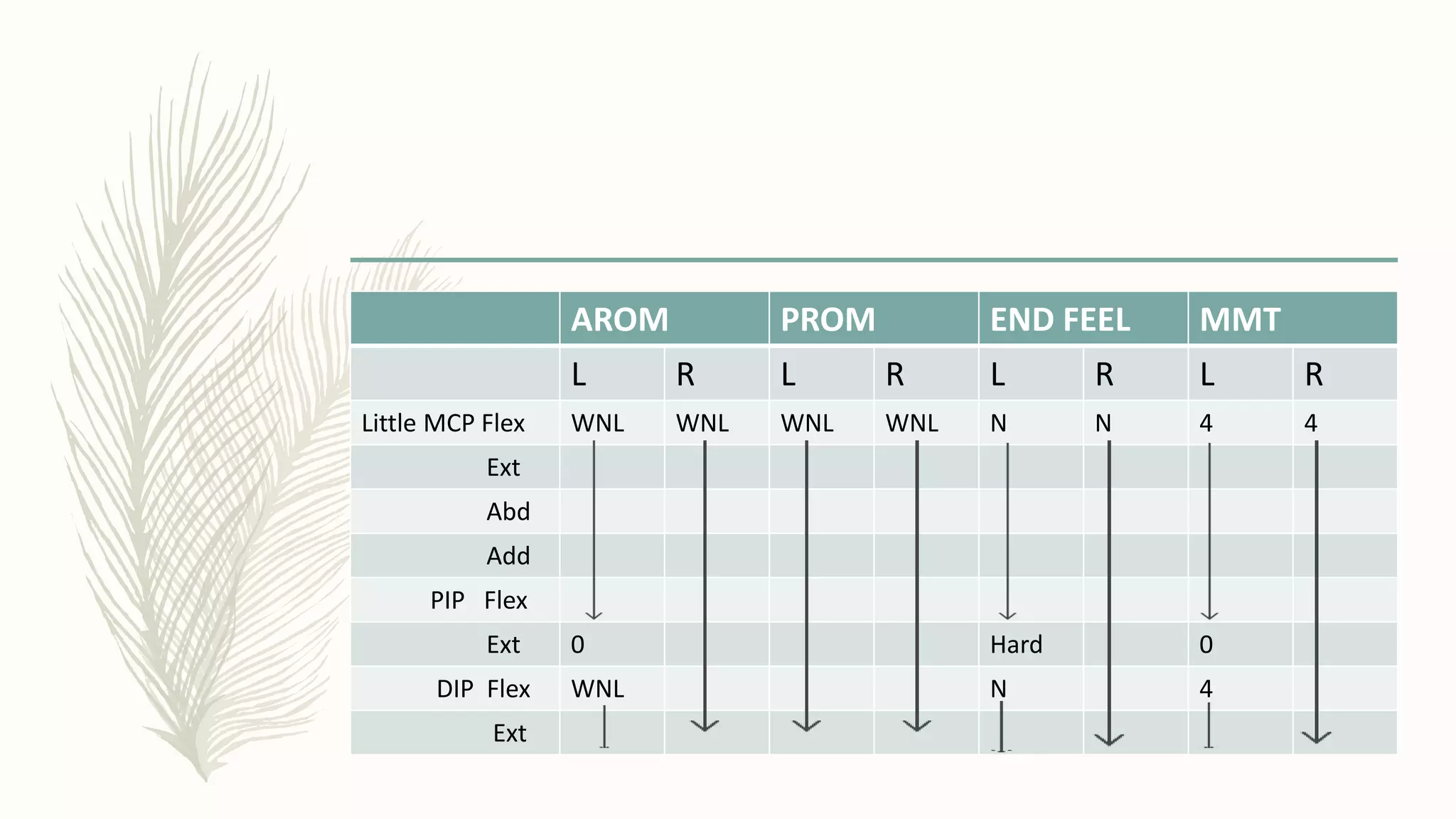
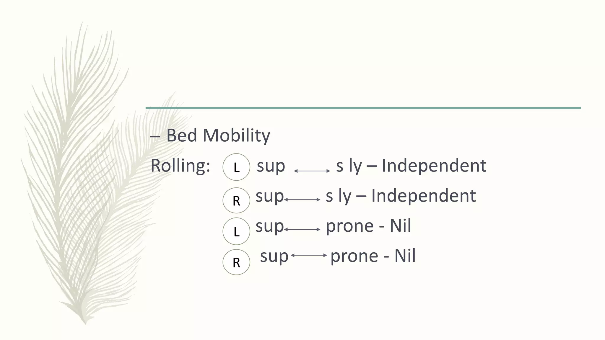
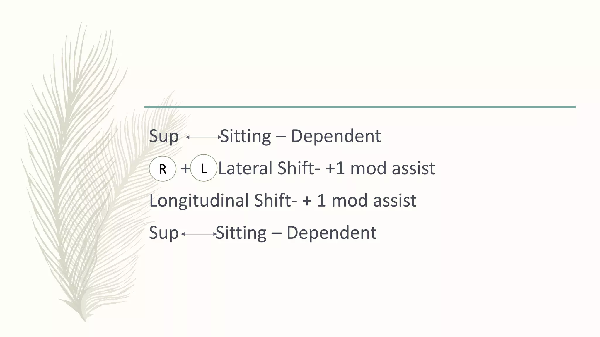
7. The Brain
– The brain is one of the largest
and most complex organs in the
human body.
It is made up of more than 100
billion nerves that communicate
in trillions of connections called
synapses.
11. Cranial Nerves
– The cranial nerves are a set of 12 paired nerves
that arise directly from the brain.
– The first two nerves arise from the cerebrum,
whereas the remaining ten emerge from the brain
stem.
15. Ascending Tracts
– Lateral Spinothalamic Tract- pain and
temperature
– Anterior Spinothalamic Tract -touch, pressure
– Dorsal White Column - conscious proprioceptive
sense, discriminative touch, vibratory sense
16. – Ventral and Dorsal Spinocerebellar Tracts - muscle
joint information from muscle spindles, tendons
and joint receptors of trunk and lower limbs
– Transmit unconscious proprioceptive information
to the cerebellum
17. Descending Tracts
– Lateral Reticulospinal Tract –Important in
reciprocal movements required for walking and
involved in postural control responses
– Lateral Corticospinal Tract –Fine skilled
movements
– Rubrospinal Tract - Innervates UE flexors
18. – Vestibulospinal Tract- relay the signal to the motor
neurons in antigravity muscles. These antigravity muscles
are extensor muscles in the legs that help maintain
upright and balanced posture.
– Tectospinal Tract- Involved in orientation of head and
eyes (turning the eyes and the head) to contralateral
visual stimuli
21. – Stroke is the rapidly developing loss of brain
function(s) due to disturbance in the blood
supply to the brain.
– Can either be Ischeamic (most common) or
Haemorrhagic
25. Anterior
Cerebral Artery
(ACA) Infarct
– Stroke in the anterior
cerebral artery results in
opposite leg weakness
– Head and eyes may
deviate toward lesion
(away from weakness)
26. – Larger infarct will include upper limb with shoulder weakness >
Hand weakness and sensory loss is also present
– Reduced Verbal expression
27. Middle Cerebral
Artery (MCA)
Infarct
– Contralateral hemiplegia
– Deviation of Head and
Eyes toward stroke (away
from hemiplegia)
– Contralateral
hemianesthesia
– Broca’s or Wernicke
Aphasia
29. Hemiplegia
Right Brain damage Left Brain damage
Paralyzed left side: hemiplegia Paralyzed right side: hemiplegia
Left sided neglect Impaired speech/ language
aphasias
Spatial –perceptual deficits Impaired right/left discrimination
Tends to deny or minimize
problems
Slow performance
Rapid performance, short
attention span
Aware of deficits: depression,
anxiety
Impulsive, safety problems Impaired comprehension related
to language and math
Impaired judgement
30. Epidemiology
– According to the latest WHO data published in may
2014 Stroke Deaths in Jamaica reached 3,243 or
18.13% of total deaths.
– The age adjusted Death Rate is 115.44 per 100,000
of population ranks Jamaica #63 in the world
31. Causes
Non- Modifiable Modifiable
Age > 65 HTN
Sex: M > F Cigarette Smoking
Race: Black> White> Asian High cholesterol
Family History of Stroke DM
Obesity
32. Pathophysiology
– Pathophysiology of Ischeamic Stroke
– Occurs when there is lack of sufficient blood flow
to perfuse cerebral tissue, due to narrowed or
blocked arteries leading to or within the brain.
33. – Narrowing is commonly the result of
atherosclerosis. As the plaques grow in size, the
blood vessel becomes narrowed and the blood
flow to the area beyond is reduced.
34. – Damaged areas of an atherosclerotic plaque can cause a
blood clot to form, which blocks the blood vessel
– blood clots or debris from elsewhere in the body,
typically the heart valves, travel through the circulatory
system and block narrower blood vessels.
35. – Pathophysiology of Haemorrhagic stroke
Haemorrhagic strokes are due to the rupture of a
blood vessels leading to compression of brain tissue
from an expanding haematoma
36. – the pressure may lead to a loss of blood supply to
affected tissue with resulting infarction, and the blood
released by brain haemorrhage appears to have direct
toxic effects on brain tissue and vasculature.
37. Signs and Symptoms
– Sudden numbness or weakness in the face, arm,
or leg, especially on one side of the body
– Sudden confusion, trouble speaking, or difficulty
understanding speech
– Sudden trouble seeing in one or both eyes
38. – Sudden trouble walking, dizziness, loss of
balance, or lack of coordination
– Sudden severe headache with no known cause
48. – Blood Test can also help with diagnosing stroke
Blood Glucose Test
Low blood glucose levels may cause symptoms
similar to those of a stroke.
49. Platelet Count
Abnormal platelet levels may be a sign of a bleeding
disorder (not enough clotting) or a thrombotic
disorder (too much clotting).
50. Prothrombin Time (PT) and Partial
Thromboplastin Time (PTT)
Measure how long it takes for your blood to clot.
51. Complications
– Complications that may occur within 72 hours
of stroke include the following:
– Cerebral swelling (edema)
– Increased intracranial pressure (ICP)
– Intracerebral haemorrhage
– Seizures
52. Complications ( con’td)
– Bedsores
– Blood clots
– Fibrosis of connective tissue resulting in decreased
mobility
– Malnutrition
– Pneumonia
– Urinary tract infections (UTIs; if a catheter is required)
55. Acute Management
– The goal for the acute management of patients
with stroke is to stabilize the patient and to
complete initial evaluation and assessment,
including imaging and laboratory studies, within a
short time frame.
56. – Critical decisions focus on the need for
intubation, blood pressure control, and
determination of risk/benefit for thrombolytic
intervention
57. Medical Management
– Anti coagulant medications
Anticoagulants such as Warfarin prevent blood clots
from forming and keep existing blood clots from
getting bigger.
58. – Anti Platelet Medications
Anti platelet medications such as Aspirin keep
platelets in the blood from sticking together.
59. – Statins
Statins lower cholesterol and the risk for another stroke
– atorvastatin (Lipitor)
– lovastatin (Altoprev, Mevacor)
– pravastatin (Pravachol)
– rosuvastatin (Crestor)
– simvastatin (Zocor)
61. – Medicines used to treat depression and pain may
also be prescribed after a stroke.
62. Surgical Management
– Hemorrhagic Stroke
Aneurysm Clipping
Aneurysm clipping is done to block off the aneurysm from the
blood vessels in the brain. This surgery helps prevent further
leaking of blood from the aneurysm.
63. – It also can help prevent the
aneurysm from
bursting again.
75. Prognosis
– Many people recover completely after a stroke.
For others, it can take many months to recover
from a stroke.
– Physical Therapy and other retraining methods
are greatly improving rehabilitation and recovery
76. – In general, the more deficits or loss of ability (in
walking or talking) individuals have when they
arrive in the emergency department, the worse
the outcome.
80. – HPC: Daughter- In- Law reports that the pt who lives
alone had a stroke on 4/9/2015. The pt attempted to
call a friend at the time that the stroke occurred but
he was unable to speak. The friend then went to the
house and called the pt but there was no response.
He then called other friends and police who came
and knocked down the door to get into the house.
81. – He was on the floor responsive and had an
incoherent speech but confused. He was taken to
KPH where he was placed on IV. His daughter- in- law
received a call the next day that he was at KPH. 3/52
after being admitted to KPH, the pt did a CT scan
which showed that he had B Ischaemic stroke. On
October 17, 2015, the pt got a 2nd stroke and seizure
while on the ward at KPH.
82. – The 2nd stroke led to L leg weakness whereas the
1st stroke had cause weakness on the R side. His
daughter-in – law felt that the hospital was not
managing him properly so she transferred him to
UHWI where he did a CT scan on the lungs and
found out that he had PE in B lungs.
83. – CT scan was also done on the brain which
revealed that he had Multiple Infarction in the
brain. He was admitted to UHWI for 3/52. He
received chest physiotherapy, warfarin and
antibiotics. While at UHWI, the PT had four
seizures. Upon D/C, the pt was followed up at
Medical Clinic but is now D/C.
84. – The daughter-in-law attempted to get PT for him
privately after being D/C from UHWI but could
not afford it and it wasn't until December 2016
after being recommended by a friend that she
come to clinic with the pt at SJGRC and referred
for out pt therapy
86. – SFH:
Occupation: Retired; Real Estate Agent and also owned a
newspaper company called “Auto- Report”
Family of stroke: Nil
Religion: Christianity
Hobbies: Reading
Drink ° Smoke °
Hand Dominance: R Hnd
87. – Home Situation: Pt lives alone
Description of the house: Pt lives in a two storey
house with modern convenience and has a step on
the outside. The yard is gravel and the pt has good
road access
89. – PT Goals: The daughter – in- law would like to
see the pt walk again
90. Objective
O/E: The pt was seen in w/c, rounded shoulders, flexed
head position and has a poor posture. Elbows mostly in
flexed position and the trunk is deviated to the R side.
Supine position: Flexed head position and L K
contracture
Standing: Nil
91. – Shoulder Subluxation: Nil
Scapular Mobility: Restriction in all movements
on the +
– Skin Condition
Color: Nil Scars: Nil Sweating: Nil
Texture: Nil Oedema: Nil
R L
92. – Memory : Long Term
– Attention Span: Fair
– Ability to Follow Instructions and Command : Good
– Communication: Oriented
– Facial Features : Nil
– Vision: Good Hearing: Good Speech: Fair
93. Coordination
– Finger to nose: Fair on both sides
– Pronation and Supination:
– Heel to Shin: Poor on both sides
– Sensation: Good for the UE + LE
94. – AROM: Cervical Extension - 66 °
Cervical Flexion- 37 °
Cervical Rotation- 32 °
Cervical Rotation - 22 °
Cervical Side Flexion- 0°
L
R
L
R
95. AROM PROM END FEEL MMT TONE Patient’s
Position
L R L R L R L R L R
Short
Sitting/Sup
Short Sitting
Supine
Short Sitting
Sh Flexion 0°-89° 0°-91° 0°-92° 0°-93° Hard Hard 4 4 0 0
Sh Ext WNL WNL WNL WNL N N 5 3+
Sh Abd 0°-89° 0°-67° 0°-76° Hard Hard 5 4
Sh Add 89°-0° 67°-0° WNL N N 5 4
Sh Int Rot 0°-80° 0°-90° WNL 3 3
Sh Ext Rot WNL 0°-24° 0°-30° Hard 3 3
Elbow
Flex
WNL WNL N 5 4
Elbow Ext 3+ 5
Forearm
Pron
½
range
4 2-
96. AROM PROM END FEEL MMT TONE Patient’s
Position
L R L R L R L R L R
Short Sitting
s ly
s ly
Supine
Supine
Forearm
Sup
WNL 1/2
range
WNL WNL N N 3+ 2- 0 0
Wrist Flex 3+ 2-
Wrist ext WNL 3+ 2-
Wrist Uln.
Dev
½
range
3+ 4
Wrist Rad
Dev
3+ 2-
Hip Flex ¾
range
Hard Hard 3- 3+
Hip ext 2 2
Hip abd 0 WNL 0 N 0 2-
Hip add 2- 1+ 1+
97. AROM PROM END FEEL MMT TONE Patient’s
Position
L R L R L R L R L R
Supine
Short Sitting,
supine
Sup
Hip Int Rot 0° WNL 0° WNL Hard N 0 0 0 0
Hip Ext Rot
Kn Flex 31°-
95°
0°-
120°
31°-
101°
N 3- 2 1 1
K ext 95°-
31°
120°-
0°
101°-
28°
Hard 3- 3- 0 0
Ankle Dorsiflex 0 0 WNL N 0 0
Ankle
Plantarflex
Ankle
Inversion
Ankle Eversion
98. AROM PROM END FEEL MMT
L R L R L R L R
Thumb CMC Flex WNL WNL WNL WNL N N 4 4
CMC Ext
CMC Abd
CMC Add
CMC Opp
Thumb MCP
IP
Index MCP Flex
MCP Ext
99. AROM PROM END FEEL MMT
L R L R L R L R
Index MCP Abd WNL WNL WNL WNL N N 4 4
MCP Add
PIP Flex
Ext
DIP Flex
Ext
100. AROM PROM END FEEL MMT
L R L R L R L R
Middle MCP Flex WNL WNL WNL WNL N N 4 4
Ext
PIP Flex
Ext
DIP Flex
Ext
101. AROM PROM END FEEL MMT
L R L R L R L R
Ring MCP Flex WNL WNL WNL WNL N N 4 4
Ext
Abd
Add
PIP Flex
Ext
DIP Flex
Ext
102. AROM PROM END FEEL MMT
L R L R L R L R
Little MCP Flex WNL WNL WNL WNL N N 4 4
Ext
Abd
Add
PIP Flex
Ext 0 Hard 0
DIP Flex WNL N 4
Ext
103. – Hand Function
Gross Grip Strength: - Good
- Good
Standard Grip Strength Test:
16 kg 12 kg
R
L
L R
112. Diagnosis Body
Structure +
Function
Activity
Limitation
Participation
Restriction
Contextual Factors
B Ischaemic
Stroke
CNS: Brain
Musculoskeletal:
↓ Ms. Strength
↓ ROM
L Knee Flexion
contracture
↓scapular mobiltiy
Amb
Bathing
Cooking
Going to
church
Social
Interaction
Personal
Factors
Family
Support(-)
Religion (+)
Environmental
Factors
w/c
accessibility:
To the
bathroom (-)
Difficulty
entering house
Neuromuscular:
Balance
Poor Coordination
ms tone
Gastrointestinal
System: Bowel and
Bladder
Donning and
Duffing
clothes
Attitude
towards
Physiotherapy:
Motivated (+)
one step
outside
Two storey
house
Good road
access
113. Assessment
– Summary
– The pt is a good candidate for Physical Therapy
since he is co-operative + follow instructions but
rehab potential is guarded due to the fact that his
stroke was > 1 year ago and PT intervention was
only just sought
114. – In addition, there is poor family support and
financial support
115. – Short Term Goals
– ↑ ROM in the neck, UE + LE in 12 rx sessions
– ↑ ms strength in bil UE + LE by 1 grade in rx
sessions
– Facilitate ↓ ms tone in affected ms in 12 rx
sessions
116. – Improve Bed Mobility with +1 minimal assist in 6
rx sessions
– Good dynamic short sitting balance in 6 rx
sessions
– Independent w/c mobility on level surfaces in 12
rx sessions
117. – Pt will transfer with +1 minimal assistance in 6 rx
sessions
– Amb in // bar in 12 rx sessions
118. – Long Term Goals
– Functional ROM for bil UE+LE in 24 rx sessions
– ↑ ms strength to grade 5 for bil UE + LE in 12 rx
sessions
– Independent bed mobility in 12 rx sessions
119. – Fair Static standing balance in 12 rx sessions
– Amb with walker to independent amb in 24 rx
sessions
– Independent transfer in 12 rx sessions
120. Plan
– Strengthening exs
– Stretching exs
– Balance Training
– Transfer Training
– Facilitation Techniques ( PNF + Icing)
121. – Tilt Table Therapy
– Gait Training
– w/c mobility training
– Weight bearing exs
– Pt and family education
122. – Summary of Rx
– The pt was seen for only 5 rx sessions over the
pass 6/52
123. – On 11/01/17, 18/01/17, 25/01/17,01/02/17 and 15/02/17, rx
focused on:
– Thermotherapy on the upper back, bil sh and ant K X 20’’
– Scapular Mobilization X 40’’
– PROM Stretches to L K Hamstrings (with 20 lb weights) + Adductor
Stretches with orange foam roll X 30’’
– UE + LE strengthening exs X 30 reps
– Arm Ergometry X 30’’
– Isometric Neck Extension exs X 10 sec hold, 30 reps
124. – Bed Mobility training X 30 reps
– Transfer training X 2 reps
134. AROM PROM END FEEL MMT TONE Patient’s
Position
L R L R L R L R L R
Short
Sitting/Sup
Short Sitting
Supine
Short Sitting
Sh Flexion 0°-89° 0°-91° 0°-92° 0°-93° Hard Hard 4 4 0 0
Sh Ext WNL WNL WNL WNL N N 5 3+
Sh Abd 0°-89° 0°-67° 0°-76° Hard Hard 5 4
Sh Add 89°-0° 67°-0° WNL N N 5 4
Sh Int Rot 0°-80° 0°-90° WNL 3 3
Sh Ext Rot WNL 0°-24° 0°-30° Hard 3 3
Elbow
Flex
WNL WNL N 5 4
Elbow Ext 3+ 5
Forearm
Pron
½
range
4 2-
135. AROM PROM END FEEL MMT TONE Patient’s
Position
L R L R L R L R L R
Short Sitting
s ly
s ly
Supine
Supine
Forearm
Sup
WNL 1/2
range
WNL WNL N N 3+ 2- 0 0
Wrist Flex 3+ 2-
Wrist ext WNL 3+ 2-
Wrist Uln.
Dev
½
range
3+ 4
Wrist Rad
Dev
3+ 2-
Hip Flex ¾
range
Hard Hard 3- 3+
Hip ext 2 2
Hip abd 0 WNL 0 N 0 2-
Hip add 2- 1+ 1+
136. AROM PROM END FEEL MMT TONE Patient’s
Position
L R L R L R L R L R
Supine
Short Sitting,
supine
Sup
Hip Int Rot 0° WNL 0° WNL Hard N 0 0 0 0
Hip Ext Rot
Kn Flex 31°-
95°
0°-
120°
31°-
101°
N 3- 2 1 1
K ext 95°-
31°
120°-
0°
101°-
28°
Hard 3- 3- 0 0
Ankle Dorsiflex 0 0 WNL N 0 0
Ankle
Plantarflex
Ankle
Inversion
Ankle Eversion
137. AROM PROM END FEEL MMT
L R L R L R L R
Thumb CMC Flex WNL WNL WNL WNL N N 4 4
CMC Ext
CMC Abd
CMC Add
CMC Opp
Thumb MCP
IP
Index MCP Flex
MCP Ext
138. AROM PROM END FEEL MMT
L R L R L R L R
Index MCP Abd WNL WNL WNL WNL N N 4 4
MCP Add
PIP Flex
Ext
DIP Flex
Ext
139. AROM PROM END FEEL MMT
L R L R L R L R
Middle MCP Flex WNL WNL WNL WNL N N 4 4
Ext
PIP Flex
Ext
DIP Flex
Ext
140. AROM PROM END FEEL MMT
L R L R L R L R
Ring MCP Flex WNL WNL WNL WNL N N 4 4
Ext
Abd
Add
PIP Flex
Ext
DIP Flex
Ext
141. AROM PROM END FEEL MMT
L R L R L R L R
Little MCP Flex WNL WNL WNL WNL N N 4 4
Ext
Abd
Add
PIP Flex
Ext 0 Hard 0
DIP Flex WNL N 4
Ext
142. – Bed Mobility
Rolling: sup s ly – Independent
sup s ly – Independent
sup prone - Nil
sup prone - Nil
L
R
L
R
143. Sup Sitting – Dependent
+ Lateral Shift- +1 mod assist
Longitudinal Shift- + 1 mod assist
Sup Sitting – Dependent
LR
144. – Balance
Balance Long Sitting Short Sitting Standing
Static Nil Fair Nil
Dynamic Nil Fair Nil
145. References
– Das, P. Stroke Physical Therapy. Retrieved From http://www.physiotherapy-
treatment.com/stroke-physical-therapy.html
– Haemorrhagic Stroke. National Stroke Association. Retrieved from
http://www.stroke.org/understand-stroke/what-stroke/hemorrhagic-stroke
– Jauch, E. (2016). Acute Management of Stroke. Medscape. Retrieved from
http://emedicine.medscape.com/article/1159752-overview
– Konkel, L. Treatment for stroke.Retrieved from
http://www.everydayhealth.com/stroke/guide/treatment/
– Stroke Education. The Stroke Network. Retrieved
fromhttp://www.strokeeducation.info/brain/brainstem/cranialnerves/
– Stroke. Mayo Clinic. Retrieved from http://www.mayoclinic.org/diseases-
conditions/stroke/symptoms-causes/dxc-20117265
146. – Walker, W.Stroke. Physiopedia. Retrieved From http://www.physio-pedia.com/Stroke
– Stroke. American Association of Neurological Surgeons.Retrieved From
http://www.aans.org/Patient%20Information/Conditions%20and%20Treatments/Stroke.aspx
– How is a Stroke Diagnosed? National Heart, Lung and Blood Institute.
https://www.nhlbi.nih.gov/health/health-topics/topics/stroke/diagnosis
– Swierzewski, S. (2000). Stroke. Remedy’s Health Communities. Retrieved from
http://www.healthcommunities.com/stroke/complications.shtml
– Stroke- Medications. WebMD.Retrievec from http://www.webmd.com/stroke/tc/stroke-medications
– World Health Rankings. Retrieved from http://www.worldlifeexpectancy.com/jamaica-stroke
like interpreting touch, vision and hearing, as well as speech, reasoning, emotions, learning, and fine control of movement.
cerebellum is located under the cerebrum. Its function is to coordinate muscle movements, maintain posture, and balance.
The cerebellum is located under the cerebrum. Its function is to coordinate muscle movements, maintain posture, and balance.
The brainstem includes the midbrain, pons, and medulla. It acts as a relay center connecting the cerebrum and cerebellum to the spinal cord. It performs many automatic functions such as breathing, heart rate, body temperature, wake and sleep cycles, digestion, sneezing, coughing, vomiting, and swallowing
The frontal lobes are responsible for problem solving and judgment and motor function.• The parietal lobes manage sensation, handwriting, and body position.• The temporal lobes are involved with memory and hearing.• The occipital lobes contain the brain's visual processing system
Twelve pairs of nerves that originate in the brain, exit the skull, and lead to the head, neck and torso(olfactory and optic)
Ascending tracts- sensory nerve endings to the cerebral cortex
From the sensory endings to the cerebral cortex
Medial R-S tract (pons) induce excitation of extensors and inhibition of flexors 2. Lateral R-S tract (medulla) induces excitation of flexors and inhibition of extensors
Blood spills into or around the brain and creates swelling and pressure, damaging cells and tissue in the brain
Embolic Stroke -blood clot or plaque fragment forms somewhere in the body (usually the heart) and travels to the brain.
Thrombotic Stroke -blood clot that forms inside one of the arteries supplying blood to the brain
Intracerebral hemorrhage – bleeding within the brain ; when a blood vessel inside the brain bursts and leaks blood into surrounding brain tissue
Subarachnoid Hemorrhage-bleeding in the area between the brain and the tissue covering the brain, known as the subarachnoid space.
harmless test that uses sound waves to create pictures of the insides of your carotid arteries. These arteries supply oxygen-rich blood to your brain.
Carotid ultrasound shows whether plaque has narrowed or blocked your carotid arteries.
Your carotid ultrasound test may include a Doppler ultrasound. Doppler ultrasound is a special test that shows the speed and direction of blood moving through your blood vessels.
records the heart's electrical activity. The test shows how fast the heart is beating and its rhythm (steady or irregular). An EKG also records the strength and timing of electrical signals as they pass through each part of the heart.
An EKG can help detect heart problems that may have led to a stroke. For example, the test can help diagnose atrial fibrillation or a previous heart attack.
is a painless test that uses sound waves to create pictures of your heart.
The test gives information about the size and shape of your heart and how well your heart's chambers and valves are working.
Echo can detect possible blood clots inside the heart and problems with the aorta. The aorta is the main artery that carries oxygen-rich blood from your heart to all parts of your body.
An AVM is a tangle of faulty arteries and veins that can rupture within the brain.) AVM repair helps prevent further bleeding in the brain.