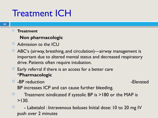
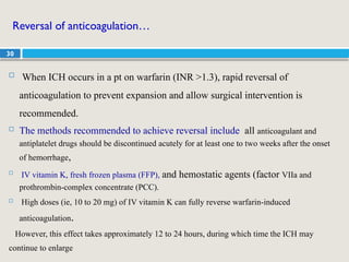
1
SEMINAR PRESENTATION
STROKE
GROUP NAME
1 DURSA SUFIYAN 0184
2 FEYSEL UMER 0187
3 BIRKI M/SANI 0192
4 EMANE ABDI 0080
5 EKRAM BEKRI 0079
6 FAYO MOHAMMED 0074
7 BEYAN AHMED 0223
8 BEYAN TAHA 0022
9 FEYSEL ABDELA 0160
10 GALMESA TEYIB 0063
11DARANE ARARSA 0157
12 HAMZA AHMEDASHIR 0212
2
Outline
 Introduction
 Epidemiology
 Classification
 Etiology & Pathophysiology
 C/presentation &diagnostic test
 Treatments
 References
3
introduction
 A stroke or cerebrovascular accident is defined as:
 Stroke involves abrupt onset of focal neurologic deficit that
lasts at least 24 hours and is presumed to be of vascular
origin.
 Stroke is a medical emergency that occurs when blood supply
to the brain is reduced due to either blockage or leakage of
blood vessel resulting into damage of brain tissue
4
Epidemiology

Around the world there are 12.2 million new stroke per year
 African Americans have stroke rates that are twice of whites
 Stroke incidence increases with age, especially after age 55
years
 Stroke mortality rates are higher in women than men, but risk of
stroke is higher in men of same age
 geographic variability exists, higher mortality termed the “stroke
belt.”
5
Classifications
6
Etiology
 Ischemic Strokes: typically resulting from thrombosis or
embolism,
 Hemorrhagic stroke: resulting from vascular rupture
 Stroke symptoms lasting < 1 h are termed a transient ischemic
attack (TIA).
 Since it involve the arteries of the brain Strokes damage brain
tissue
7
Risk Factors
• Non-modifiable risk factors
– increased age
– male gender
– race (African American,
Asian, Hispanic)
– family history of stroke
– low birth weight
• Major modifiable risk factors
– hypertension
– cardiac disease
(especially atrial
fibrillation)
– diabetes mellitus
– dyslipidemia
– cigarette smoking
8
Pathophysiology of ischemic stroke
 Ischemic stroke (87% of all strokes) results from occlusion of a
cerebral artery that strokes are due reduces cerebral blood flow.
 Ischemic either local thrombus formation or emboli from a distant site.
 Carotid atherosclerotic plaques rupture, resulting in collagen exposure,
platelet aggregation, and thrombus formation. The clot may cause
local occlusion or and travel distally, occluding a cerebral vessel.
 Atherosclerosis of large intracranial or extracranial arteries or small
artery disease can result in ischemic stroke.
9
Patho…….
 Emboli can arise from the heart in patients with atrial fibrillation,
valvular heart disease, or other prothrombotic heart problems
 In cardiogenic embolism, stasis of blood flow in the atria or
ventricles leads to formation of local clots that can dislodge and
travel through the aorta to the cerebral circulation
 Thrombus formation and embolism result in arterial occlusion,
decreasing cerebral blood flow and causing ischemia and ultimately
infarction distal to the occlusion.
10
Phathophysiology of hemorrhagic stroke
 Hemorrhagic strokes (13% of strokes) Is due to bleeding into the
brain by rupture of a blood tissue
 Brain tissue swelling and injury is a result of inflammation
 lead to increase (ICP) and herniation blood vessel repture by mass
effect
 SAH may result from trauma or rupture of an intracranial aneurysm or
arteriovenous malformation (AVM
 ICH occurs when a ruptured blood vessel within the brain causes a
hematoma
11
Con….
 Blood in the brain parenchyma causes mechanical compression of
vulnerable tissue and subsequent activation of inflammation and
neurotoxins.
 Presence of blood in the brain parenchyma causes damage to the
surrounding tissue through the mechanical effect it produces (mass
effect) and the neurotoxicity of the blood components and their
degradation products
12
Clinical presentation
• The patient may not be able to give a reliable history because of
cognitive or language deficits
• The patient may experience
– weakness on one side of the body
– inability to speak
– Double vision ..posterior circulation movement
– the headache can be severe and accompanied with vomiting
– Falling …dysarthria, altered level of consciousness
13
Diagnostic tests
 CT scan of the head will reveal an area of hyperintensity
(white) identifying that a hemorrhage has occurred.
 CT scan will either be normal or hypo intense (dark) in an
area where an infarction has occurred
 MRI of the head will reveal areas of ischemia earlier and with
better resolution than a CT scan.
 ECG will determine whether the pt has Atrial fibrillation,
which is a major risk factor for stroke
14
Diagnosis
 Laboratory tests
 Laboratory tests for hypercoagulable states should be done
only when the cause of the stroke cannot be determined
 Protein C, protein S, and anti thrombin III
 Antiphospholipid antibodies
15
Treatment of stroke
 Goals of treatment:
 reduce the ongoing neurologic injury and decrease
mortality and long-term disability
 prevent complications secondary to immobility and
neurologic dysfunction
 prevent stroke recurrence
16
Treatment
General Approach
 The initial approach is
 to ensure adequate respiratory and cardiac support standpoint
 to determine quickly whether the lesion is ischemic or
hemorrhagic based on a CT scan.
 Ischemic stroke patients presenting within hours of symptom
onset should be evaluated for reperfusion therapy
17
• Blood Pressure Control
– Elevated BP should remain untreated in the acute period
(first 7 days) after ischemic stroke;
• because of the risk of decreasing cerebral blood flow and
worsening symptoms.
– The pressure should be lowered if it exceeds 220/120 mm Hg or
there is evidence of aortic dissection, acute myocardial infarction,
pulmonary edema, or hypertensive encephalopathy.
18
If BP is treated, short-acting parenteral agents, such as labetalol and
nicardipine, or nitroprusside, are favored
19
Non pharmacologic treatment
 Ischemic Stroke
 Surgical interventions in the acute ischemic stroke patient are
limited.
 But in certain cases of ischemic cerebral edema owing to a large
infarction, craniotomy to release some of the rising pressure has
been tried.
 Haemorrhagic Stroke
 As in patients with SAH owing to a ruptured intracranial
aneurysm, in arteriovenous malformations (AVMs)
 surgical intervention to either clip or ablate the offending
vascular abnormality substantially reduces mortality owing to
rebleeding
20
Pharmacological treatment
 Acute treatment
a) IV tissue plasminogen activator (tPA)_ alteplase -- 0.9mg/kg/hr
with 10% given as initial bolus over 1 minute
• Within 4.5 hours of onset
• avoidance of antithrombotic therapy for 24 hours,
b) Aspirin 160–325 mg daily within 48 hours of onset.
 never be given within 24 hours of the administration of tPA
 shown to reduce long-term death and disability
21
Pharmacological treatment
 Inclusion criteria for alteplase use in schemic stroke
 age 18 years or older
 diagnosis of ischemic stroke cause neurologic deficit
 time onset well established less than 4.5 hrs
 Exclusion criteria for alteplase use in ischemic stroke
 High clinical suspension SAH even with normal CT
 Evidence ICH non contrast head CT
 Active internal bleeding
 patients has received heparin within 48 hrs &elevated APTT
 major &minor surgery within 14days
 lumbar puncture with in 7 days
 recently acute myocardial infraction
 SBP >185 mm hg or DBP mm hg at time of treatment
22
Pharmacological treatment
 Secondary prevention of ischemic stroke : Use antiplatelet
therapy in non cardio embolic stroke.
1st
line agents
 ASA 50-325mg daily
 ASA 25mg +ER dipyridamole 200mg twice daily
Alternatives ; clopidogrel 75 mg daily
 For patients with atrial fibrillation and a presumed cardiac source of
embolism, oral anticoagulation with a vitamin K antagonist
(warfarin), apixaban , dabigatran, or rivaroxaban is recommended.
23
Pharmacological treatment
Statins
 The statins reduce the risk of stroke in patients with CAD and
elevated plasma lipids.
 high-intensity statin therapy(e.g. atorvastatin 80 mg daily) to achieve
a reduction of LDL of at least 50% for secondary stroke prevention
 Heparin for Prophylaxis of DVT recommended for the prevention of
DVT in hospitalized patients with decreased mobility
 UFH 5000 IU daily SC
24
Blood pressure lowering
 Elevated blood pressure is very common in ischemic stroke patients, and
treatment of hypertension in these patients is associated with a decreased risk of
stroke recurrence.
 AHA/ASA guidelines recommend an ACE inhibitor and a diuretic for the
reduction of BP in pts with stroke or TIA.
 Early blood pressure lowering can worsen symptoms, however; therefore,
recommendations are limited to pts outside of the acute stroke period (first 7 days).
 Despite this, there is support for carefully restarting antihypertensive therapy after
24 hrs in the acute stroke pts
25
Hemorrhagic stroke
26
●Hemorrhagic stroke
 Two major categories of hemorrhagic stroke:
o ICH—bleeding into brain parenchyma
o SAH—bleeding into the CSF; outside brain parenchyma
Intracerebral Hemorrhage (ICH)
27
 Intracerebral Hemorrhage is bleeding in to the brain parenchyma. It is
associated with a high mortality rate (50% at 30 days)
 Clinical features
Abrupt onset of a focal neurologic deficit that worsens steadily over 30 to
90 minutes.
- Altered level of consciousness, stupor, or coma
-Headache, vomiting
Pupillary findings in ICH and corresponding level of involvement
o Pinpoint pupils—pons
o Poorly reactive pupils—thalamus
o Dilated pupils—putamen
 Complications
 -Raised intracranial pressure
 - Seizures
 - Rebleeding
Treatment ICH
28
 Treatment
Non pharmacologic
 Admission to the ICU
 ABC‘s (airway, breathing, and circulation)—airway management is
important due to altered mental status and decreased respiratory
drive. Patients often require intubation.
 Early referral if there is an access for a better care
*Pharmacologic
 -BP reduction -Elevated
BP increases ICP and can cause further bleeding.
 Treatment isindicated if systolic BP is >180 or the MAP is
>130.
 - Labetalol : Intravenous boluses Initial dose: 10 to 20 mg IV
push over 2 minutes
29
 Until target is achieved give double the initial dose in
15 minutes (maximum dose: 80 mg/dose)
- A total maximum dose: 300 mg OR
□Labetalol: Continuous IV infusion
- Initial: 0.5 to 2 mg/minute.
-Titrate the dose to a maximum of 10 mg/minute.
- A total maximum dose: 300mg
30
Reversal of anticoagulation…
 When ICH occurs in a pt on warfarin (INR >1.3), rapid reversal of
anticoagulation to prevent expansion and allow surgical intervention is
recommended.
 The methods recommended to achieve reversal include all anticoagulant and
antiplatelet drugs should be discontinued acutely for at least one to two weeks after the onset
of hemorrhage,

IV vitamin K, fresh frozen plasma (FFP), and hemostatic agents (factor VIIa and
prothrombin-complex concentrate (PCC).
 High doses (ie, 10 to 20 mg) of IV vitamin K can fully reverse warfarin-induced
anticoagulation.
However, this effect takes approximately 12 to 24 hours, during which time the ICH may
continue to enlarge
31
Intracranial pressure control
 Increased ICP due to ICH can result from the hematoma itself and
from surrounding edema, and may contribute to brain injury and
neurologic deterioration.
 Approach to the management of elevated ICP
 Elevate the head of the bed to 30 degrees, once hypovolemia is
excluded.
 Analgesia and sedation, particularly in unstable, intubated pts
 Glucocorticoids should not generally be used to lower the lCP in
patients with ICH.
A randomized trial found that dexamethasone did not improve outcome but did
increase complication rates, primarily infection
32
 Aggressive measures — Monitoring and treatment of ICP
should be considered for pts with GCS <8
 IV mannitol is the treatment of choice to lower increased ICP
-Administered as an initial bolus of 1 g/kg, followed by
infusions of 0.25 to 0.5 g/kg every 6 to 8 hours. Administer over
30 to 60 minutes. It should not be given over a long period (as
continuous infusion)
 Goal of therapy is to achieve plasma hyperosmolality (300 to
310 mosmol/kg) while maintaining an adequate plasma volume
 Barbiturate anesthesia can be used if mannitol fails to
lower ICP to an acceptable range.
33
Intracranial pressure control
 Severe elevations in BP may worsen ICH by representing a
continued force for bleeding
 If SBP is >200 mm Hg or MAP is >150 mm Hg, then consider
aggressive reduction of BP with continuous IV meds monitor BP q 5
minutes
 If SBP is >180 mm Hg or MAP is >130 mm Hg and there is
suspicion of increased ICP, reducing BP using intermittent or
continuous IV meds to keep CPP >60 to 80 mm Hg
 If SBP is >180 mm Hg or MAP is >130 mm Hg and no suspicion of
elevated ICP, then consider a modest reduction of BP(eg, MAP of
110 mm Hg or target BP of 160/90 mm Hg) using intermittent or
continuous IV medications to control BP, and clinically re examine
the patient every 15 minutes.
34
Resumption Antiplatelets
 The timing of antiplatelet use after ICH is largely empiric.
 There is risk of rebleeding and hematoma expansion in the first several
hours. At 10 days, rebleeding is unlikely.
 Guidelines state that antiplatelets should be discontinued for at least one
to two weeks.
 Some experts have argued that aspirin can be used safely as soon as 48
hours after ICH in those who require prophylaxis for venous
thromboembolism.
 If aspirin is used after ICH, a lower dose (30 to 160 mg daily) is both
effective and safer than higher doses
Subarachnoid Hemorrhage
35
Treatment
Non pharmacologic
 -Bed rest in a quiet and dark room.
- Early referral if neuro-radio-surgical intervention is available at the next
level hospital
Pharmacologic
-Stool softeners to avoid straining (increases ICP and risk of re-rupture).
-Analgesia for headache (acetaminophen 1 gm Po QID).
-IV fluids for hydration.
-Control of HTN—lower the BP gradually because the elevation in BP may
be a compensation for the decrease in cerebral perfusion pressure
(secondary to increased ICP or cerebral arterial narrowing).
-Calcium channel blocker (Nimodipine) for vasospasm lowers the incidence
of cerebral infarction by one-third.
- Prophylactic anticonvulsant
36
Evaluation Of Therapeutic Outcomes
Patients with acute stroke should be monitored intensely
for the development of neurologic worsening,
 complications, and adverse effects from treatments
37
Reference
1. Dipiro pharmacotherapy a pathophysiology
seven edition
2. Standard treatment guidelines 4th
Edition,2021
38
Thank you!

Stroke seminar presentation Pharmacotherapy

  • 1.
    1 SEMINAR PRESENTATION STROKE GROUP NAME 1DURSA SUFIYAN 0184 2 FEYSEL UMER 0187 3 BIRKI M/SANI 0192 4 EMANE ABDI 0080 5 EKRAM BEKRI 0079 6 FAYO MOHAMMED 0074 7 BEYAN AHMED 0223 8 BEYAN TAHA 0022 9 FEYSEL ABDELA 0160 10 GALMESA TEYIB 0063 11DARANE ARARSA 0157 12 HAMZA AHMEDASHIR 0212
  • 2.
    2 Outline  Introduction  Epidemiology Classification  Etiology & Pathophysiology  C/presentation &diagnostic test  Treatments  References
  • 3.
    3 introduction  A strokeor cerebrovascular accident is defined as:  Stroke involves abrupt onset of focal neurologic deficit that lasts at least 24 hours and is presumed to be of vascular origin.  Stroke is a medical emergency that occurs when blood supply to the brain is reduced due to either blockage or leakage of blood vessel resulting into damage of brain tissue
  • 4.
    4 Epidemiology  Around the worldthere are 12.2 million new stroke per year  African Americans have stroke rates that are twice of whites  Stroke incidence increases with age, especially after age 55 years  Stroke mortality rates are higher in women than men, but risk of stroke is higher in men of same age  geographic variability exists, higher mortality termed the “stroke belt.”
  • 5.
  • 6.
    6 Etiology  Ischemic Strokes:typically resulting from thrombosis or embolism,  Hemorrhagic stroke: resulting from vascular rupture  Stroke symptoms lasting < 1 h are termed a transient ischemic attack (TIA).  Since it involve the arteries of the brain Strokes damage brain tissue
  • 7.
    7 Risk Factors • Non-modifiablerisk factors – increased age – male gender – race (African American, Asian, Hispanic) – family history of stroke – low birth weight • Major modifiable risk factors – hypertension – cardiac disease (especially atrial fibrillation) – diabetes mellitus – dyslipidemia – cigarette smoking
  • 8.
    8 Pathophysiology of ischemicstroke  Ischemic stroke (87% of all strokes) results from occlusion of a cerebral artery that strokes are due reduces cerebral blood flow.  Ischemic either local thrombus formation or emboli from a distant site.  Carotid atherosclerotic plaques rupture, resulting in collagen exposure, platelet aggregation, and thrombus formation. The clot may cause local occlusion or and travel distally, occluding a cerebral vessel.  Atherosclerosis of large intracranial or extracranial arteries or small artery disease can result in ischemic stroke.
  • 9.
    9 Patho…….  Emboli canarise from the heart in patients with atrial fibrillation, valvular heart disease, or other prothrombotic heart problems  In cardiogenic embolism, stasis of blood flow in the atria or ventricles leads to formation of local clots that can dislodge and travel through the aorta to the cerebral circulation  Thrombus formation and embolism result in arterial occlusion, decreasing cerebral blood flow and causing ischemia and ultimately infarction distal to the occlusion.
  • 10.
    10 Phathophysiology of hemorrhagicstroke  Hemorrhagic strokes (13% of strokes) Is due to bleeding into the brain by rupture of a blood tissue  Brain tissue swelling and injury is a result of inflammation  lead to increase (ICP) and herniation blood vessel repture by mass effect  SAH may result from trauma or rupture of an intracranial aneurysm or arteriovenous malformation (AVM  ICH occurs when a ruptured blood vessel within the brain causes a hematoma
  • 11.
    11 Con….  Blood inthe brain parenchyma causes mechanical compression of vulnerable tissue and subsequent activation of inflammation and neurotoxins.  Presence of blood in the brain parenchyma causes damage to the surrounding tissue through the mechanical effect it produces (mass effect) and the neurotoxicity of the blood components and their degradation products
  • 12.
    12 Clinical presentation • Thepatient may not be able to give a reliable history because of cognitive or language deficits • The patient may experience – weakness on one side of the body – inability to speak – Double vision ..posterior circulation movement – the headache can be severe and accompanied with vomiting – Falling …dysarthria, altered level of consciousness
  • 13.
    13 Diagnostic tests  CTscan of the head will reveal an area of hyperintensity (white) identifying that a hemorrhage has occurred.  CT scan will either be normal or hypo intense (dark) in an area where an infarction has occurred  MRI of the head will reveal areas of ischemia earlier and with better resolution than a CT scan.  ECG will determine whether the pt has Atrial fibrillation, which is a major risk factor for stroke
  • 14.
    14 Diagnosis  Laboratory tests Laboratory tests for hypercoagulable states should be done only when the cause of the stroke cannot be determined  Protein C, protein S, and anti thrombin III  Antiphospholipid antibodies
  • 15.
    15 Treatment of stroke Goals of treatment:  reduce the ongoing neurologic injury and decrease mortality and long-term disability  prevent complications secondary to immobility and neurologic dysfunction  prevent stroke recurrence
  • 16.
    16 Treatment General Approach  Theinitial approach is  to ensure adequate respiratory and cardiac support standpoint  to determine quickly whether the lesion is ischemic or hemorrhagic based on a CT scan.  Ischemic stroke patients presenting within hours of symptom onset should be evaluated for reperfusion therapy
  • 17.
    17 • Blood PressureControl – Elevated BP should remain untreated in the acute period (first 7 days) after ischemic stroke; • because of the risk of decreasing cerebral blood flow and worsening symptoms. – The pressure should be lowered if it exceeds 220/120 mm Hg or there is evidence of aortic dissection, acute myocardial infarction, pulmonary edema, or hypertensive encephalopathy.
  • 18.
    18 If BP istreated, short-acting parenteral agents, such as labetalol and nicardipine, or nitroprusside, are favored
  • 19.
    19 Non pharmacologic treatment Ischemic Stroke  Surgical interventions in the acute ischemic stroke patient are limited.  But in certain cases of ischemic cerebral edema owing to a large infarction, craniotomy to release some of the rising pressure has been tried.  Haemorrhagic Stroke  As in patients with SAH owing to a ruptured intracranial aneurysm, in arteriovenous malformations (AVMs)  surgical intervention to either clip or ablate the offending vascular abnormality substantially reduces mortality owing to rebleeding
  • 20.
    20 Pharmacological treatment  Acutetreatment a) IV tissue plasminogen activator (tPA)_ alteplase -- 0.9mg/kg/hr with 10% given as initial bolus over 1 minute • Within 4.5 hours of onset • avoidance of antithrombotic therapy for 24 hours, b) Aspirin 160–325 mg daily within 48 hours of onset.  never be given within 24 hours of the administration of tPA  shown to reduce long-term death and disability
  • 21.
    21 Pharmacological treatment  Inclusioncriteria for alteplase use in schemic stroke  age 18 years or older  diagnosis of ischemic stroke cause neurologic deficit  time onset well established less than 4.5 hrs  Exclusion criteria for alteplase use in ischemic stroke  High clinical suspension SAH even with normal CT  Evidence ICH non contrast head CT  Active internal bleeding  patients has received heparin within 48 hrs &elevated APTT  major &minor surgery within 14days  lumbar puncture with in 7 days  recently acute myocardial infraction  SBP >185 mm hg or DBP mm hg at time of treatment
  • 22.
    22 Pharmacological treatment  Secondaryprevention of ischemic stroke : Use antiplatelet therapy in non cardio embolic stroke. 1st line agents  ASA 50-325mg daily  ASA 25mg +ER dipyridamole 200mg twice daily Alternatives ; clopidogrel 75 mg daily  For patients with atrial fibrillation and a presumed cardiac source of embolism, oral anticoagulation with a vitamin K antagonist (warfarin), apixaban , dabigatran, or rivaroxaban is recommended.
  • 23.
    23 Pharmacological treatment Statins  Thestatins reduce the risk of stroke in patients with CAD and elevated plasma lipids.  high-intensity statin therapy(e.g. atorvastatin 80 mg daily) to achieve a reduction of LDL of at least 50% for secondary stroke prevention  Heparin for Prophylaxis of DVT recommended for the prevention of DVT in hospitalized patients with decreased mobility  UFH 5000 IU daily SC
  • 24.
    24 Blood pressure lowering Elevated blood pressure is very common in ischemic stroke patients, and treatment of hypertension in these patients is associated with a decreased risk of stroke recurrence.  AHA/ASA guidelines recommend an ACE inhibitor and a diuretic for the reduction of BP in pts with stroke or TIA.  Early blood pressure lowering can worsen symptoms, however; therefore, recommendations are limited to pts outside of the acute stroke period (first 7 days).  Despite this, there is support for carefully restarting antihypertensive therapy after 24 hrs in the acute stroke pts
  • 25.
  • 26.
    Hemorrhagic stroke 26 ●Hemorrhagic stroke Two major categories of hemorrhagic stroke: o ICH—bleeding into brain parenchyma o SAH—bleeding into the CSF; outside brain parenchyma
  • 27.
    Intracerebral Hemorrhage (ICH) 27 Intracerebral Hemorrhage is bleeding in to the brain parenchyma. It is associated with a high mortality rate (50% at 30 days)  Clinical features Abrupt onset of a focal neurologic deficit that worsens steadily over 30 to 90 minutes. - Altered level of consciousness, stupor, or coma -Headache, vomiting Pupillary findings in ICH and corresponding level of involvement o Pinpoint pupils—pons o Poorly reactive pupils—thalamus o Dilated pupils—putamen  Complications  -Raised intracranial pressure  - Seizures  - Rebleeding
  • 28.
    Treatment ICH 28  Treatment Nonpharmacologic  Admission to the ICU  ABC‘s (airway, breathing, and circulation)—airway management is important due to altered mental status and decreased respiratory drive. Patients often require intubation.  Early referral if there is an access for a better care *Pharmacologic  -BP reduction -Elevated BP increases ICP and can cause further bleeding.  Treatment isindicated if systolic BP is >180 or the MAP is >130.  - Labetalol : Intravenous boluses Initial dose: 10 to 20 mg IV push over 2 minutes
  • 29.
    29  Until targetis achieved give double the initial dose in 15 minutes (maximum dose: 80 mg/dose) - A total maximum dose: 300 mg OR □Labetalol: Continuous IV infusion - Initial: 0.5 to 2 mg/minute. -Titrate the dose to a maximum of 10 mg/minute. - A total maximum dose: 300mg
  • 30.
    30 Reversal of anticoagulation… When ICH occurs in a pt on warfarin (INR >1.3), rapid reversal of anticoagulation to prevent expansion and allow surgical intervention is recommended.  The methods recommended to achieve reversal include all anticoagulant and antiplatelet drugs should be discontinued acutely for at least one to two weeks after the onset of hemorrhage,  IV vitamin K, fresh frozen plasma (FFP), and hemostatic agents (factor VIIa and prothrombin-complex concentrate (PCC).  High doses (ie, 10 to 20 mg) of IV vitamin K can fully reverse warfarin-induced anticoagulation. However, this effect takes approximately 12 to 24 hours, during which time the ICH may continue to enlarge
  • 31.
    31 Intracranial pressure control Increased ICP due to ICH can result from the hematoma itself and from surrounding edema, and may contribute to brain injury and neurologic deterioration.  Approach to the management of elevated ICP  Elevate the head of the bed to 30 degrees, once hypovolemia is excluded.  Analgesia and sedation, particularly in unstable, intubated pts  Glucocorticoids should not generally be used to lower the lCP in patients with ICH. A randomized trial found that dexamethasone did not improve outcome but did increase complication rates, primarily infection
  • 32.
    32  Aggressive measures— Monitoring and treatment of ICP should be considered for pts with GCS <8  IV mannitol is the treatment of choice to lower increased ICP -Administered as an initial bolus of 1 g/kg, followed by infusions of 0.25 to 0.5 g/kg every 6 to 8 hours. Administer over 30 to 60 minutes. It should not be given over a long period (as continuous infusion)  Goal of therapy is to achieve plasma hyperosmolality (300 to 310 mosmol/kg) while maintaining an adequate plasma volume  Barbiturate anesthesia can be used if mannitol fails to lower ICP to an acceptable range.
  • 33.
    33 Intracranial pressure control Severe elevations in BP may worsen ICH by representing a continued force for bleeding  If SBP is >200 mm Hg or MAP is >150 mm Hg, then consider aggressive reduction of BP with continuous IV meds monitor BP q 5 minutes  If SBP is >180 mm Hg or MAP is >130 mm Hg and there is suspicion of increased ICP, reducing BP using intermittent or continuous IV meds to keep CPP >60 to 80 mm Hg  If SBP is >180 mm Hg or MAP is >130 mm Hg and no suspicion of elevated ICP, then consider a modest reduction of BP(eg, MAP of 110 mm Hg or target BP of 160/90 mm Hg) using intermittent or continuous IV medications to control BP, and clinically re examine the patient every 15 minutes.
  • 34.
    34 Resumption Antiplatelets  Thetiming of antiplatelet use after ICH is largely empiric.  There is risk of rebleeding and hematoma expansion in the first several hours. At 10 days, rebleeding is unlikely.  Guidelines state that antiplatelets should be discontinued for at least one to two weeks.  Some experts have argued that aspirin can be used safely as soon as 48 hours after ICH in those who require prophylaxis for venous thromboembolism.  If aspirin is used after ICH, a lower dose (30 to 160 mg daily) is both effective and safer than higher doses
  • 35.
    Subarachnoid Hemorrhage 35 Treatment Non pharmacologic -Bed rest in a quiet and dark room. - Early referral if neuro-radio-surgical intervention is available at the next level hospital Pharmacologic -Stool softeners to avoid straining (increases ICP and risk of re-rupture). -Analgesia for headache (acetaminophen 1 gm Po QID). -IV fluids for hydration. -Control of HTN—lower the BP gradually because the elevation in BP may be a compensation for the decrease in cerebral perfusion pressure (secondary to increased ICP or cerebral arterial narrowing). -Calcium channel blocker (Nimodipine) for vasospasm lowers the incidence of cerebral infarction by one-third. - Prophylactic anticonvulsant
  • 36.
    36 Evaluation Of TherapeuticOutcomes Patients with acute stroke should be monitored intensely for the development of neurologic worsening,  complications, and adverse effects from treatments
  • 37.
    37 Reference 1. Dipiro pharmacotherapya pathophysiology seven edition 2. Standard treatment guidelines 4th Edition,2021
  • 38.