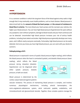
ITRA, Jamnagar​
Cardiovascular System
disorders
Prepared By:
Vaidehi Vadhvana, Ph.D. Scholar
Pharmacology Department
Vaidehi Vadhvana
The cardiovascular system, also known as the circulatory system, is a complex network
of organs, vessels, and cells responsible for transporting oxygen, nutrients, hormones, and
waste products throughout the body.​
Heart Disease: Types, Causes, and Symptoms
There are many types of heart disease, and each one has its own symptoms and treatment. For
some, lifestyle changes and medicine can make a huge difference in improving your health. For
others, you may need surgery to make your ticker work well again. Cardiovascular diseases are
conditions that affect the structures or function of your heart, such as:
CAD and vascular disease are due to hardening of
the arteries (atherosclerosis). CAD happens when
the arteries in the heart are narrowed or blocked.
It’s the most common kind of heart disease and
causes most heart attacks as well as angina (chest
pain). Vascular disease is a problem in other blood
vessels which reduce blood flow and affect the
function of your heart.
Heart Arrhythmias Cause the heart to beat too slowly, too quickly, or in a disorganized fashion.
Millions of Canadians experience heart rhythm disorders that disrupt blood flow. There are
many types of arrhythmias – some have no symptoms or warning signs; others can be sudden
and fatal.
Structural heart disease refers to abnormalities of the heart’s structure – including its valves,
walls, muscles, or blood vessels near the heart. It can be present at birth (congenital) or
acquired after birth through infection, wear and tear, or other factors.
1
Vaidehi Vadhvana
Heart Failure is a serious condition that develops after the heart becomes damaged or
weakened. The two most common causes of heart failure are heart attack and high BP. There is
no cure, but early diagnosis, lifestyle
changes and medication can help people
lead an active life, stay out of the hospital
and live longer.
Heart Valve Disease refers to any of
several conditions that prevent one or
more of the valves (mitral, tricuspid, aortic
and pulmonic valves) in the heart from
functioning adequately to assure proper circulation. The valves make sure blood flows in only
one direction through the heart.
Pericardial Disease Any disease (mostly inflammation) of the pericardium, the sac that
surrounds your heart. Cause either due to virus, RA
or injury. Pericarditis often follows open heart
surgery.
Cardiomyopathy (Heart Muscle Disease) is a
disease of heart muscle or myocardium. It gets
stretched, thickened, or stiff. So, the heart may get
too weak to pump well. Cause due to certain drugs
or toxins, viral infections, and chemotherapy. No
exact cause.
Congenital Heart Disease develops in babies that’s
still in the womb. Symptoms may persists after birth
or in adult age. Septal abnormalities i.e. holes in the wall that separates the left and right sides
of the heart. So, the procedure to patch the hole is done. Another type of abnormality is called
2
Vaidehi Vadhvana
pulmonary stenosis. A narrow valve causes a decrease in the flow of blood to your lungs. A
procedure or surgery can open or replace the valve.
Heart attack or Myocardial Infarction happens when a part of the heart muscle doesn’t get
enough blood. The more time that passes without treatment to restore blood flow, the greater
the damage to the heart muscle.
CAD is the main cause of heart attack. A less common cause is a severe spasm, or sudden
contraction, of a coronary artery that can stop blood flow to the heart muscle, Heart muscle
disease (cardiomyopathy), Peripheral vascular disease, Rheumatic heart disease, Stroke,
Vascular disease (blood vessel disease), Angina pectoris, Hypertensive heart disease, Aorta
disease and Marfan syndrome, Deep vein thrombosis and pulmonary embolism.
Causes;
Heart disease can be caused by:
1.​ Medical conditions;
o High BP
o High Cholesterol levels
o Diabetes
o High blood pressure during pregnancy
o Sleep apnea
· Lifestyle risk factors;
o Unhealthy diet
o Not enough physical activity
o Unhealthy weight
o Smoking
o Too much alcohol
o Stress
o Birth Control or hormone replacement therapy
3
Vaidehi Vadhvana
There are many risk factors associated with coronary heart disease and stroke. Some risk
factors, such as family history, cannot be modified, while other risk factors, like high blood
pressure, can be modified with treatment.
1.​ Non modifiable” risk factors; “Cannot be changed”. These are:
1.​ Age – risk increases as you get older
2.​ Gender - before the age of 60 men are at greater risk than women
3.​ Family history – your risk may increase if close blood relatives experienced early
heart disease.
2. Modifiable risk factors: Risk factors that can change:
1.​ Raised or altered levels of blood cholesterol
2.​ Raised triglycerides with low HDL-cholesterol
3.​ High blood pressure
4.​ Diabetes
5.​ Smoking
6.​ Being overweight/obesity
7.​ Being inactive
8.​ Excessive alcohol
9.​ Excessive stress
List of few Cardiac Disorders:
1.​ Hypertension
2.​ Angina pectoris
3.​ Cardiac Arrhythmia
4.​ Cardiac failure
4
Vaidehi Vadhvana
HYPERTENSION
It is a common condition in which the long-term force of the blood against artery walls is high
enough that it may eventually cause health problems, such as heart disease. Blood pressure is
determined both by the amount of blood the heart pumps and the amount of resistance to
blood flow in arteries. The more blood your heart pumps and the narrower your arteries, the
higher your blood pressure. You can have high blood pressure (hypertension) for years without
any symptoms. Even without symptoms, damage to blood vessels and your heart continues and
can be detected. Uncontrolled high blood pressure increases your risk of serious health
problems, including heart attack and stroke. High blood pressure generally develops over many
years, and it affects nearly everyone eventually. Fortunately, high blood pressure can be easily
detected. And once you know you have high blood pressure, you can work with your doctor to
control it.
Pathophysiology of HT:
Blood pressure is expressed in terms of systolic blood pressure (higher reading), which reflects
the blood pressure when the heart is contracted (systole), and diastolic blood pressure (lower
reading), which reflects the blood
pressure during relaxation (diastole).
Hypertension can be diagnosed when
either systolic pressure, diastolic
pressure, or both are raised.
Blood pressure is determined by the
cardiac output balanced against systemic
vascular resistance. The process of maintaining blood pressure is complex, and involves
numerous physiological mechanisms, including arterial baroreceptors, the
renin–angiotensin–aldosterone system, atrial natriuretic peptide, endothelins, and
mineralocorticoid and glucocorticoid steroids. Together, these complex systems manage the
5
Vaidehi Vadhvana
degree of vasodilation or vasoconstriction within the systemic circulation, and the retention or
excretion of sodium and water, to maintain an adequate circulating blood volume. Dysfunction
in any of these processes can lead to the development of hypertension. This may be through
increased cardiac output, increased systemic vascular resistance, or both.
Blood vessels become less elastic and more rigid as patients age, which reduces vasodilatation
and increases systemic vascular resistance, leading to a higher systolic blood pressure (often
with a normal diastolic pressure). In contrast, hypertension in younger patients tends to be
associated with increased cardiac output, which can be caused by environmental or genetic
factors.
Symptoms:
Most people with high B.P have no signs or symptoms, even if B.P readings reach dangerously
high levels. A few people with high B.P may have headaches, shortness of breath or nosebleeds,
6
Vaidehi Vadhvana
but these signs and symptoms aren't specific and usually don't occur until high B.P has reached
a severe or life-threatening stage.
Causes:
1.​ Primary (essential) hypertension:
For most adults, there's no identifiable cause of high B.P. It tends to develop gradually over
many years.
2.​ Secondary hypertension:
Some people have high B.P caused by an underlying condition. It tends to appear suddenly and
cause higher B.P than does primary hypertension. Various conditions and medications can lead
to secondary hypertension, including:
1. ​ Kidney problems
2. ​ Adrenal gland tumors
3. ​ Thyroid problems
4. ​ Certain defects in blood vessels you're born with (congenital)
5. ​ Certain medications, such as birth control pills, cold remedies, decongestants,
over-the-counter pain relievers and some prescription drugs.
6. ​ Illegal drugs, such as cocaine and amphetamines
7. ​ Alcohol abuse or chronic alcohol use.
Diagnosis:
●​ To measure the B.P, your doctor or a specialist will usually place an inflatable arm cuff
around the arm and measure the B.P using a pressure-measuring gauge.
●​ A B.P reading, given in millimeters of mercury (mm Hg), has two numbers.
7
Vaidehi Vadhvana
●​ The first, or upper, number measures the pressure in the arteries when the heart beats
(systolic pressure). The second, or lower, number measures the pressure in the arteries
between beats (diastolic pressure).
Resistant hypertension:
When B.P is difficult to control. If your blood pressure remains stubbornly high despite taking
at least three different types of high blood pressure drugs, one of which usually should be a
diuretic, you may have resistant hypertension. People who have controlled high blood pressure
but are taking four different types of medications at the same time to achieve that control also
are considered to have resistant hypertension.
Blood pressure measurements fall into four general categories:
1.​ Normal blood pressure. Your blood pressure is normal if it's below 120/80 mm Hg.
2.​ Elevated blood pressure. Elevated blood pressure is a systolic pressure ranging from
120 to 129 mm Hg and a diastolic pressure below 80 mm Hg. It tends to get worse over
time unless steps are taken to control B.P.
3.​ Stage 1 hypertension: It is a systolic pressure ranging from 130 to 139 mm Hg or a
diastolic pressure ranging from 80 to 89 mm Hg.
4.​ Stage 2 hypertension: More severe hypertension, stage 2 hypertension is a systolic
pressure of 140 mm Hg or higher or a diastolic pressure of 90 mm Hg or higher.
Both numbers in a blood pressure reading are important. But after age 60, the systolic reading
is even more significant. Isolated systolic hypertension is a condition in which the diastolic
pressure is normal (less than 90 mm Hg) but systolic pressure is high (greater than 140 mm Hg).
This is a common type of high blood pressure among people older than 60.
8
Vaidehi Vadhvana
Complications:
The excessive pressure on your artery walls caused by high blood pressure can damage your
blood vessels, as well as organs in your body. The higher your blood pressure and the longer it
goes uncontrolled, the greater the damage. Uncontrolled high blood pressure can lead to:
1.​ Heart attack or stroke. High blood pressure can cause hardening and thickening of the
arteries (atherosclerosis), which can lead to a heart attack, stroke or other
complications.
2.​ Aneurysm. Increased blood pressure can cause blood vessels to weaken and bulge,
forming an aneurysm. If an aneurysm ruptures, it can be life-threatening.
3.​ Heart failure. To pump blood against the higher pressure in the vessels, heart muscle
thickens. Eventually, the thickened muscle may have a hard time pumping enough
blood to meet the body's needs, which can lead to heart failure.
4.​ Weakened and narrowed blood vessels in your kidneys. This can prevent these
organs from functioning normally.
5.​ Thickened, narrowed or torn blood vessels in the eyes. This can result in vision loss.
6.​ Metabolic syndrome. This syndrome is a cluster of disorders of the body's metabolism,
including increased waist circumference; high triglycerides; low high-density lipoprotein
(HDL) cholesterol, the "good" cholesterol; high blood pressure; and high insulin levels.
These conditions make it more likely to develop diabetes, heart disease and stroke.
7.​ Trouble with memory or understanding. Uncontrolled high blood pressure may also
affect your ability to think, remember and learn. Trouble with memory or understanding
concepts is more common in people with high B.P.
9
Vaidehi Vadhvana
Angina pectoris
Angina Pectoris is a medical condition characterized by chest pain or discomfort caused by
reduced blood flow to the heart muscle. It is typically
triggered by physical exertion or stress and is often described
as a tightness, squeezing, or burning sensation in the chest.
In more severe cases, angina can be a sign of underlying
heart disease and may require medical intervention. This
document will explore the causes, symptoms, diagnosis, and
treatment options for angina pectoris.​
Pathophysiology;
Angina, also known as angina pectoris, is chest pain or pressure, usually due to not enough
blood flow to the heart muscle. Angina results when there is an imbalance between the heart's
oxygen demand and supply. This imbalance can result from an increase in demand (e.g., during
exercise) without a proportional increase in supply (e.g., due to obstruction or atherosclerosis
of the coronary arteries). However, the pathophysiology of angina in females varies significantly
as compared to males. Non-obstructive coronary disease is more common in females. It is the
reduction of coronary flow due to transient platelet aggregation on apparently normal
endothelium, Coronary artery spasms, or Coronary thrombosis.
Classification:
1.Stable angina:
Also known as 'effort angina', this refers to the classic type of angina related to myocardial
ischemia. A typical presentation of stable angina is that of chest discomfort and associated
symptoms precipitated by some activity (running, walking, etc.) with minimal or non-existent
10
Vaidehi Vadhvana
symptoms at rest or after administration of sublingual nitroglycerin. Symptoms typically abate
several minutes after activity and recur when activity resumes. Other recognized precipitants of
stable angina include cold weather, heavy meals, and emotional stress.
2. Unstable angina
UA, also "crescendo angina"; this is a form of acute coronary syndrome that is defined as angina
pectoris that changes or worsens. It has at least one of these 3 features: It occurs at rest (or with
minimal exertion), usually lasting more than 10 minutes. It is severe and of new onset (i.e.,
Within the prior 4–6 weeks). It occurs with a crescendo pattern (i.e., Distinctly more severe,
prolonged, or frequent than before).
What differentiates stable angina from unstable angina (other than symptoms):
In stable angina, the developing atheroma is protected with a fibrous cap. This cap may rupture
in unstable angina, allowing blood clots to precipitate and further decrease the area of the
coronary vessel's lumen. This explains why, in many cases, unstable angina develops
independently of activity.
Signs and symptoms:
●​ Angina pectoris can be quite painful, but many patients with angina complain of chest
discomfort rather than actual pain.
●​ The discomfort is usually described as;
●​ Pressure, heaviness, tightness, squeezing, burning, or choking sensation.
●​ Apart from chest discomfort, anginal pains may also be experienced in the epigastrium
(upper central abdomen), back, neck area, jaw, or shoulders. This is explained by the
concept of referred pain.
Referred pain: It is due to the fact that the spinal level that receives visceral sensation from the
heart simultaneously receives cutaneous sensation from parts of the skin specified by that
11
Vaidehi Vadhvana
spinal nerve's dermatome, without an ability to discriminate between the two. Typical locations
for referred pain are arms (often inner left arm), shoulders, and neck into the jaw.
Angina is typically precipitated by exertion or emotional stress. It is exacerbated by having a full
stomach and by cold temperatures. Pain may be accompanied by breathlessness, sweating, and
nausea in some cases. In this case, the pulse rate and the blood pressure increase. Chest pain
lasting only a few seconds is normally not angina.
Major risk factors for angina include; Cigarette smoking, Diabetes, High cholesterol, High B.P,
Sedentary lifestyle, and family history of premature heart disease.
CARDIAC ARRHYTHMIA
Cardiac Dysrhythmia or Irregular Heartbeat
Cardiac arrhythmias are disturbances in the normal rhythm of the heart, caused by issues with
the electrical impulses that coordinate heartbeats. These disruptions can lead to the heart
beating too fast, too slow, or irregularly. Understanding the types, causes, symptoms, diagnosis,
and treatment of cardiac arrhythmias is crucial for effective management.
Types of Cardiac Arrhythmias
1.​ Tachycardia:
a.​ Sinus Tachycardia: Elevated
heart rate originating from
the sinoatrial node.
b.​ Atrial Fibrillation (AFib):
Rapid, irregular beating of
the atria.
12
Vaidehi Vadhvana
c.​ Atrial Flutter:Similar to AFib but with more organized and regular electrical
impulses.
d.​ Supraventricular Tachycardia (SVT):Fast heart rate originating above the
ventricles.
e.​ Ventricular Tachycardia (VT): Rapid heart rate originating from the ventricles.
f.​ Ventricular Fibrillation (VF): Chaotic electrical activity in the ventricles, often
life-threatening.
2. Bradycardia:
a.​ Sinus Bradycardia: Slow heart rate due to slow impulses from the sinoatrial node.
b.​ Heart Block: Delay or blockage in the transmission of electrical impulses from the
atria to the ventricles.
3. Premature Beats:
a.​ Premature Atrial Contractions (PACs): Extra beats originating from the atria.
b.​ Premature Ventricular Contractions (PVCs): Extra beats originating from the
ventricles.
Causes and Risk Factors
1.​ Structural Heart Issues:
●​ - Cardiomyopathy
●​ - Coronary artery disease
●​ - Heart failure
●​ - Valve disorders
2.​ Non-Structural Factors:
●​ - Electrolyte imbalances (e.g., potassium, magnesium)
●​ - Medication side effects
●​ - Excessive alcohol or caffeine intake
13
Vaidehi Vadhvana
●​ - Recreational drug use
●​ - Thyroid disorders
●​ - Stress and anxiety
3.​ Genetic Predisposition:
●​ - Familial arrhythmia syndromes (e.g., Long QT Syndrome, Brugada Syndrome)
Symptoms
1.​ Common Symptoms:
●​ - Palpitations
●​ - Dizziness or lightheadedness
●​ - Shortness of breath
●​ - Chest pain
●​ - Fatigue
●​ - Syncope (fainting)
2.​ Severe Symptoms:
●​ - Severe chest pain
●​ - Sudden collapse
●​ - Cardiac arrest
Diagnosis
1.​ Medical History and Physical Examination:
●​ - Comprehensive review of symptoms and medical history
●​ - Physical examination to detect abnormal heart sounds or rhythms
2. Diagnostic Tests:
●​ Electrocardiogram (ECG):Primary tool for diagnosing arrhythmias.
●​ Holter Monitor:Continuous ECG recording for 24-48 hours.
●​ Event Recorder: Similar to Holter monitor but used for longer periods.
14
Vaidehi Vadhvana
●​ Echocardiogram: Ultrasound imaging of the heart.
●​ Electrophysiological Study (EPS): Invasive test to map electrical activity.
●​ Stress Test: Monitoring heart activity during physical exertion.
Treatment
1.​ Lifestyle Modifications:
●​ - Avoiding caffeine, alcohol, and smoking
●​ - Managing stress
●​ - Maintaining a healthy diet and weight
●​ - Regular physical activity
2.​ Medications:
●​ Antiarrhythmic Drugs: Control heart rate and rhythm.
●​ Beta-Blockers: Reduce heart rate and workload.
●​ Calcium Channel Blockers: Control heart rate.
●​ Anticoagulants: Prevent blood clots in atrial fibrillation.
3.​ Non-Pharmacological Interventions:
●​ Cardioversion: Electrical shock to restore normal rhythm.
●​ Ablation Therapy: Destroying small areas of heart tissue causing arrhythmias.
●​ Pacemaker: Device to regulate heart rate.
●​ Implantable Cardioverter-Defibrillator (ICD): Device to correct life-threatening
arrhythmias.
Prognosis and Management
1.​ Prognosis:
●​ - Depends on the type and severity of arrhythmia.
●​ - Can range from benign to life-threatening conditions.
2.​ Long-term Management:
●​ - Regular follow-up with a cardiologist
15
Vaidehi Vadhvana
●​ - Monitoring and adjusting treatment plans
●​ - Education and support for patients and families
Cardiac arrhythmias are complex conditions with a variety of types, causes, and treatments.
Early diagnosis and appropriate management are critical in preventing complications and
improving patient outcomes. Ongoing research and advances in medical technology continue
to enhance the understanding and treatment of these heart rhythm disorders.
Heart failure
Heart failure (HF), often referred to as congestive heart failure (CHF), is a physiological state
in which cardiac output is insufficient to meet the needs of the body and lungs. The term
"congestive heart failure" is often used, as one of the common symptoms is congestion, or
build-up of fluid in a person's tissues and veins in the lungs or other parts of the body.
Specifically, congestion takes the form of water retention and swelling(edema), both as
peripheral edema (causing swollen limbs and feet) and as pulmonary edema (causing
breathing difficulty), as well as ascites (swollen abdomen). This is a common problem in old
age as a result of cardiovascular disease, but it can happen at any age, even in fetuses.
Signs and symptoms:
Commonly include; shortness of breath, excessive tiredness, and
leg swelling. The shortness of breath is usually worse with exercise,
while lying down, and may wake the person at night, chest pain. A
limited ability to exercise is also a common feature.
Common causes of heart failure;
16
Vaidehi Vadhvana
Include CAD including a previous MI (heart attack), high B.P, atrial fibrillation, valvular heart
disease, excess alcohol use, infection, and cardiomyopathy of an unknown cause.
These cause heart failure by changing either the structure or the functioning of the heart.
Types:
1.​ Due to Left Ventricular Dysfunction
2.​ Heart Failure with normal ejection fraction
The severity of disease is usually graded by the degree of problems with exercise.Heart failure
is not the same as MI (in which part of the heart muscle dies) or cardiac arrest (in which blood
flow stops altogether).
Other diseases that may have symptoms similar to heart failure include obesity, kidney failure,
liver problems, anemia, and thyroid disease.
Diagnosis:
The condition is diagnosed based on the history of the symptoms and a physical examination
with confirmation by ECG. Blood tests, ECG, and chest radiography may be useful to determine
the underlying cause.
Treatment:
Treatment depends on the severity and cause of the disease. In people with chronic stable mild
heart failure, treatment commonly consists of lifestyle modifications such as stopping smoking,
physical exercise,and dietary changes, as well as medications.
In those with heart failure due to left ventricular dysfunction, angiotensin converting
enzyme inhibitors or angiotensin receptor blockers along with beta blockers are
17
Vaidehi Vadhvana
recommended. For those with severe disease, aldosterone antagonists, or hydralazine with a
nitrate may be used. Diuretics are useful for preventing fluid retention.
Sometimes, depending on the cause, an implanted device such as a pacemaker or an
implantable cardiac defibrillator may be recommended. In some moderate or severe cases,
cardiac resynchronization therapy (CRT) or cardiac contractility modulation may be of benefit.
A ventricular assist device or occasionally a heart transplant may be recommended in those
with severe disease that persists despite all other measures.
MYOCARDIAL INFARCTION (MI)
MI refers to tissue death (infarction) of the heart muscle (myocardium). It is a type of acute
coronary syndrome, which describes a sudden or short-term change in symptoms related to
blood flow to the heart. Unlike other causes of acute coronary syndromes, such as unstable
angina, a MI occurs when there is cell death, as measured by a blood test for biomarkers (the
cardiac protein troponin or the cardiac enzyme CK-MB).
Myocardial infarction (MI), commonly known as a heart attack, occurs when blood flow
decreases or stops to a part of the heart, causing damage to the heart muscle. The most
common symptom is chest pain or discomfort which may travel into the shoulder, arm, back,
neck, or jaw. Often it occurs in the center or left side of the chest and lasts for more than a few
minutes.
18
Vaidehi Vadhvana
The discomfort may occasionally feel like heartburn. Other symptoms may include shortness of
breath, nausea, feeling faint, a cold sweat, or feeling tired. An MI may cause heart failure, an
irregular heartbeat, cardiogenic shock, or cardiac arrest. Most MIs occur due to coronary artery
disease.
Risk factors: High blood pressure, smoking, diabetes, lack of exercise, obesity, high blood
cholesterol, poor diet, excessive alcohol intake, older age, among others. The complete
blockage of a coronary artery caused by a rupture of an atherosclerotic plaque is usually the
underlying mechanism of an MI. MIs are less commonly caused by coronary artery spasms,
which may be due to cocaine, significant emotional stress, and extreme cold.
Diagnosis:
A number of tests are useful to help with diagnosis, including ECGs, blood tests, and coronary
angiography. An ECG, which is a recording of the heart's electrical activity, may confirm an ST
elevation MI (STEMI) if ST elevation is present. Commonly used blood tests include troponin
and less often creatine kinase MB.
Angiography:
Angiography is the X-ray imaging of blood vessels which is done by injecting contrast agents
into the bloodstream through a thin plastic tube (catheter) which is placed directly in the blood
vessel. X-ray images are called angiograms.
Electrophysiology:
An ECG/EKG may be used to identify arrhythmias, ischemic heart disease, right and left
ventricular hypertrophy, and presence of conduction delay or abnormalities
CONCLUSION:
19
Vaidehi Vadhvana
You will not necessarily develop cardiovascular disease if you have a risk factor. But the more
risk factors you have the greater the likelihood that you will, unless you take action to modify
your risk factors and work to prevent them compromising your heart health.
20

Cardiovascular System disorders: Pathophysiological aspects

  • 1.
    ITRA, Jamnagar​ Cardiovascular System disorders PreparedBy: Vaidehi Vadhvana, Ph.D. Scholar Pharmacology Department
  • 2.
    Vaidehi Vadhvana The cardiovascularsystem, also known as the circulatory system, is a complex network of organs, vessels, and cells responsible for transporting oxygen, nutrients, hormones, and waste products throughout the body.​ Heart Disease: Types, Causes, and Symptoms There are many types of heart disease, and each one has its own symptoms and treatment. For some, lifestyle changes and medicine can make a huge difference in improving your health. For others, you may need surgery to make your ticker work well again. Cardiovascular diseases are conditions that affect the structures or function of your heart, such as: CAD and vascular disease are due to hardening of the arteries (atherosclerosis). CAD happens when the arteries in the heart are narrowed or blocked. It’s the most common kind of heart disease and causes most heart attacks as well as angina (chest pain). Vascular disease is a problem in other blood vessels which reduce blood flow and affect the function of your heart. Heart Arrhythmias Cause the heart to beat too slowly, too quickly, or in a disorganized fashion. Millions of Canadians experience heart rhythm disorders that disrupt blood flow. There are many types of arrhythmias – some have no symptoms or warning signs; others can be sudden and fatal. Structural heart disease refers to abnormalities of the heart’s structure – including its valves, walls, muscles, or blood vessels near the heart. It can be present at birth (congenital) or acquired after birth through infection, wear and tear, or other factors. 1
  • 3.
    Vaidehi Vadhvana Heart Failureis a serious condition that develops after the heart becomes damaged or weakened. The two most common causes of heart failure are heart attack and high BP. There is no cure, but early diagnosis, lifestyle changes and medication can help people lead an active life, stay out of the hospital and live longer. Heart Valve Disease refers to any of several conditions that prevent one or more of the valves (mitral, tricuspid, aortic and pulmonic valves) in the heart from functioning adequately to assure proper circulation. The valves make sure blood flows in only one direction through the heart. Pericardial Disease Any disease (mostly inflammation) of the pericardium, the sac that surrounds your heart. Cause either due to virus, RA or injury. Pericarditis often follows open heart surgery. Cardiomyopathy (Heart Muscle Disease) is a disease of heart muscle or myocardium. It gets stretched, thickened, or stiff. So, the heart may get too weak to pump well. Cause due to certain drugs or toxins, viral infections, and chemotherapy. No exact cause. Congenital Heart Disease develops in babies that’s still in the womb. Symptoms may persists after birth or in adult age. Septal abnormalities i.e. holes in the wall that separates the left and right sides of the heart. So, the procedure to patch the hole is done. Another type of abnormality is called 2
  • 4.
    Vaidehi Vadhvana pulmonary stenosis.A narrow valve causes a decrease in the flow of blood to your lungs. A procedure or surgery can open or replace the valve. Heart attack or Myocardial Infarction happens when a part of the heart muscle doesn’t get enough blood. The more time that passes without treatment to restore blood flow, the greater the damage to the heart muscle. CAD is the main cause of heart attack. A less common cause is a severe spasm, or sudden contraction, of a coronary artery that can stop blood flow to the heart muscle, Heart muscle disease (cardiomyopathy), Peripheral vascular disease, Rheumatic heart disease, Stroke, Vascular disease (blood vessel disease), Angina pectoris, Hypertensive heart disease, Aorta disease and Marfan syndrome, Deep vein thrombosis and pulmonary embolism. Causes; Heart disease can be caused by: 1.​ Medical conditions; o High BP o High Cholesterol levels o Diabetes o High blood pressure during pregnancy o Sleep apnea · Lifestyle risk factors; o Unhealthy diet o Not enough physical activity o Unhealthy weight o Smoking o Too much alcohol o Stress o Birth Control or hormone replacement therapy 3
  • 5.
    Vaidehi Vadhvana There aremany risk factors associated with coronary heart disease and stroke. Some risk factors, such as family history, cannot be modified, while other risk factors, like high blood pressure, can be modified with treatment. 1.​ Non modifiable” risk factors; “Cannot be changed”. These are: 1.​ Age – risk increases as you get older 2.​ Gender - before the age of 60 men are at greater risk than women 3.​ Family history – your risk may increase if close blood relatives experienced early heart disease. 2. Modifiable risk factors: Risk factors that can change: 1.​ Raised or altered levels of blood cholesterol 2.​ Raised triglycerides with low HDL-cholesterol 3.​ High blood pressure 4.​ Diabetes 5.​ Smoking 6.​ Being overweight/obesity 7.​ Being inactive 8.​ Excessive alcohol 9.​ Excessive stress List of few Cardiac Disorders: 1.​ Hypertension 2.​ Angina pectoris 3.​ Cardiac Arrhythmia 4.​ Cardiac failure 4
  • 6.
    Vaidehi Vadhvana HYPERTENSION It isa common condition in which the long-term force of the blood against artery walls is high enough that it may eventually cause health problems, such as heart disease. Blood pressure is determined both by the amount of blood the heart pumps and the amount of resistance to blood flow in arteries. The more blood your heart pumps and the narrower your arteries, the higher your blood pressure. You can have high blood pressure (hypertension) for years without any symptoms. Even without symptoms, damage to blood vessels and your heart continues and can be detected. Uncontrolled high blood pressure increases your risk of serious health problems, including heart attack and stroke. High blood pressure generally develops over many years, and it affects nearly everyone eventually. Fortunately, high blood pressure can be easily detected. And once you know you have high blood pressure, you can work with your doctor to control it. Pathophysiology of HT: Blood pressure is expressed in terms of systolic blood pressure (higher reading), which reflects the blood pressure when the heart is contracted (systole), and diastolic blood pressure (lower reading), which reflects the blood pressure during relaxation (diastole). Hypertension can be diagnosed when either systolic pressure, diastolic pressure, or both are raised. Blood pressure is determined by the cardiac output balanced against systemic vascular resistance. The process of maintaining blood pressure is complex, and involves numerous physiological mechanisms, including arterial baroreceptors, the renin–angiotensin–aldosterone system, atrial natriuretic peptide, endothelins, and mineralocorticoid and glucocorticoid steroids. Together, these complex systems manage the 5
  • 7.
    Vaidehi Vadhvana degree ofvasodilation or vasoconstriction within the systemic circulation, and the retention or excretion of sodium and water, to maintain an adequate circulating blood volume. Dysfunction in any of these processes can lead to the development of hypertension. This may be through increased cardiac output, increased systemic vascular resistance, or both. Blood vessels become less elastic and more rigid as patients age, which reduces vasodilatation and increases systemic vascular resistance, leading to a higher systolic blood pressure (often with a normal diastolic pressure). In contrast, hypertension in younger patients tends to be associated with increased cardiac output, which can be caused by environmental or genetic factors. Symptoms: Most people with high B.P have no signs or symptoms, even if B.P readings reach dangerously high levels. A few people with high B.P may have headaches, shortness of breath or nosebleeds, 6
  • 8.
    Vaidehi Vadhvana but thesesigns and symptoms aren't specific and usually don't occur until high B.P has reached a severe or life-threatening stage. Causes: 1.​ Primary (essential) hypertension: For most adults, there's no identifiable cause of high B.P. It tends to develop gradually over many years. 2.​ Secondary hypertension: Some people have high B.P caused by an underlying condition. It tends to appear suddenly and cause higher B.P than does primary hypertension. Various conditions and medications can lead to secondary hypertension, including: 1. ​ Kidney problems 2. ​ Adrenal gland tumors 3. ​ Thyroid problems 4. ​ Certain defects in blood vessels you're born with (congenital) 5. ​ Certain medications, such as birth control pills, cold remedies, decongestants, over-the-counter pain relievers and some prescription drugs. 6. ​ Illegal drugs, such as cocaine and amphetamines 7. ​ Alcohol abuse or chronic alcohol use. Diagnosis: ●​ To measure the B.P, your doctor or a specialist will usually place an inflatable arm cuff around the arm and measure the B.P using a pressure-measuring gauge. ●​ A B.P reading, given in millimeters of mercury (mm Hg), has two numbers. 7
  • 9.
    Vaidehi Vadhvana ●​ Thefirst, or upper, number measures the pressure in the arteries when the heart beats (systolic pressure). The second, or lower, number measures the pressure in the arteries between beats (diastolic pressure). Resistant hypertension: When B.P is difficult to control. If your blood pressure remains stubbornly high despite taking at least three different types of high blood pressure drugs, one of which usually should be a diuretic, you may have resistant hypertension. People who have controlled high blood pressure but are taking four different types of medications at the same time to achieve that control also are considered to have resistant hypertension. Blood pressure measurements fall into four general categories: 1.​ Normal blood pressure. Your blood pressure is normal if it's below 120/80 mm Hg. 2.​ Elevated blood pressure. Elevated blood pressure is a systolic pressure ranging from 120 to 129 mm Hg and a diastolic pressure below 80 mm Hg. It tends to get worse over time unless steps are taken to control B.P. 3.​ Stage 1 hypertension: It is a systolic pressure ranging from 130 to 139 mm Hg or a diastolic pressure ranging from 80 to 89 mm Hg. 4.​ Stage 2 hypertension: More severe hypertension, stage 2 hypertension is a systolic pressure of 140 mm Hg or higher or a diastolic pressure of 90 mm Hg or higher. Both numbers in a blood pressure reading are important. But after age 60, the systolic reading is even more significant. Isolated systolic hypertension is a condition in which the diastolic pressure is normal (less than 90 mm Hg) but systolic pressure is high (greater than 140 mm Hg). This is a common type of high blood pressure among people older than 60. 8
  • 10.
    Vaidehi Vadhvana Complications: The excessivepressure on your artery walls caused by high blood pressure can damage your blood vessels, as well as organs in your body. The higher your blood pressure and the longer it goes uncontrolled, the greater the damage. Uncontrolled high blood pressure can lead to: 1.​ Heart attack or stroke. High blood pressure can cause hardening and thickening of the arteries (atherosclerosis), which can lead to a heart attack, stroke or other complications. 2.​ Aneurysm. Increased blood pressure can cause blood vessels to weaken and bulge, forming an aneurysm. If an aneurysm ruptures, it can be life-threatening. 3.​ Heart failure. To pump blood against the higher pressure in the vessels, heart muscle thickens. Eventually, the thickened muscle may have a hard time pumping enough blood to meet the body's needs, which can lead to heart failure. 4.​ Weakened and narrowed blood vessels in your kidneys. This can prevent these organs from functioning normally. 5.​ Thickened, narrowed or torn blood vessels in the eyes. This can result in vision loss. 6.​ Metabolic syndrome. This syndrome is a cluster of disorders of the body's metabolism, including increased waist circumference; high triglycerides; low high-density lipoprotein (HDL) cholesterol, the "good" cholesterol; high blood pressure; and high insulin levels. These conditions make it more likely to develop diabetes, heart disease and stroke. 7.​ Trouble with memory or understanding. Uncontrolled high blood pressure may also affect your ability to think, remember and learn. Trouble with memory or understanding concepts is more common in people with high B.P. 9
  • 11.
    Vaidehi Vadhvana Angina pectoris AnginaPectoris is a medical condition characterized by chest pain or discomfort caused by reduced blood flow to the heart muscle. It is typically triggered by physical exertion or stress and is often described as a tightness, squeezing, or burning sensation in the chest. In more severe cases, angina can be a sign of underlying heart disease and may require medical intervention. This document will explore the causes, symptoms, diagnosis, and treatment options for angina pectoris.​ Pathophysiology; Angina, also known as angina pectoris, is chest pain or pressure, usually due to not enough blood flow to the heart muscle. Angina results when there is an imbalance between the heart's oxygen demand and supply. This imbalance can result from an increase in demand (e.g., during exercise) without a proportional increase in supply (e.g., due to obstruction or atherosclerosis of the coronary arteries). However, the pathophysiology of angina in females varies significantly as compared to males. Non-obstructive coronary disease is more common in females. It is the reduction of coronary flow due to transient platelet aggregation on apparently normal endothelium, Coronary artery spasms, or Coronary thrombosis. Classification: 1.Stable angina: Also known as 'effort angina', this refers to the classic type of angina related to myocardial ischemia. A typical presentation of stable angina is that of chest discomfort and associated symptoms precipitated by some activity (running, walking, etc.) with minimal or non-existent 10
  • 12.
    Vaidehi Vadhvana symptoms atrest or after administration of sublingual nitroglycerin. Symptoms typically abate several minutes after activity and recur when activity resumes. Other recognized precipitants of stable angina include cold weather, heavy meals, and emotional stress. 2. Unstable angina UA, also "crescendo angina"; this is a form of acute coronary syndrome that is defined as angina pectoris that changes or worsens. It has at least one of these 3 features: It occurs at rest (or with minimal exertion), usually lasting more than 10 minutes. It is severe and of new onset (i.e., Within the prior 4–6 weeks). It occurs with a crescendo pattern (i.e., Distinctly more severe, prolonged, or frequent than before). What differentiates stable angina from unstable angina (other than symptoms): In stable angina, the developing atheroma is protected with a fibrous cap. This cap may rupture in unstable angina, allowing blood clots to precipitate and further decrease the area of the coronary vessel's lumen. This explains why, in many cases, unstable angina develops independently of activity. Signs and symptoms: ●​ Angina pectoris can be quite painful, but many patients with angina complain of chest discomfort rather than actual pain. ●​ The discomfort is usually described as; ●​ Pressure, heaviness, tightness, squeezing, burning, or choking sensation. ●​ Apart from chest discomfort, anginal pains may also be experienced in the epigastrium (upper central abdomen), back, neck area, jaw, or shoulders. This is explained by the concept of referred pain. Referred pain: It is due to the fact that the spinal level that receives visceral sensation from the heart simultaneously receives cutaneous sensation from parts of the skin specified by that 11
  • 13.
    Vaidehi Vadhvana spinal nerve'sdermatome, without an ability to discriminate between the two. Typical locations for referred pain are arms (often inner left arm), shoulders, and neck into the jaw. Angina is typically precipitated by exertion or emotional stress. It is exacerbated by having a full stomach and by cold temperatures. Pain may be accompanied by breathlessness, sweating, and nausea in some cases. In this case, the pulse rate and the blood pressure increase. Chest pain lasting only a few seconds is normally not angina. Major risk factors for angina include; Cigarette smoking, Diabetes, High cholesterol, High B.P, Sedentary lifestyle, and family history of premature heart disease. CARDIAC ARRHYTHMIA Cardiac Dysrhythmia or Irregular Heartbeat Cardiac arrhythmias are disturbances in the normal rhythm of the heart, caused by issues with the electrical impulses that coordinate heartbeats. These disruptions can lead to the heart beating too fast, too slow, or irregularly. Understanding the types, causes, symptoms, diagnosis, and treatment of cardiac arrhythmias is crucial for effective management. Types of Cardiac Arrhythmias 1.​ Tachycardia: a.​ Sinus Tachycardia: Elevated heart rate originating from the sinoatrial node. b.​ Atrial Fibrillation (AFib): Rapid, irregular beating of the atria. 12
  • 14.
    Vaidehi Vadhvana c.​ AtrialFlutter:Similar to AFib but with more organized and regular electrical impulses. d.​ Supraventricular Tachycardia (SVT):Fast heart rate originating above the ventricles. e.​ Ventricular Tachycardia (VT): Rapid heart rate originating from the ventricles. f.​ Ventricular Fibrillation (VF): Chaotic electrical activity in the ventricles, often life-threatening. 2. Bradycardia: a.​ Sinus Bradycardia: Slow heart rate due to slow impulses from the sinoatrial node. b.​ Heart Block: Delay or blockage in the transmission of electrical impulses from the atria to the ventricles. 3. Premature Beats: a.​ Premature Atrial Contractions (PACs): Extra beats originating from the atria. b.​ Premature Ventricular Contractions (PVCs): Extra beats originating from the ventricles. Causes and Risk Factors 1.​ Structural Heart Issues: ●​ - Cardiomyopathy ●​ - Coronary artery disease ●​ - Heart failure ●​ - Valve disorders 2.​ Non-Structural Factors: ●​ - Electrolyte imbalances (e.g., potassium, magnesium) ●​ - Medication side effects ●​ - Excessive alcohol or caffeine intake 13
  • 15.
    Vaidehi Vadhvana ●​ -Recreational drug use ●​ - Thyroid disorders ●​ - Stress and anxiety 3.​ Genetic Predisposition: ●​ - Familial arrhythmia syndromes (e.g., Long QT Syndrome, Brugada Syndrome) Symptoms 1.​ Common Symptoms: ●​ - Palpitations ●​ - Dizziness or lightheadedness ●​ - Shortness of breath ●​ - Chest pain ●​ - Fatigue ●​ - Syncope (fainting) 2.​ Severe Symptoms: ●​ - Severe chest pain ●​ - Sudden collapse ●​ - Cardiac arrest Diagnosis 1.​ Medical History and Physical Examination: ●​ - Comprehensive review of symptoms and medical history ●​ - Physical examination to detect abnormal heart sounds or rhythms 2. Diagnostic Tests: ●​ Electrocardiogram (ECG):Primary tool for diagnosing arrhythmias. ●​ Holter Monitor:Continuous ECG recording for 24-48 hours. ●​ Event Recorder: Similar to Holter monitor but used for longer periods. 14
  • 16.
    Vaidehi Vadhvana ●​ Echocardiogram:Ultrasound imaging of the heart. ●​ Electrophysiological Study (EPS): Invasive test to map electrical activity. ●​ Stress Test: Monitoring heart activity during physical exertion. Treatment 1.​ Lifestyle Modifications: ●​ - Avoiding caffeine, alcohol, and smoking ●​ - Managing stress ●​ - Maintaining a healthy diet and weight ●​ - Regular physical activity 2.​ Medications: ●​ Antiarrhythmic Drugs: Control heart rate and rhythm. ●​ Beta-Blockers: Reduce heart rate and workload. ●​ Calcium Channel Blockers: Control heart rate. ●​ Anticoagulants: Prevent blood clots in atrial fibrillation. 3.​ Non-Pharmacological Interventions: ●​ Cardioversion: Electrical shock to restore normal rhythm. ●​ Ablation Therapy: Destroying small areas of heart tissue causing arrhythmias. ●​ Pacemaker: Device to regulate heart rate. ●​ Implantable Cardioverter-Defibrillator (ICD): Device to correct life-threatening arrhythmias. Prognosis and Management 1.​ Prognosis: ●​ - Depends on the type and severity of arrhythmia. ●​ - Can range from benign to life-threatening conditions. 2.​ Long-term Management: ●​ - Regular follow-up with a cardiologist 15
  • 17.
    Vaidehi Vadhvana ●​ -Monitoring and adjusting treatment plans ●​ - Education and support for patients and families Cardiac arrhythmias are complex conditions with a variety of types, causes, and treatments. Early diagnosis and appropriate management are critical in preventing complications and improving patient outcomes. Ongoing research and advances in medical technology continue to enhance the understanding and treatment of these heart rhythm disorders. Heart failure Heart failure (HF), often referred to as congestive heart failure (CHF), is a physiological state in which cardiac output is insufficient to meet the needs of the body and lungs. The term "congestive heart failure" is often used, as one of the common symptoms is congestion, or build-up of fluid in a person's tissues and veins in the lungs or other parts of the body. Specifically, congestion takes the form of water retention and swelling(edema), both as peripheral edema (causing swollen limbs and feet) and as pulmonary edema (causing breathing difficulty), as well as ascites (swollen abdomen). This is a common problem in old age as a result of cardiovascular disease, but it can happen at any age, even in fetuses. Signs and symptoms: Commonly include; shortness of breath, excessive tiredness, and leg swelling. The shortness of breath is usually worse with exercise, while lying down, and may wake the person at night, chest pain. A limited ability to exercise is also a common feature. Common causes of heart failure; 16
  • 18.
    Vaidehi Vadhvana Include CADincluding a previous MI (heart attack), high B.P, atrial fibrillation, valvular heart disease, excess alcohol use, infection, and cardiomyopathy of an unknown cause. These cause heart failure by changing either the structure or the functioning of the heart. Types: 1.​ Due to Left Ventricular Dysfunction 2.​ Heart Failure with normal ejection fraction The severity of disease is usually graded by the degree of problems with exercise.Heart failure is not the same as MI (in which part of the heart muscle dies) or cardiac arrest (in which blood flow stops altogether). Other diseases that may have symptoms similar to heart failure include obesity, kidney failure, liver problems, anemia, and thyroid disease. Diagnosis: The condition is diagnosed based on the history of the symptoms and a physical examination with confirmation by ECG. Blood tests, ECG, and chest radiography may be useful to determine the underlying cause. Treatment: Treatment depends on the severity and cause of the disease. In people with chronic stable mild heart failure, treatment commonly consists of lifestyle modifications such as stopping smoking, physical exercise,and dietary changes, as well as medications. In those with heart failure due to left ventricular dysfunction, angiotensin converting enzyme inhibitors or angiotensin receptor blockers along with beta blockers are 17
  • 19.
    Vaidehi Vadhvana recommended. Forthose with severe disease, aldosterone antagonists, or hydralazine with a nitrate may be used. Diuretics are useful for preventing fluid retention. Sometimes, depending on the cause, an implanted device such as a pacemaker or an implantable cardiac defibrillator may be recommended. In some moderate or severe cases, cardiac resynchronization therapy (CRT) or cardiac contractility modulation may be of benefit. A ventricular assist device or occasionally a heart transplant may be recommended in those with severe disease that persists despite all other measures. MYOCARDIAL INFARCTION (MI) MI refers to tissue death (infarction) of the heart muscle (myocardium). It is a type of acute coronary syndrome, which describes a sudden or short-term change in symptoms related to blood flow to the heart. Unlike other causes of acute coronary syndromes, such as unstable angina, a MI occurs when there is cell death, as measured by a blood test for biomarkers (the cardiac protein troponin or the cardiac enzyme CK-MB). Myocardial infarction (MI), commonly known as a heart attack, occurs when blood flow decreases or stops to a part of the heart, causing damage to the heart muscle. The most common symptom is chest pain or discomfort which may travel into the shoulder, arm, back, neck, or jaw. Often it occurs in the center or left side of the chest and lasts for more than a few minutes. 18
  • 20.
    Vaidehi Vadhvana The discomfortmay occasionally feel like heartburn. Other symptoms may include shortness of breath, nausea, feeling faint, a cold sweat, or feeling tired. An MI may cause heart failure, an irregular heartbeat, cardiogenic shock, or cardiac arrest. Most MIs occur due to coronary artery disease. Risk factors: High blood pressure, smoking, diabetes, lack of exercise, obesity, high blood cholesterol, poor diet, excessive alcohol intake, older age, among others. The complete blockage of a coronary artery caused by a rupture of an atherosclerotic plaque is usually the underlying mechanism of an MI. MIs are less commonly caused by coronary artery spasms, which may be due to cocaine, significant emotional stress, and extreme cold. Diagnosis: A number of tests are useful to help with diagnosis, including ECGs, blood tests, and coronary angiography. An ECG, which is a recording of the heart's electrical activity, may confirm an ST elevation MI (STEMI) if ST elevation is present. Commonly used blood tests include troponin and less often creatine kinase MB. Angiography: Angiography is the X-ray imaging of blood vessels which is done by injecting contrast agents into the bloodstream through a thin plastic tube (catheter) which is placed directly in the blood vessel. X-ray images are called angiograms. Electrophysiology: An ECG/EKG may be used to identify arrhythmias, ischemic heart disease, right and left ventricular hypertrophy, and presence of conduction delay or abnormalities CONCLUSION: 19
  • 21.
    Vaidehi Vadhvana You willnot necessarily develop cardiovascular disease if you have a risk factor. But the more risk factors you have the greater the likelihood that you will, unless you take action to modify your risk factors and work to prevent them compromising your heart health. 20