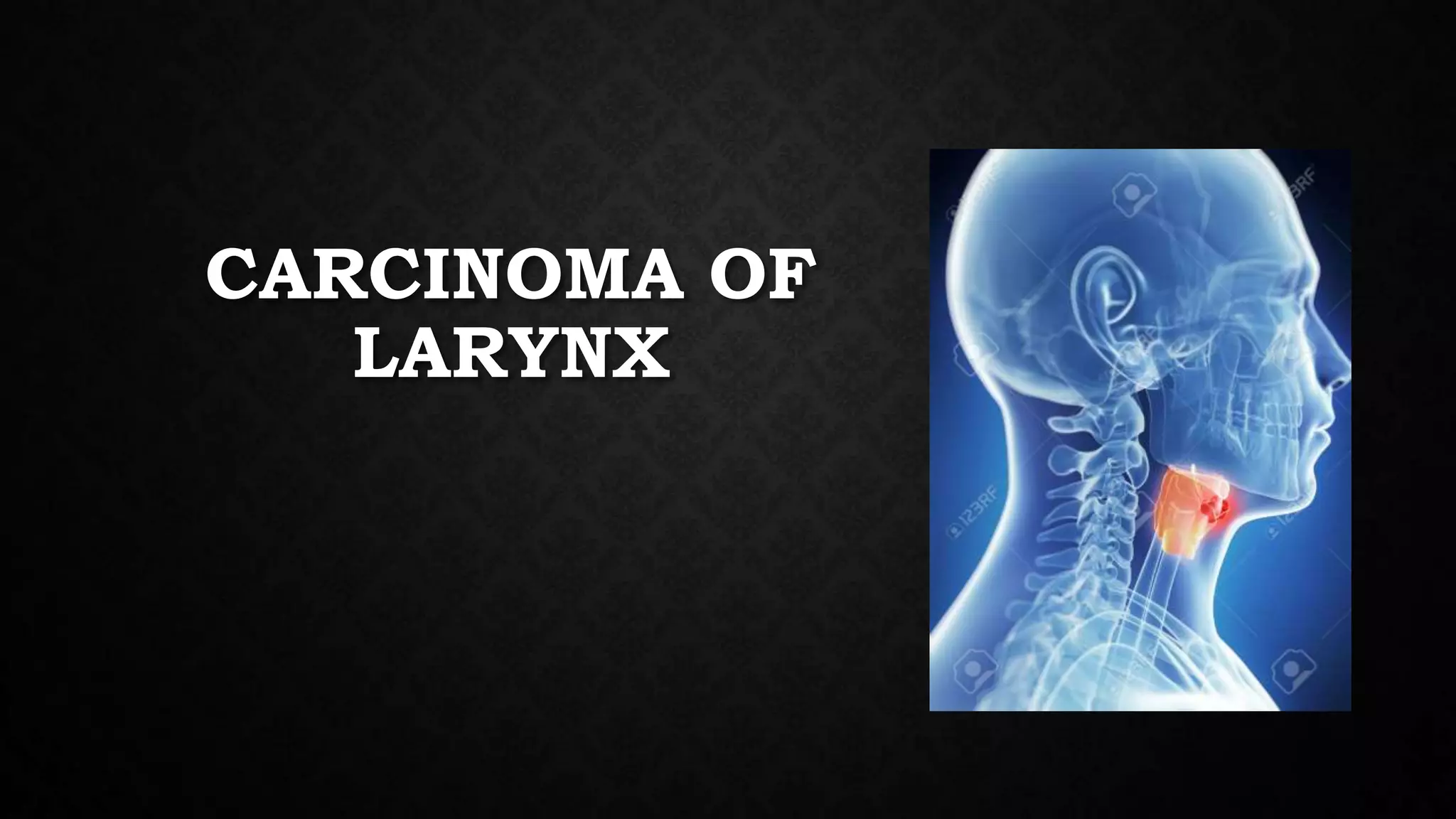
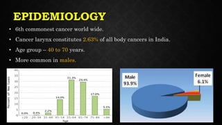
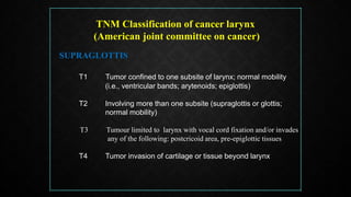
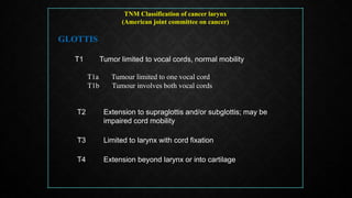
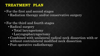
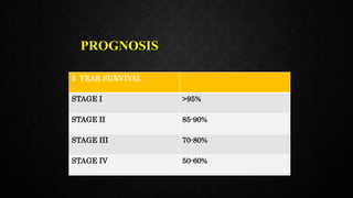
CARCINOMA OF
LARYNX
EPIDEMIOLOGY
• 6th commonest cancer world wide.
• Cancer larynx constitutes 2.63% of all body cancers in India.
• Age group – 40 to 70 years.
• More common in males.
ETIOLOGY
• TARGET - Mnemonic
• Tobacco – Benzopyrene is a carcinogen.
Less than 5% cancer occurs in non-smoker
• Alcohol – synergistic with tobacco
• Radiation
• Genetic
• Environmental exposure - Asbestos, mustard gas.
• Tumors – solitary papilloma, leukoplakia, erythroplakia..
CLASSIFICATION OF SITE
HISTOPATHOLOGY
• 90-95% are squamous cell carcinoma
with various grades of differentiation
• 5-10% lesion includes
• Verrucous carcinomas
• Spindle cell carcinomas
• Malignant salivary gland tumors
• Sarcomas
TNM Classification of cancer larynx
(American joint committee on cancer)
SUPRAGLOTTIS
T1 Tumor confined to one subsite of larynx; normal mobility
(i.e., ventricular bands; arytenoids; epiglottis)
T2 Involving more than one subsite (supraglottis or glottis;
normal mobility)
T3 Tumour limited to larynx with vocal cord fixation and/or invades
any of the following: postcricoid area, pre-epiglottic tissues
T4 Tumor invasion of cartilage or tissue beyond larynx
TNM Classification of cancer larynx
(American joint committee on cancer)
GLOTTIS
T1 Tumor limited to vocal cords, normal mobility
T1a Tumour limited to one vocal cord
T1b Tumour involves both vocal cords
T2 Extension to supraglottis and/or subglottis; may be
impaired cord mobility
T3 Limited to larynx with cord fixation
T4 Extension beyond larynx or into cartilage
TNM Classification of cancer larynx
(American joint committee on cancer)
SUBGLOTTIS
T1 Tumour limited to the subglottis
T2 Tumour extends to vocal cord(s) with normal or impaired
mobility
T3 Tumour limited to larynx with vocal cord fixation
T4 Extension beyond larynx or into cartilage
Regional Lymph Nodes (N)
Nx Cannot be assessed
N0 No regional metastasis
N1 Single positive ipsilateral node, less than 3 cm
N2 Nodes less than 6 cm
N2a Single ipsilateral node 3-6 cm
N2b Many ipsilateral nodes less than 6 cm
N2c Bilateral and contralateral node less than 6 cm
N3 Node(s) greater than 6 cm
Distant Metastasis (M)
Mx Distant metastasis cannot be assessed
M0 No distant metastasis
M1 Distant metastasis
Stage Grouping
0 Tis N0 M0
I T1 N0 M0
II T2 N0 M0
III T3 N0 M0
T1 N1 M0
T2 N1 M0
T3 N1 M0
IVA T4 N0 M0
Any T N2 M0
IVB Any T N3 M0
IVC Any T Any N M1
Histopathologic Grades
Grade 1 : Well-differentiated
Grade 2 : Moderately differentiated
Grade 3 : Poorly differentiated
SUPRAGLOTTIC CANCER
• EPIGLOTTIS
• VENTRICULAR BAND
• ARYEPIGLOTTIC FOLD
• VENTRICLE
SITE
SPREAD OF TUMOR
LOCAL LYMPHATIC
SYMPTOMS
• Muffled voice ( hot potato speech)
• Hoarseness of voice
• Foreign body sensation
• Dysphagia
• Pain throat and referred otalgia(via vagus)
• Neck swelling
• Aspiration
• Stridor
GLOTTIC CANCER
• Most commonly involved
• Free edge and upper surface of vocal
cord is the most frequent site
SYMPTOMS
• Persistence Hoarseness – cardinal feature
• Cough due to aspiration
• Dyspnea
• Stridor
• Hemoptysis (rare)
SUBGLOTTIC CARCINOMA
• Rare (1-5%)
• Invades cricothyriod membrane, thyroid gland and strap muscles of
neck
• Lymphatic metastasis
• Prelaryngeal
• Paratracheal
• Lower jugular nodes
• Symptoms
• Stridor or laryngeal obstruction
• Hoarseness (late feature)
DIAGNOSIS
1. HISTORY - any patient in cancer age group having persistent or
gradually increasing hoarseness for 3 weeks must have laryngeal
examination to exclude cancer.
2. Indirect Laryngoscopy
• Appearance of lesion
• Vocal cord mobility
• Extent of disease
• Lesions of suprahyoid epiglottis are usually exophytic
& infrahyoid epiglottis lesions are ulcerative.
• Lesions of vocal cord may appear as raised nodule,
ulcer or thickening.
• Lesions of anterior commissure may appear as
granulation tissue.
• Lesions of subglottic region appear as a raised
submucosal nodule.
3. Video laryngoscopy.
4. Examination of neck.
(i) Extralaryngeal spread of disease
(ii) nodal metastasis.
5. Radiography
• X-RAY chest
• Soft tissue lateral view
• CT /MRI scan
6. DIRECT LARYNGOSCOPY
7. MICROLARYNGOSCOPY
8. SUPRAVITAL STAIN AND BIOPSY
TREATMENT
1. Radiotherapy
2. Surgery
(a) Conservation laryngeal surgery
(b) Total laryngectomy
3. Combined therapy. Surgery with pre- or postoperative
radiotherapy
4. Endoscopic CO2 laser excision
5. Organ preservation
TREATMENT PLAN
• For the first and second stages
• Radiation therapy and/or conservative surgery
• For the third and fourth stages
• Radical surgery
• Total laryngectomy
• Laryngopharyngectomy
• Combined with unilateral radical neck dissection with or
without contralateral modified neck dissection
• Post operative radiotherapy
RADIOTHERAPY
CURATIVE – 6500 grays/30 fraction for 6 weeks.
90% cure rate, if vocal mobility is not impaired.
SURGERY INDICATIONS
• Fixed cords
• Subglottic extension
• Cartilage invasion
• Nodal metastases.
CONSERVATIVE SURGERY
ADVANTAGES
• Preserves voice
• Avoids permanent tracheostome.
INCLUSION UNDER CONSERVATIVE SURGERY
• (i) Excision of vocal cord after splitting the larynx (cordectomy via
laryngofissure).
• (ii) Excision of vocal cord and anterior commissure region (partial frontolateral
laryngectomy).
• (iii) Excision of supraglottis, i.e. epiglottis, aryepiglottic folds, false cords and
ventricle (partial horizontal laryngectomy).
TOTAL LARYNGECTOMY
• The entire larynx including the hyoid bone, pre-epiglottic space, strap muscles and one or
more rings of trachea are removed.
• Pharyngeal wall is repaired and lower tracheal stump sutured to the skin for breathing.
INDICATIONS
• (i) T3 lesions (i.e. with cord fixed)
• (ii) All T4 lesions
• (iii) Invasion of thyroid or cricoid cartilage
• (iv) Bilateral arytenoid cartilage involvement
• (v) Lesions of posterior commissure
• (vi) Failure after radiotherapy
• (vii) Transglottic cancers,
• Combined therapy.
Surgical ablation may be combine with pre-
or postoperative radiation to decrease the
incidence of recurrence.
• Endoscopic resection with CO2 laser
T1 lesions of the supra- or infrahyoid
epiglottis with or without neck nodes are treated
with CO2 laser.
Laser excision has the advantages of lower
cost, lower duration of treatment and morbidity.
PROGNOSIS
5 YEAR SURVIVAL
STAGE I >95%
STAGE II 85-90%
STAGE III 70-80%
STAGE IV 50-60%
VOCAL REHABILITATION AFTER TOTAL
LARYNGECTOMY
ELECTROLARYNX
Vibrating disc is held against the soft
tissues of the neck and a low-pitched
sound is produced in the hypopharynx
which is further modulated into speech by
tongue, lips, teeth and palate

Carcinoma larynx

  • 1.
  • 2.
    EPIDEMIOLOGY • 6th commonestcancer world wide. • Cancer larynx constitutes 2.63% of all body cancers in India. • Age group – 40 to 70 years. • More common in males.
  • 3.
    ETIOLOGY • TARGET -Mnemonic • Tobacco – Benzopyrene is a carcinogen. Less than 5% cancer occurs in non-smoker • Alcohol – synergistic with tobacco • Radiation • Genetic • Environmental exposure - Asbestos, mustard gas. • Tumors – solitary papilloma, leukoplakia, erythroplakia..
  • 4.
  • 6.
    HISTOPATHOLOGY • 90-95% aresquamous cell carcinoma with various grades of differentiation • 5-10% lesion includes • Verrucous carcinomas • Spindle cell carcinomas • Malignant salivary gland tumors • Sarcomas
  • 7.
    TNM Classification ofcancer larynx (American joint committee on cancer) SUPRAGLOTTIS T1 Tumor confined to one subsite of larynx; normal mobility (i.e., ventricular bands; arytenoids; epiglottis) T2 Involving more than one subsite (supraglottis or glottis; normal mobility) T3 Tumour limited to larynx with vocal cord fixation and/or invades any of the following: postcricoid area, pre-epiglottic tissues T4 Tumor invasion of cartilage or tissue beyond larynx
  • 8.
    TNM Classification ofcancer larynx (American joint committee on cancer) GLOTTIS T1 Tumor limited to vocal cords, normal mobility T1a Tumour limited to one vocal cord T1b Tumour involves both vocal cords T2 Extension to supraglottis and/or subglottis; may be impaired cord mobility T3 Limited to larynx with cord fixation T4 Extension beyond larynx or into cartilage
  • 9.
    TNM Classification ofcancer larynx (American joint committee on cancer) SUBGLOTTIS T1 Tumour limited to the subglottis T2 Tumour extends to vocal cord(s) with normal or impaired mobility T3 Tumour limited to larynx with vocal cord fixation T4 Extension beyond larynx or into cartilage
  • 10.
    Regional Lymph Nodes(N) Nx Cannot be assessed N0 No regional metastasis N1 Single positive ipsilateral node, less than 3 cm N2 Nodes less than 6 cm N2a Single ipsilateral node 3-6 cm N2b Many ipsilateral nodes less than 6 cm N2c Bilateral and contralateral node less than 6 cm N3 Node(s) greater than 6 cm
  • 11.
    Distant Metastasis (M) MxDistant metastasis cannot be assessed M0 No distant metastasis M1 Distant metastasis Stage Grouping 0 Tis N0 M0 I T1 N0 M0 II T2 N0 M0 III T3 N0 M0 T1 N1 M0 T2 N1 M0 T3 N1 M0 IVA T4 N0 M0 Any T N2 M0 IVB Any T N3 M0 IVC Any T Any N M1 Histopathologic Grades Grade 1 : Well-differentiated Grade 2 : Moderately differentiated Grade 3 : Poorly differentiated
  • 13.
    SUPRAGLOTTIC CANCER • EPIGLOTTIS •VENTRICULAR BAND • ARYEPIGLOTTIC FOLD • VENTRICLE SITE SPREAD OF TUMOR LOCAL LYMPHATIC
  • 14.
    SYMPTOMS • Muffled voice( hot potato speech) • Hoarseness of voice • Foreign body sensation • Dysphagia • Pain throat and referred otalgia(via vagus) • Neck swelling • Aspiration • Stridor
  • 15.
    GLOTTIC CANCER • Mostcommonly involved • Free edge and upper surface of vocal cord is the most frequent site
  • 16.
    SYMPTOMS • Persistence Hoarseness– cardinal feature • Cough due to aspiration • Dyspnea • Stridor • Hemoptysis (rare)
  • 17.
    SUBGLOTTIC CARCINOMA • Rare(1-5%) • Invades cricothyriod membrane, thyroid gland and strap muscles of neck • Lymphatic metastasis • Prelaryngeal • Paratracheal • Lower jugular nodes • Symptoms • Stridor or laryngeal obstruction • Hoarseness (late feature)
  • 18.
    DIAGNOSIS 1. HISTORY -any patient in cancer age group having persistent or gradually increasing hoarseness for 3 weeks must have laryngeal examination to exclude cancer. 2. Indirect Laryngoscopy • Appearance of lesion • Vocal cord mobility • Extent of disease • Lesions of suprahyoid epiglottis are usually exophytic & infrahyoid epiglottis lesions are ulcerative. • Lesions of vocal cord may appear as raised nodule, ulcer or thickening. • Lesions of anterior commissure may appear as granulation tissue. • Lesions of subglottic region appear as a raised submucosal nodule.
  • 19.
    3. Video laryngoscopy. 4.Examination of neck. (i) Extralaryngeal spread of disease (ii) nodal metastasis. 5. Radiography • X-RAY chest • Soft tissue lateral view • CT /MRI scan
  • 20.
    6. DIRECT LARYNGOSCOPY 7.MICROLARYNGOSCOPY 8. SUPRAVITAL STAIN AND BIOPSY
  • 21.
    TREATMENT 1. Radiotherapy 2. Surgery (a)Conservation laryngeal surgery (b) Total laryngectomy 3. Combined therapy. Surgery with pre- or postoperative radiotherapy 4. Endoscopic CO2 laser excision 5. Organ preservation
  • 22.
    TREATMENT PLAN • Forthe first and second stages • Radiation therapy and/or conservative surgery • For the third and fourth stages • Radical surgery • Total laryngectomy • Laryngopharyngectomy • Combined with unilateral radical neck dissection with or without contralateral modified neck dissection • Post operative radiotherapy
  • 23.
    RADIOTHERAPY CURATIVE – 6500grays/30 fraction for 6 weeks. 90% cure rate, if vocal mobility is not impaired. SURGERY INDICATIONS • Fixed cords • Subglottic extension • Cartilage invasion • Nodal metastases.
  • 24.
    CONSERVATIVE SURGERY ADVANTAGES • Preservesvoice • Avoids permanent tracheostome. INCLUSION UNDER CONSERVATIVE SURGERY • (i) Excision of vocal cord after splitting the larynx (cordectomy via laryngofissure). • (ii) Excision of vocal cord and anterior commissure region (partial frontolateral laryngectomy). • (iii) Excision of supraglottis, i.e. epiglottis, aryepiglottic folds, false cords and ventricle (partial horizontal laryngectomy).
  • 25.
    TOTAL LARYNGECTOMY • Theentire larynx including the hyoid bone, pre-epiglottic space, strap muscles and one or more rings of trachea are removed. • Pharyngeal wall is repaired and lower tracheal stump sutured to the skin for breathing. INDICATIONS • (i) T3 lesions (i.e. with cord fixed) • (ii) All T4 lesions • (iii) Invasion of thyroid or cricoid cartilage • (iv) Bilateral arytenoid cartilage involvement • (v) Lesions of posterior commissure • (vi) Failure after radiotherapy • (vii) Transglottic cancers,
  • 26.
    • Combined therapy. Surgicalablation may be combine with pre- or postoperative radiation to decrease the incidence of recurrence. • Endoscopic resection with CO2 laser T1 lesions of the supra- or infrahyoid epiglottis with or without neck nodes are treated with CO2 laser. Laser excision has the advantages of lower cost, lower duration of treatment and morbidity.
  • 27.
    PROGNOSIS 5 YEAR SURVIVAL STAGEI >95% STAGE II 85-90% STAGE III 70-80% STAGE IV 50-60%
  • 28.
    VOCAL REHABILITATION AFTERTOTAL LARYNGECTOMY
  • 29.
    ELECTROLARYNX Vibrating disc isheld against the soft tissues of the neck and a low-pitched sound is produced in the hypopharynx which is further modulated into speech by tongue, lips, teeth and palate

Editor's Notes

  • #7 On the basis of histology ,more then 90% are sq cell ca with various grades
  • #12 Although there are many staging systems in practice, but mostly tumour staging is carried out with AJCC(american joint committee on cancer) and UICC (uninion of international cancer control) is based on TNM classification system
  • #18 Sub glottic carcinoma is a rare upto 2 %
  • #23 Treatment of choice For 1st & 2nd stages is radiotherapy and conservatie surgery ,for 3rd & 4th stages total laryngectomy is the mainstay of treatment , usually combined with post- operative radio therapy of residual disease.
  • #28 Prognosis depends on the tumour stage