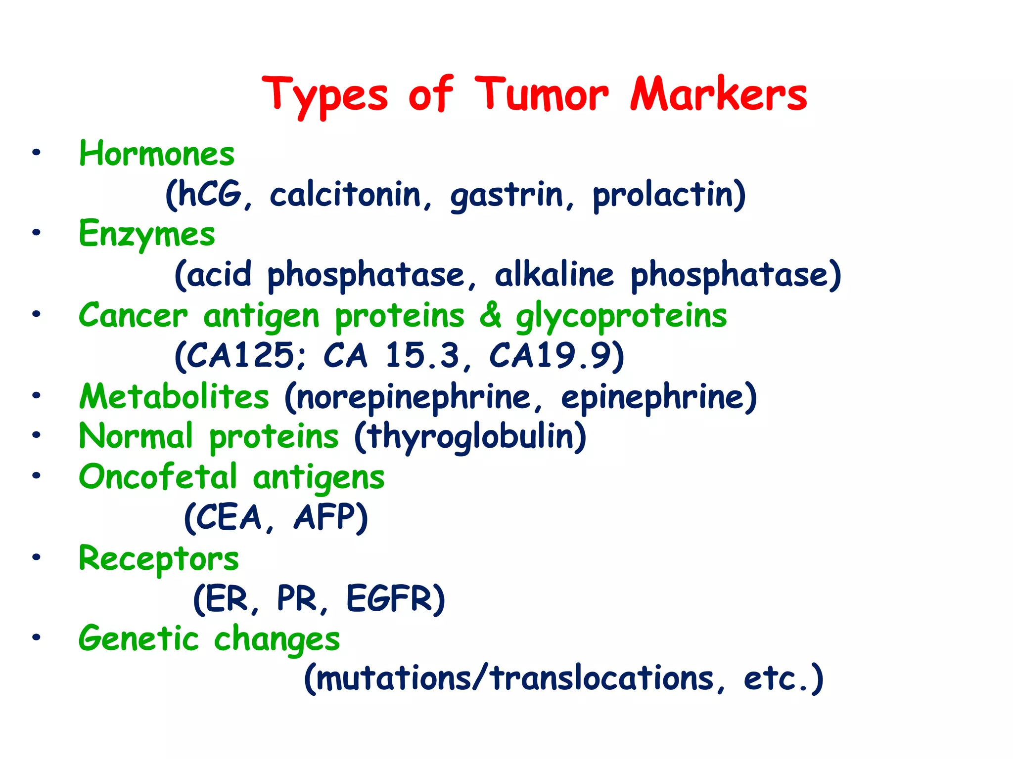
CANCER
&
TUMOR MARKERS
Kshema Thakur, PhD
Molecular Biology & Biochemistry
Cancer mortality is second largest cause of death in the world
after cardiovascular disease
INDIA
Estimated number of people living with the disease: around 2.25
million
Every year, new cancer patients registered: Over 11 lakh
Cancer-related deaths: over 7.8 lakh
One woman dies of cervical cancer every 8 minutes in India.
For every two women diagnosed with breast cancer, one woman
dies.
Risk of dying from cancer before the age of 75 years is 7.34%
in males and 6.28% in females.
CANCER STATISTICS
(http://cancerindia.org.in/cancer-statistics/)
Men women
1 Lip, oral cavity Breast
2 Lung Lip, oral cavity
3 Stomach Cervix
4 Colorectum Lung
5 Oesophagus Gastric
Tumour: accumulation of cellular mass caused
by excessive cell proliferation.
diseases resulting from tumours are called
neoplasias (new growth)
1. Benign:
Self limiting
growth. E.g.
moles, warts
2. Malignant:
Having high
propensity of
invasion and
spread
Diseases related to
malignant tumours
only are called
cancer.
BASICS
CANCER
De-differentiation
Uncontrolled cell growth
Loss in cell cycle check points
Loss of contact inhibition
Loss of apoptosis
Loss in anchorage dependence
Metabolic alterations
Angiogenesis
Malignant transformation
Multifactorial: caused by many
contributing factors making them
complex. Such as multiple genes or
involving multiple tissues/organs
Overview of changes caused by
cancer in a cell
BIOCHEMICAL ALTERATIONS IN CANCER
1. Anomalies in glycolysis and TCA are observed. Cancer
cells use excess of glucose and less oxygen with large
production of lactic acid
2. Rate of synthesis or DNA and RNA are considerably
higher than normal cells indicative of rapid cell
multiplication
3. Increase in ribonucleotide reductase and decreased
catabolism of pyrimidines
4. Membrane permeability and cell surface charge are
altered
5. Activities of certain enzymes such as proteases
decreases
BIOCHEMICAL ALTERATIONS IN CANCER
(contd.)
6. The isoenzymes typical of fetal cells make appearance in
cancer cells
7. Cell membrane antigens are altered. New antigens
appear while some normal ones are lost. These antigens
are mainly constituted by surface glycoproteins and
glycolipids
8. Can grow in absence of normal mitotic growth factor.
They tend to secret their growth factor which in normal
cells is strictly coordinated
9. Loss of tumour suppressor genes is observed
10.Alteration in transport proteins
CELL’S CONTROLS
Oncogenes
Genes that have the capacity to transform the normal
cells into malignant cells
Generated by somatic mutations such as point
mutations, gene amplification, gene traslocation, gene
truncation etc.
Leads to either overproduction of a normal cellular
structural protein or synthesis of an abnormal protein
product called “oncoprotein”
alb, erb-B, erb-A, myc, sis, src, ras
Proto-oncogenes
These are the oncogenes present in normal cell. Can be understood
as inactive oncogenes.
Around 80 proto-oncogenes are presently known.
Oncosuppressors/tumor suppressors/anti-oncogene
Cellular genes that inhibit cell proliferation or tumour
development
Thus have key roles in cell cycle determination and apoptosis
Mutation in tumour suppressor genes result in loss of inhibition
of cell division – a classic halmark of cancereous growth
Even one functional copy of oncosuppressor can protect the cell
Mutations in oncosuppressor are more responsible for tumours
than protooncogenes
The most common types are RB1 Gene and p53 gene
RB, WT, FAP, DCC, p53, BRAC, VHL
CELL’S CONTROLS
(contd.)
How do tumor suppressors work?
RB1 gene
Regulates cell growth and keeps cells from dividing
too fast or in an uncontrolled way.
pRB interacts with other proteins to influence cell
survival, the self-destruction of cells (apoptosis), and
the process by which cells mature to carry out special
functions (differentiation).
Role of mutations in Retinoblastoma
The p53 gene has a crucial role in cell cycle regulation.
Is called guardian of genome.
If a person inherits only one functional copy of the p53 gene
from their parents, they are predisposed to cancer and usually
develop several independent tumors in a variety of tissues in early
adulthood. This condition is rare, and is known as Li-Fraumeni
syndrome.
How do tumor suppressors work?
(contd.)
IMMUNE SYSTEM
T-cells
Natural killer (NK) cells
Antibody dependent complement
mediated lysis
Antibody dependent cell mediated
cytolysis (ADCC)
Macrophages
CARCINOGENS
Chemical carcinogens (oncogen)
Also called mutagens. Benzopyrenes, cholanthrenes,
dimethyl nitrosamine
Tobacco/cigarette
70/7000 chemicals that cause cancer. Benzopyrenes,
nicotine, carbon monoxide, nitrogen dioxide, carbon soot
Physical carcinogens
X-rays, UV-rays, Gamma rays
Oncogenic viruses
Epstein-Barr virus, Human papilloma carcinoma virus,
hepatitis-B virus
Aflatoxins
Compounds synthesized by Aspergillus flavus. Grows on
rice, wheat and groundnut
Oncofetal antigens
ONCOGENIC VIRUSES (contd.)
How do viruses transform cells?
 Virus infection provides a “hit” towards the genesis of
cancer by following:
 Act as a “mutagen”
 Other cofactors (genetic, immunological or
environmental) may be needed for a development
of cancer
 Cell transformation is accompanied by the persistence
of all or part of the viral genome and continual
expression of a limited number of viral genes.
 Viral oncogenes are expressed that alter normal cellular
gene expression and signal transduction pathways
ONCOGENIC VIRUSES
ONCOGENIC VIRUSES
ONCOGENIC VIRUSES
ONCOGENIC VIRUSES
ONCOGENIC VIRUSES
PROGRESSION OF
CANCER
1. Uncontrolled cell division
2. Tumor lump (benign)
3. Angiogenesis
4. Invasion to the adjacent tissue
5. Spread to other tissues
6. Secondary cancer/recurrence
Months
Years
TUMOR
MARKERS
Tumour marker is a substance most
commonly proteins, produced by the
body in response to the cancer or by
the cancerous tissue itself and that can
be detected in blood, urine or other
tissues.
Some markers are specific for a
particular cancer while others are seen
in several types.
TUMOUR MARKER
Roles of a tumor marker
1.Screening
2.Early detection of cancer
3.Diagnosing cancer
4.Staging of cancer
5.Determining the prognosis of certain
cancers
6.Determining the effectiveness of
certain treatment
7.Detecting recurrent cancer
8.To assess response to treatment
9.Follow up of a cancer patient
Characteristics of an ideal tumor marker
• Specificity for a single type of cancer
•High sensitivity and specificity for cancerous
growth
•Correlation of marker level with tumor size
•Homogeneous (i.e., minimal post-translational
modifications)
•Short half-life in circulation
•Detectable in occult disease
Determination of tumour markers
• Immunochemistry
- radio immune assay – RIA, IRMA
- enzyme immune assay - ELISA, EIA,MEIA
- fluorescence assay - FPIA, TRACE
- chemiluminiscence assay - CLIA
Types of Tumor Markers
• Hormones
(hCG, calcitonin, gastrin, prolactin)
• Enzymes
(acid phosphatase, alkaline phosphatase)
• Cancer antigen proteins & glycoproteins
(CA125; CA 15.3, CA19.9)
• Metabolites (norepinephrine, epinephrine)
• Normal proteins (thyroglobulin)
• Oncofetal antigens
(CEA, AFP)
• Receptors
(ER, PR, EGFR)
• Genetic changes
(mutations/translocations, etc.)
Tumor markers in routine use
Marker Cancer
CA15-3, BR 27.29
CEA, CA 19-9
CA 72.4, CA 19-9, CEA
NSE, CYFA 21.1
PSA, PAP
Breast
Colorectal
Gastric
Lung
Prostate
OvarianCA 125
Calcitonin, thyroglobulin
hCG
CA 19-9, CEA
AFP, CA 19-1
Thyroid
Trophoblastic
Pancreatic
Hepatocellular
BoneBAP, Osteocalcin, NTx
Catecholamines, metabolites Pheochromocytoma
Fecal occult blood Colon cancer
Benign conditions which may
have elevated AFP levels
Hepatitis
Cirrhosis
Biliary tract obstruction
Alcoholic liver disease
Ataxia telangiectasia
Hereditary tyrosinemia
Malignancies with elevated levels
Mainly seen in three malignancies:
1.Non-seminomatous germ cell tumour (NSGCT)
2.Hepatocellular carcinoma
3.Hepatoblastoma
Occasionally elevated in adenocarcinomas
Also elevated due to chemotherapy induced liver
damage!
AFP
Estrogen receptors/progesterone receptor
 Breast cancer tissues are commonly tested for these
markers by immunohistochemistry.
 Breast cancers that contain estrogen receptors are
often referred to as “ER +ve”, while those with
progesterone receptors are “PR +ive” (>10 fmol is +ve).
 Their main use is as a predictor of prognosis (survival
outlook).
 About 7 out of 10 breast cancers test positive for
atleast one of these markers.
 These cancers tend to have a better prognosis than
cancers without these receptors and are much likely to
respond to hormonal therapy such as tamoxifen or
aromatase inhibitors.
 >60% of ER +ve and <10% of ER –ve tumors respond.
 Degree of positivity is proportional to grade & histology
 Increased expression in a wide range of solid
tumors- colorectal, breast, lung (NSCLC), head
and neck and pancreatic cancers
 Increased expression has been correlated with
disease progression, poor treatment outcome and
poor survival
 Targeted therapies – geftinib, erlotinib,
cetuximab, ABX-EGF are directed against these
receptors
Epidermal growth factor receptor
Inherited mutations of tumour suppressor
genes
p53, RB1: useful for screening and
identification of families or individuals at high
risk of developing various cancers
BRCA1 and BRCA2: breast cancer
BRCA1: also at risk of ovarian, colon and
prostate cancer
BRCA2: risk of breast cancer in men
ANTI-CANCER DRUGS
METHOTRAXATE
6-MERCAPTOPURINE
6-THIOGUANINE
CYCLOPHOSPHAMIDE
MITOMYCIN-C
ACTINOMYCIN-D
ADRIAMYCIN (DOXORUBICIN)
CISPLATIN
ETOPOSIDE
TAMOXIFEN
Following points are to be considered before ordering
investigation of serum tumour markers
 A single test can never be relied on
 Always a panel of markers be recommended
 When testing the same marker for the same patient
in serial testing, insist on use of same lab and test
kit.
 Half life of the marker must be considered before
interpreting the results.
 It is important to consider clearance of marker in
the blood circulation.
Take home message
Sources:
1. http://cancerindia.org.in/cancer-statistics/)
2. Biochemistry
U. Satyanarayan & U. Chakrapani
3. World wibe web
Thank you !

Cancer and tumor markers

  • 1.
    CANCER & TUMOR MARKERS Kshema Thakur,PhD Molecular Biology & Biochemistry
  • 2.
    Cancer mortality issecond largest cause of death in the world after cardiovascular disease INDIA Estimated number of people living with the disease: around 2.25 million Every year, new cancer patients registered: Over 11 lakh Cancer-related deaths: over 7.8 lakh One woman dies of cervical cancer every 8 minutes in India. For every two women diagnosed with breast cancer, one woman dies. Risk of dying from cancer before the age of 75 years is 7.34% in males and 6.28% in females. CANCER STATISTICS (http://cancerindia.org.in/cancer-statistics/) Men women 1 Lip, oral cavity Breast 2 Lung Lip, oral cavity 3 Stomach Cervix 4 Colorectum Lung 5 Oesophagus Gastric
  • 3.
    Tumour: accumulation ofcellular mass caused by excessive cell proliferation. diseases resulting from tumours are called neoplasias (new growth) 1. Benign: Self limiting growth. E.g. moles, warts 2. Malignant: Having high propensity of invasion and spread Diseases related to malignant tumours only are called cancer. BASICS
  • 5.
    CANCER De-differentiation Uncontrolled cell growth Lossin cell cycle check points Loss of contact inhibition Loss of apoptosis Loss in anchorage dependence Metabolic alterations Angiogenesis Malignant transformation Multifactorial: caused by many contributing factors making them complex. Such as multiple genes or involving multiple tissues/organs
  • 6.
    Overview of changescaused by cancer in a cell
  • 7.
    BIOCHEMICAL ALTERATIONS INCANCER 1. Anomalies in glycolysis and TCA are observed. Cancer cells use excess of glucose and less oxygen with large production of lactic acid 2. Rate of synthesis or DNA and RNA are considerably higher than normal cells indicative of rapid cell multiplication 3. Increase in ribonucleotide reductase and decreased catabolism of pyrimidines 4. Membrane permeability and cell surface charge are altered 5. Activities of certain enzymes such as proteases decreases
  • 8.
    BIOCHEMICAL ALTERATIONS INCANCER (contd.) 6. The isoenzymes typical of fetal cells make appearance in cancer cells 7. Cell membrane antigens are altered. New antigens appear while some normal ones are lost. These antigens are mainly constituted by surface glycoproteins and glycolipids 8. Can grow in absence of normal mitotic growth factor. They tend to secret their growth factor which in normal cells is strictly coordinated 9. Loss of tumour suppressor genes is observed 10.Alteration in transport proteins
  • 9.
    CELL’S CONTROLS Oncogenes Genes thathave the capacity to transform the normal cells into malignant cells Generated by somatic mutations such as point mutations, gene amplification, gene traslocation, gene truncation etc. Leads to either overproduction of a normal cellular structural protein or synthesis of an abnormal protein product called “oncoprotein” alb, erb-B, erb-A, myc, sis, src, ras Proto-oncogenes These are the oncogenes present in normal cell. Can be understood as inactive oncogenes. Around 80 proto-oncogenes are presently known.
  • 10.
    Oncosuppressors/tumor suppressors/anti-oncogene Cellular genesthat inhibit cell proliferation or tumour development Thus have key roles in cell cycle determination and apoptosis Mutation in tumour suppressor genes result in loss of inhibition of cell division – a classic halmark of cancereous growth Even one functional copy of oncosuppressor can protect the cell Mutations in oncosuppressor are more responsible for tumours than protooncogenes The most common types are RB1 Gene and p53 gene RB, WT, FAP, DCC, p53, BRAC, VHL CELL’S CONTROLS (contd.)
  • 11.
    How do tumorsuppressors work? RB1 gene Regulates cell growth and keeps cells from dividing too fast or in an uncontrolled way. pRB interacts with other proteins to influence cell survival, the self-destruction of cells (apoptosis), and the process by which cells mature to carry out special functions (differentiation).
  • 12.
    Role of mutationsin Retinoblastoma
  • 13.
    The p53 genehas a crucial role in cell cycle regulation. Is called guardian of genome. If a person inherits only one functional copy of the p53 gene from their parents, they are predisposed to cancer and usually develop several independent tumors in a variety of tissues in early adulthood. This condition is rare, and is known as Li-Fraumeni syndrome. How do tumor suppressors work? (contd.)
  • 14.
    IMMUNE SYSTEM T-cells Natural killer(NK) cells Antibody dependent complement mediated lysis Antibody dependent cell mediated cytolysis (ADCC) Macrophages
  • 15.
    CARCINOGENS Chemical carcinogens (oncogen) Alsocalled mutagens. Benzopyrenes, cholanthrenes, dimethyl nitrosamine Tobacco/cigarette 70/7000 chemicals that cause cancer. Benzopyrenes, nicotine, carbon monoxide, nitrogen dioxide, carbon soot Physical carcinogens X-rays, UV-rays, Gamma rays Oncogenic viruses Epstein-Barr virus, Human papilloma carcinoma virus, hepatitis-B virus Aflatoxins Compounds synthesized by Aspergillus flavus. Grows on rice, wheat and groundnut Oncofetal antigens
  • 16.
    ONCOGENIC VIRUSES (contd.) Howdo viruses transform cells?  Virus infection provides a “hit” towards the genesis of cancer by following:  Act as a “mutagen”  Other cofactors (genetic, immunological or environmental) may be needed for a development of cancer  Cell transformation is accompanied by the persistence of all or part of the viral genome and continual expression of a limited number of viral genes.  Viral oncogenes are expressed that alter normal cellular gene expression and signal transduction pathways
  • 17.
  • 18.
  • 19.
  • 20.
  • 21.
  • 22.
    PROGRESSION OF CANCER 1. Uncontrolledcell division 2. Tumor lump (benign) 3. Angiogenesis 4. Invasion to the adjacent tissue 5. Spread to other tissues 6. Secondary cancer/recurrence Months Years
  • 24.
  • 25.
    Tumour marker isa substance most commonly proteins, produced by the body in response to the cancer or by the cancerous tissue itself and that can be detected in blood, urine or other tissues. Some markers are specific for a particular cancer while others are seen in several types. TUMOUR MARKER
  • 26.
    Roles of atumor marker 1.Screening 2.Early detection of cancer 3.Diagnosing cancer 4.Staging of cancer 5.Determining the prognosis of certain cancers 6.Determining the effectiveness of certain treatment 7.Detecting recurrent cancer 8.To assess response to treatment 9.Follow up of a cancer patient
  • 27.
    Characteristics of anideal tumor marker • Specificity for a single type of cancer •High sensitivity and specificity for cancerous growth •Correlation of marker level with tumor size •Homogeneous (i.e., minimal post-translational modifications) •Short half-life in circulation •Detectable in occult disease
  • 28.
    Determination of tumourmarkers • Immunochemistry - radio immune assay – RIA, IRMA - enzyme immune assay - ELISA, EIA,MEIA - fluorescence assay - FPIA, TRACE - chemiluminiscence assay - CLIA
  • 29.
    Types of TumorMarkers • Hormones (hCG, calcitonin, gastrin, prolactin) • Enzymes (acid phosphatase, alkaline phosphatase) • Cancer antigen proteins & glycoproteins (CA125; CA 15.3, CA19.9) • Metabolites (norepinephrine, epinephrine) • Normal proteins (thyroglobulin) • Oncofetal antigens (CEA, AFP) • Receptors (ER, PR, EGFR) • Genetic changes (mutations/translocations, etc.)
  • 30.
    Tumor markers inroutine use Marker Cancer CA15-3, BR 27.29 CEA, CA 19-9 CA 72.4, CA 19-9, CEA NSE, CYFA 21.1 PSA, PAP Breast Colorectal Gastric Lung Prostate OvarianCA 125 Calcitonin, thyroglobulin hCG CA 19-9, CEA AFP, CA 19-1 Thyroid Trophoblastic Pancreatic Hepatocellular BoneBAP, Osteocalcin, NTx Catecholamines, metabolites Pheochromocytoma Fecal occult blood Colon cancer
  • 34.
    Benign conditions whichmay have elevated AFP levels Hepatitis Cirrhosis Biliary tract obstruction Alcoholic liver disease Ataxia telangiectasia Hereditary tyrosinemia
  • 35.
    Malignancies with elevatedlevels Mainly seen in three malignancies: 1.Non-seminomatous germ cell tumour (NSGCT) 2.Hepatocellular carcinoma 3.Hepatoblastoma Occasionally elevated in adenocarcinomas Also elevated due to chemotherapy induced liver damage! AFP
  • 40.
    Estrogen receptors/progesterone receptor Breast cancer tissues are commonly tested for these markers by immunohistochemistry.  Breast cancers that contain estrogen receptors are often referred to as “ER +ve”, while those with progesterone receptors are “PR +ive” (>10 fmol is +ve).  Their main use is as a predictor of prognosis (survival outlook).  About 7 out of 10 breast cancers test positive for atleast one of these markers.  These cancers tend to have a better prognosis than cancers without these receptors and are much likely to respond to hormonal therapy such as tamoxifen or aromatase inhibitors.  >60% of ER +ve and <10% of ER –ve tumors respond.  Degree of positivity is proportional to grade & histology
  • 41.
     Increased expressionin a wide range of solid tumors- colorectal, breast, lung (NSCLC), head and neck and pancreatic cancers  Increased expression has been correlated with disease progression, poor treatment outcome and poor survival  Targeted therapies – geftinib, erlotinib, cetuximab, ABX-EGF are directed against these receptors Epidermal growth factor receptor
  • 42.
    Inherited mutations oftumour suppressor genes p53, RB1: useful for screening and identification of families or individuals at high risk of developing various cancers BRCA1 and BRCA2: breast cancer BRCA1: also at risk of ovarian, colon and prostate cancer BRCA2: risk of breast cancer in men
  • 43.
  • 44.
    Following points areto be considered before ordering investigation of serum tumour markers  A single test can never be relied on  Always a panel of markers be recommended  When testing the same marker for the same patient in serial testing, insist on use of same lab and test kit.  Half life of the marker must be considered before interpreting the results.  It is important to consider clearance of marker in the blood circulation. Take home message
  • 47.
  • 48.