Downloaded 1,601 times


![Cells as Biomarkers
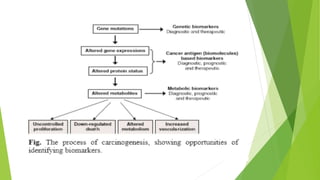
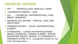
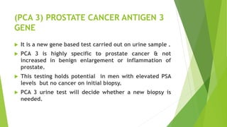
Circulating Tumor Cells [CTCs] :
They provide early and reliable indication of disease
progression and survival of patients on systemic therapy
for metastatic breast cancer as early as 3-4 weeks after
initiation of therapy.
Superior to standard tumor markers in predicting
prognosis.
Can be used as an early predictor of treatment efficacy
and extremely in sparing patients from futile therapy.
Can be detected by immunocytometry.](https://image.slidesharecdn.com/newertumormarkersbydr-140409083143-phpapp02/85/Newer-Tumour-Markers-45-320.jpg)
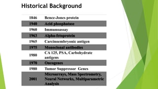
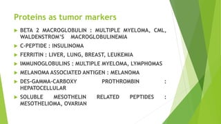
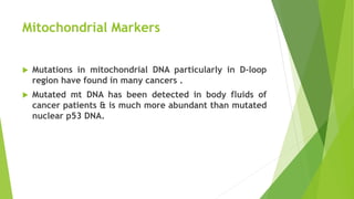
![T-regulatory cells [CD4+,CD25+,Foxp3] :
These are important in inducing and maintaining
peripheral self-tolerance and thus preventing immune
pathologies.
Increased T regulatory activity is associated with poor
response to tumor antigens and contribute to immune
dysfunction resulting in tumor growth.
T-regulatory cells may serve as surrogate immune
marker of cancer progression and perhaps prognosis.
It is also useful as a predictor of response to therapies.
CD 90 IS A DIAGNOSTIC MARKER TO DIFFERENTIATE
BETWEEN MALIGNANT PLEURAL MESOTHELIOMA & LUNG
CARCINOMA WITH IMMUNO HISTOCHEMISTRY .](https://image.slidesharecdn.com/newertumormarkersbydr-140409083143-phpapp02/85/Newer-Tumour-Markers-46-320.jpg)
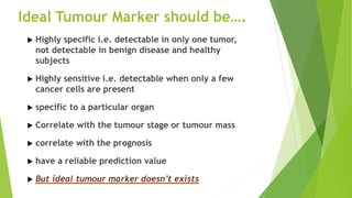
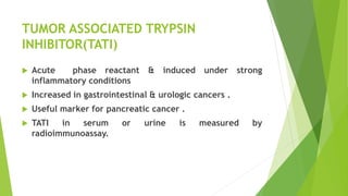
![Cancer Stem Cells [CSC] :
Subpopulation of cancer cells which resemble the
developmental hierarchy of the normal tissue from which
the tumor arose.
Evidence for existence of CSC initially came from studies of
AML.
CSC are now demonstrated in many solid tumors including
glioblastoma, medulloblastoma, breast cancer, melanoma
and prostate cancer.
CSC are resistant to chemotherapy and radiation therapy.
Eradication is the critical determinant in achieving cure.
Identifying and characterizing CSC for every possible tumor
is of paramount importance and will likely lead to new
therapeutic avenues.](https://image.slidesharecdn.com/newertumormarkersbydr-140409083143-phpapp02/85/Newer-Tumour-Markers-47-320.jpg)
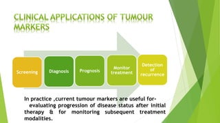
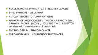
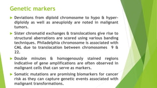
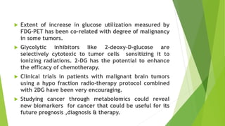
![Metabolic Biomarkers
Bio-energetic index of cell has been suggested for
classification and prognosis of cancer, besides
predicting the response to therapy.
Positron emission tomography allows non invasive and
quantitative analysis of various biologic process.
It uses a glucose analogue [2-deoxy-D-glucose] labelled
with positron emitter Fluorine 18.
FDG that is partially metabolized and trapped as its
phosphate [2-DG-6-P] in the tumor tissue, thus,
localizing the tumor](https://image.slidesharecdn.com/newertumormarkersbydr-140409083143-phpapp02/85/Newer-Tumour-Markers-52-320.jpg)
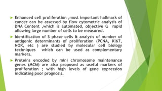
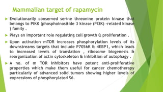
![Therapeutic Biomarkers
Targeted therapies display greater selectivity for tumor cells.
Eg : Small molecule drugs that inhibit the activity of tyrosine
kinases [ Eg : Imatinib and Erlotinib targeting ABL & EGFR].
Antibody bevacizumab targets a growth factor that stimulates
tumor blood vessel growth.](https://image.slidesharecdn.com/newertumormarkersbydr-140409083143-phpapp02/85/Newer-Tumour-Markers-54-320.jpg)
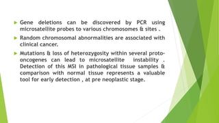
![Telomerase
One of the best markers for human cancer associated with
only malignant tumors.
Telomerase enzyme ensures the maintenance of telomere and
thereby protecting the cell from degradation and cell death.
In cancer cells telomerase shuttling system is impaired.
The TRAP (telomeric repeat amplification protocol) assay is
used for detection of telomerase activity.
It has been a target for anti-cancer therapeutics that turn off
telomerase and there by inhibit tumor growth.
Currently two clinical trials : one using a vaccine [GRNVAC1]
and the other a lapidated drug [GRN163L] are under way to
evaluate the efficacy of telomerase inhibitors.](https://image.slidesharecdn.com/newertumormarkersbydr-140409083143-phpapp02/85/Newer-Tumour-Markers-55-320.jpg)
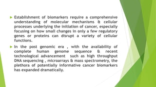
![Histone deacetylases [HDACs]
HDACs are associated with oncogenesis by regulating the
expression of certain tumor suppressor genes leading to
excessive proliferation and tumorogenesis.
They have recently been the attractive targets for cancer
therapeutics.
HDAC inhibitors are currently under clinical investigation
in a number of hematological malignancies and solid
tumors.](https://image.slidesharecdn.com/newertumormarkersbydr-140409083143-phpapp02/85/Newer-Tumour-Markers-56-320.jpg)
![PIN1
Peptidyl-Prolyl-Isomerase [PPIase], PIN1 regulates post
phosphorylation event which is in the form Cis and Trans
isomerization of phosphoserine/threonine – proline peptide
bonds at selective sides.
Over expression of PIN1 has been reported in human breast
cancer cell lines and tissues, and its expression closely
correlates with the level of cyclin D1 in tumors.
PIN1 opens a new target for the development of specific
therapeutics as phosphorylated p53 is known substrates of
PIN1.
Inhibition of PIN1 induces mitotic arrest and apoptosis in
tumor cell lines
Recent studies with PIN1 inhibitor Juglone delays the growth
of various tumor cell lines.](https://image.slidesharecdn.com/newertumormarkersbydr-140409083143-phpapp02/85/Newer-Tumour-Markers-58-320.jpg)


![REFERENCES-
Cancer Biomarkers – Current Perspectives. [Indian J Med Res 132, August
2010, pp 129-149.
Teitz Textbook of clinical chemistry and molecular diagnostics.
MMP13 is potentially a new tumor marker for breast cancer diagnostic.
[PubMed – indexed for MEDLINE]
BioPlat- a software for human cancer biomarker discovery.[Bioinformatics.
02/2014]
Involvement of adiponectin and leptin in breast cancer: clinical and in vitro
studies.[ENDOCR RELAT CANCER, 2009 December;16(4): 1197-210]
Detecting cancer through markers (Dr. Mohini Bhargava- Express Healthcare)
Cancer biomarker discovery : current status & future perspectives.
( International journal of Radiation Biology ).
Stress fuels cancer spread by triggering master gene :ATF 3.](https://image.slidesharecdn.com/newertumormarkersbydr-140409083143-phpapp02/85/Newer-Tumour-Markers-66-320.jpg)

This document discusses newer tumor markers that can be used for cancer diagnosis, prognosis, and monitoring treatment. It describes various types of biochemical entities that serve as tumor markers, including nucleic acids, proteins, sugars, lipids, and whole tumor cells. Specific examples of tumor markers are discussed, such as enzymes, hormones, oncofetal antigens, tumor-associated proteins, carbohydrate antigens, and genetic markers. The ideal properties of tumor markers and their clinical applications are also summarized.


![HEREDITARY BREAST and OVARY CANCER [HBOC] SYNDROME, Dr BUI DAC CHI.](https://cdn.slidesharecdn.com/ss_thumbnails/hboc1-150312034459-conversion-gate01-thumbnail.jpg?width=640&height=640&fit=bounds)


![Enzyme_Tumour_Markers[1] [Read-Only].pdf](https://cdn.slidesharecdn.com/ss_thumbnails/enzymetumourmarkers1read-only-260123213318-97319479-thumbnail.jpg?width=640&height=640&fit=bounds)


![CTEV [ clubfoot] DR ARUN LAL ,DR MOHAMED ASHRAF travancore medical college k...](https://cdn.slidesharecdn.com/ss_thumbnails/ctevclubfootdrarunlaldrmohamedashraftravancoremedicalcollegekollamkeralaindia-260208063247-18fc466c-thumbnail.jpg?width=640&height=640&fit=bounds)


