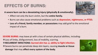
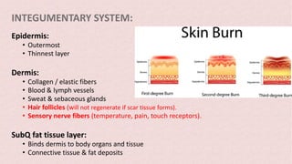
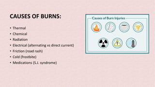
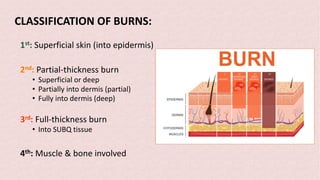
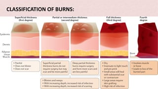
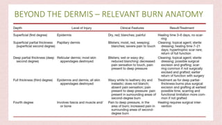
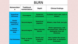
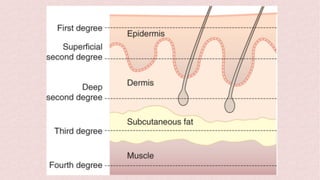
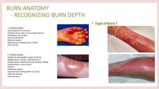
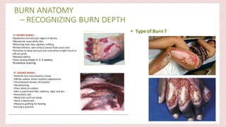
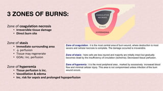
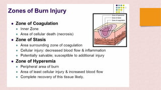
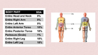
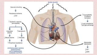
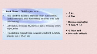
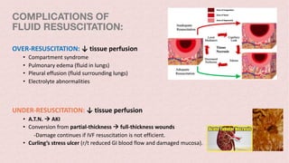
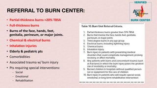
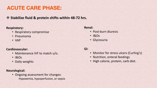
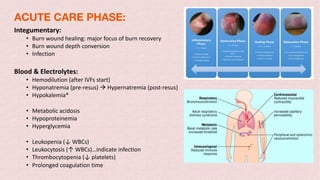
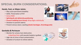
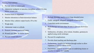
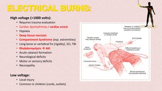
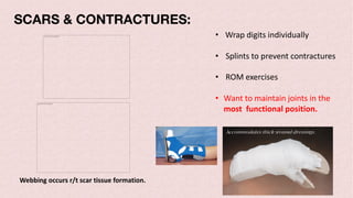
BURNS
Linda H. Warren
EdD RN MSN CCRN
The picture can't be displayed.
OBJECTIVES:
• Review anatomy (elderly)
• Causes of burns
• Burn classification & severity
• Extent of Injury
v TBSA*
v Categories and Zones
Physiological Responses
v Hypovolemic stage
• Relative hypovolemia r/t fluid shifts.
v Diuretic stage
• r/t IVF resuscitation & overhydration.
Treatment
v Emergent
v Acute (hospitalization)
v Rehabilitation
EFFECTS OF BURNS:
A severe burn can be a devastating injury (physically & emotionally).
• Affect not only the burn victim, but the entire family.
• Burns can also cause emotional problems such as depression, nightmares, or PTSD.
• Loss of a friend, family member, or possessions may add grief to the emotional
impact of a burn.
SEVERE BURNS: may leave pt with a loss of certain physical abilities, including:
vLoss of limbs, disfigurement, loss of mobility, scarring.
vRECURRENT INFECTIONS: burned skin has ↓ ability to fight infection.
vSevere burns can penetrate deep skin layers, causing muscle or tissue
damage that may affect every system of the body.
INTEGUMENTARY SYSTEM:
Epidermis:
• Outermost
• Thinnest layer
Dermis:
• Collagen / elastic fibers
• Blood & lymph vessels
• Sweat & sebaceous glands
• Hair follicles (will not regenerate if scar tissue forms).
• Sensory nerve fibers (temperature, pain, touch receptors).
SubQ fat tissue layer:
• Binds dermis to body organs and tissue
• Connective tissue & fat deposits
EFFECTS OF AGING ON SKIN:
• Less resilience to mechanical trauma.
• Atrophy of dermal & subQ tissues
• ↓ microcirculation
• ↓ immunological response
• ↓ physiological reserve
• Co-morbidities
• ↑ morbidity and mortality
v Tissue changes predispose elderly pts to
deeper wounds & slower healing.
v Massive amts of IVF can effect renal and
cardiac fx in pts with CHF or renal disease.
SKIN FUNCTIONS:
• Protection
• Temperature regulation
• Regulates fluid loss
• Vitamin D synthesis
• Sensory perception
• Person’s identity
• Body image
• Cosmetic appearance
CAUSES OF BURNS:
• Thermal
• Chemical
• Radiation
• Electrical (alternating vs direct current)
• Friction (road rash)
• Cold (frostbite)
• Medications (S.J. syndrome)
CLASSIFICATION OF BURNS:
1st: Superficial skin (into epidermis)
2nd: Partial-thickness burn
• Superficial or deep
• Partially into dermis (partial)
• Fully into dermis (deep)
3rd: Full-thickness burn
• Into SUBQ tissue
4th: Muscle & bone involved
CLASSIFICATION OF BURNS:
1st Degree
• Superficial
• Classic sunburn
• Scald injury
• Localized to epidermis
The picture can't be displayed.
2nd Degree (Superficial)
• Extends into upper dermis.
• Blisters, blanching, painful (ex: severe sunburn)
• Weepy “wet” appearance
• Regeneration of hair follicles / sweat glands within 7-14 days
The picture can't be displayed.
2nd Degree (Deep)
• Partial-thickness, extends deep into dermis.
• Nerves & blood vessels damaged.
• Pale, mottled, dry skin.
• ↓ sensation
The picture can't be displayed.
3rd Degree (Full-Thickness)
The picture can't be displayed.
• Destroys epidermis, dermis, & accessory structures
• Extends into SUBQ tissue
• Dry, brown, yellow, or black in color
• Non-pliable à feels leathery
• May need skin graft
• ↓ sensation
3 ZONES OF BURNS:
Zone of coagulation necrosis
• Irreversible tissue damage
• Direct burn site
Zone of stasis
• Immediate surrounding area
• ↓ perfusion
• Tissue may regenerate
• GOAL: inc. perfusion
Zone of hyperemia
• Tissue perfusion is inc.
• Vasodilation & edema
• Inc. risk for sepsis and prolonged hypoperfusion
The picture can't be displayed.
3 ZONES OF BURNS:
Extent of Injury (TBSA)
**RULE OF NINES: The picture can't be displayed.
LUND-BROWDER
The picture can't be displayed.
RULE OF PALMS:
The picture can't be displayed.
• Used for scattered burns.
• Not measured with nurse’s
palm, but with pt’s palm.
• Entire hand up to the wrist
= 1% TBSA
• Lung Injury can occur with…
- Smoke
- Chemical Toxins
- Byproducts of incomplete combustion (treated wood, sutt)
• Exposure to Toxic Gases:
- Carbon monoxide poisoning
- Cyanide poisoning
• Supraglottic Injury:
- Thermal injuries most common cause.
- Injury to pharynx & larynx à airway obstruction
• Subglottic Injury:
- Chemical injury most common cause.
- Damages ciliary function
- Erythema & edema à bronchospasms
- Mucosal ulceration
INHALATION INJURIES:
The picture can't be displayed.
• Exposure to smoke in a confined area.
• Facial burns / singed nasal hair
• Soot around mouth / nose
• Carbonaceous sputum (black)
• Hypoxemia (agitation, tachycardia, restless, dysrhythmias, confusion)
• Abnormal breath sounds
• Respiratory distress (↑ WOB, sternal retractions, dysphagia)
• ↑ carboxyHgb levels
• Abnormal ABGs (↑ RR à alkalosis, ↓ RR à acidosis)
• Inactivation of surfactant results in:
- Alveolar collapse
- Pneumonia
- Pulmonary edema
INHALATION INJURIES:
CLINICAL MANIFESTATIONS
TOXIC GASES:
Carbon Monoxide (CO) Poisoning:
• Binds to Hgb more rapidly than oxygen.
• Cells starved of O2 even with supp. oxygen
• Tissue Hypoxia
Cyanide Poisoning:
• Binds to respiratory enzymes in the mitochondria
• Inhibits cell metabolism
• Impaired oxygen utilization
Carbon Monoxide Poisoning
• Carboxyhemoglobin Levels (COHgb)
• Pulse ox unable to differentiate between oxyHgb/ COHgb à INACCURATE PULSE OX READING
COHgb <10-15%
• No Symptoms
• Headache or visual acuity
COHgb 15-40%
• CNS dysfunction
• Restless, confused, dizzy
• Impaired dexterity
• N/V
COHgb 40-60%
• Loss of consciousness
• Tachycardia / Tachypnea
• Seizures
• Cherry red or cyanotic skin.
COHgb >60%
• Coma / death
• Falsely elevated SpO2 readings
in pt’s with CO poisoning.
• Need to put pts on
supplemental oxygen.
If pt given 100% O2 or placed in
hyperbaric chamber… half-life of
COHgb is cut substantially.
PATHO. OF BURNS:
• Initial Hemostatic response
-Coagulation & microvascular constriction
• Resuscitative Phase
-Vasodilation & capillary leaking (weeping)
• Epithelization
-Restoration of fluid balance, temp. regulation,
& microbial barrier FX (skin integrity).
• Fibrogenesis
-Associated with wound appearance & tissue strength.
• Capillaries leak à Third spacing
• F&E Imbalances: Na+, K+, Ca+, lactate
- Hypovolemia
- pH: acidosis (r/t muscle breakdown)
- Hyperkalemia**
- Hyponatremia
• Hemoconcentration:
- ↑ blood viscosity
- ↓ perfusion
- May show high H+H prior to IVF resuscitation.
- After starting IVF… H+H will ↓ drastically.
- Tissue hypoxia
PATHO. OF BURNS:
Multi-Organ Effects of Burns
The picture can't be displayed.
Curling’s ulcer
Rhabdomyolysis à AKI
Electrolyte Disturbances:
Initial Resuscitation (0-36 hrs after injury):
• Begins at time of injury (not EMS arrival).
• Depends on burn severity.
• Hyperkalemia à cells damaged à release of K+ from cells à dysrhythmias
• Hyponatremia à capillary leakage à edema
• Pts will require serial bloodwork: BMP (q2-4h)
- CENTRAL ACCESS for frequent lab tests.
- IO access may be required.
Post-Resuscitation:
• Hypernatremia
• Hypokalemia
• Hypocalcemia
• Hypomagnesemia
• Hypophosphatemia
Electrolyte Disturbances: Post-Resuscitation
• Hypernatremia: Fluid shifts intravascularly
- Change IVF to isotonic or hypotonic (shifts fluid into cells)
- Isotonic: NS, LRs, D5W
- Hypotonic: 0.45% NS, 0.33% NS, 2.5 D5W
• Hypokalemia: K+ shifts back into cells & urinary losses of K+
- Monitor EKG for dysrhythmias
• Hypocalcemia: Ca+ shift back into cells & urinary losses of Ca+
• Hypomagnesemia: Coexistent with hypokalemia & hypocalcemia
• Hypophosphatemia: Mobilization of fluid & nutritional status
- Phosphorus always shows us pt’s NUTRITIONAL STATUS***
- Low phosphorus = poor nutrition
vIVF Resuscitation: Based on burn size (TBSA)
vFluid of choice: Lactated Ringer’s
Formula is used for…
• Burns covering >20% TBSA
• 2nd degree burns
• 3rd degree burns
Adult Formula: 4ml x weight (kg) x TBSA burned
• Initial 24-hr fluid requirement:
- Half of the volume to be infused given in the first 8 hrs.
- Remaining half given over next 16 hrs.
- Based on initial time of burn event (NOT arrival to ED).
- Titrate infusion rate to urinary output.
- Want ↑ u/o to prevent AKI (100-150 mL/hr)
PARKLAND FORMULA:
Children with burns >20%:
• 2ml x weight (kg) x TBSA burned
• Add calculated maintenance fluid:
0–10 kg à 100cc/kg
11–20 kg à 50 cc/kg (+1000)
>21 kg à 20 cc/kg (+1500)
• Divide total by 24 à hourly maintenance rate (cc/hr)
Lactated Ringer’s (LRs)***
- First half given over 8 hrs.
- Second half given over 16 hrs.
- Titrate infusion rate to urinary output.
- Contains buffers that help with metabolic acidosis & ↑ lactic acid levels.
2
PARKLAND FORMULA:
RESUSCITATIVE PHASE: (pre-hospital)
• Extricate the patient safely
• STOP the burning process!!!
ü Cool with tepid water
ü Never apply ice! (vasoconstriction à further tissue damage)
ü Remove all jewelry
ü Clothing adhering to skin is NOT removed.
ü If clothing is not adhering, remove it.
• Electrical Injuries
ü Remove patient from source.
ü Protect rescuer (wood), use PPE
• Chemical Injuries:
ü Brush powdered chemicals off prior to removing clothes & lavaging.
ü Remove all clothing
ü Lavage with clean water prior to & during transport.
ü Remove contact lenses.
ü Irrigate eyes: inner à outer canthus (prevent cross-contamination)
ü Neutralizing agents not recommended
vIdentify life-threatening injuries (secondary injuries: internal organs, frx)
vRapid transport to hospital – Minimize time on scene.
PRIMARY SURVERY:
Airway with c-spine precautions:
• Assess for inhalation injuries
• Facial burns
• Stridor
Breathing:
• NRB 100%
• Intubation
Circulation:
• Remove clothing and jewelry
• Two large bore IVs through non-burned skin.
• Administer LRs.
• Monitor for signs of hypovolemia.
• Prevent heat loss à cover with clean sheets & blankets
RESUSCITATIVE PHASE: (pre-hospital)
1. Administer High Flow 100% Oxygen at 15 L/min (NRB or ETT)
2. Assess Breathing: Look for signs and symptoms of inhalation injury & deterioration.
3. Assess for Deep Circumferential Chest Burns
• Burns can restrict chest expansion à altered respirations.
• Burnt skin loses elasticity quickly à tourniquet effect when combined with rapidly accumulating edema.
• Respiratory insufficiency or ischemia of an extremity are a risk.
• Escharotomy may be required.
4. Assess for COHb Poisoning:
• Consider CO poisoning in pts who sustain burns in an enclosed area.
• CO has a 280x greater affinity for Hgb than oxygen does.
• O2 administration is pivotal to help O2 bind to Hgb & unbound CO can be exhaled.
• SpO2 is unreliable indicator of CO poisoning.
• Pulse-ox interprets CO as oxygen à pt can be hypoxic with high SpO2 readings
-Supplemental O2 should be given regardless of SpO2
Assess for symptoms of high COHb levels:
• Hx of altered LOC
• Cherry pink skin
• Anxiety, restlessness
• HR
• BP à arterial line
• Urine output:
- Insert a Foley
- Monitor hourly u/o
- Simple way to assess fluid balance
• Distal pulses
• Capillary refill
• Color and turgor of unburned skin
• ABGs
• Serum lactate
ASSESS FLUID STATUS:
PREVENT HYPOTHERMIA:
1. Cover the burn ASAP
2. Stop cooling the wound if core temperature <35oC
3. Cover the patient with blankets:
• Warm blankets
• Space blankets
• Forced air warming blanket (Bair Hugger)
4. Warm IV fluids
5. Keep the room warm
6. Check temp. regularly
7. Remove any wet dressings, sheets etc. (evaporative heat loss)
q Exposure in confined area
q Facial burns / singed nasal hair
q Soot around mouth / nose
q Carbonaceous sputum (black)
• Massive edema develops rapidly à requires ETT
• Longer they wait, the narrower the airway gets.
• If airway is completely closed off…need to trach pt.
Electrical injuries:
• HIGH RISK of ATN r/t myoglobin release à
rhabdomyolysis à AKI
• Require LARGE VOLUMES of IVF replacement.
BURN SHOCK:
• Shock from intravascular volume loss r/t sudden F&E shifts
immediately following a burn injury.
• Combination of distributive & hypovolemic shock.
• ↑ capillary permeability, third-spacing à relative hypovolemia
• ↓ oncotic pressure:
- Leaking of proteins (fluid follows protein)
- Draws intravascular fluid into interstitium (tissues).
- Relative hypovolemia (fluids moves intravascularly à interstitially)
• Edema formation
- Due to ↓ oncotic pressure
- Natural inflammatory response
- Risk of compartment-like syndrome à escharotomy or fasciotomy
• ↑ K+
• ↓ Na+
• Hemoconcentration
• ↑ Hgb, ↑ Hct
• ↑ lactic acid
• Metabolic acidosis
COMPLICATIONS OF
FLUID RESUSCITATION:
OVER-RESUSCITATION: ↓ tissue perfusion
• Compartment syndrome
• Pulmonary edema (fluid in lungs)
• Pleural effusion (fluid surrounding lungs)
• Electrolyte abnormalities
UNDER-RESUSCITATION: ↓ tissue perfusion
• A.T.N. à AKI
• Conversion from partial-thickness à full-thickness wounds
-Damage continues if IVF resuscitation is not efficient.
• Curling’s stress ulcer (r/t reduced GI blood flow and damaged mucosa).
PHASES of BURN PROGRESSION:
The picture can't be displayed.
REFERRAL TO BURN CENTER:
• Partial-thickness burns >20% TBSA
• Full-thickness burns
• Burns of the face, hands, feet,
genitalia, perineum, or major joints.
• Chemical & electrical burns
• Inhalation injuries
• Elderly & pediatric pts
• Comorbidities
• Associated trauma w/ burn injury
• Pts requiring special interventions:
- Social
- Emotional
- Rehabilitation
v Stabilize fluid & protein shifts within 48-72 hrs.
Respiratory:
• Respiratory compromise
• Pneumonia
• VAP
Cardiovascular:
• Maintenance IVF to match u/o.
• I&Os
• Daily weights
Neurological:
• Ongoing assessment for changes:
-Hypoxemia, hypoperfusion, or sepsis
Renal:
• Post-burn diuresis
• I&Os
• Glycosuria
GI:
• Monitor for stress ulcers (Curling’s)
• Nutrition, enteral feedings
• High calorie, protein, carb diet.
ACUTE CARE PHASE:
Integumentary:
• Burn wound healing: major focus of burn recovery
• Burn wound depth conversion
• Infection
Blood & Electrolytes:
• Hemodilution (after IVFs start)
• Hyponatremia (pre-resus) à Hypernatremia (post-resus)
• Hypokalemia*
• Metabolic acidosis
• Hypoproteinemia
• Hyperglycemia
• Leukopenia (↓ WBCs)
• Leukocytosis (↑ WBCs)…indicate infection
• Thrombocytopenia (↓ platelets)
• Prolonged coagulation time
ACUTE CARE PHASE:
SPECIAL BURN CONSIDERATIONS:
Facial Burns:
• Potential for inhalation injuries
• Edema à compromised airway à ETT
• Elevate HOB
• Prevent excessive bleeding & damage to new tissue growth.
• Topical antimicrobial agent
Ear Burns:
• Chondritis (r/t poor vascular supply of ear)
• Prevent pressure on area
• Sulfamylon (mafenide acetate): antimicrobial of choice
Eye Burns:
• Remove contact lenses
• Exam by ophthalmologist
• Irrigate / Irrigate / Irrigate
• Protect cornea & conjunctive from exposure / drying out (eyelid retraction)
• Ophthalmic ointments and artificial tears.
All will go to burn centers!
Hands, Feet, or Major Joints:
• May cause permanent disability
• GOAL: Preservation of FX!!
• Active ROM
• Splinting & anti-deformity positioning
• Prevent webbing (scar tissue): Wrap digits individually
• Elevation to prevent edema
• Prevent venous stasis / pooling (elastic bandages, thromboguards)
Genitalia & Perineum
• Monitor for urinary tract obstruction.
-Indwelling Foley until wounds healed or grafted.
• High risk of urine or fecal contamination.
• Elevation of scrotum to relieve edema.
SPECIAL BURN CONSIDERATIONS:
High voltage (>1000 volts):
• Requires trauma evaluation
• Cardiac dysrhythmias / cardiac arrest
• Hypoxia
• Deep tissue necrosis
• Compartment Syndrome (esp. extremities)
• Long bone or vertebral frx (rigidity), SCI, TBI
• Rhabdomyolysis à AKI
• Acute cataract formation
• Neurological deficits
• Motor or sensory deficits
• Neuropathy
Low voltage:
• Local Injury
• Common in children (cords, outlets)
• Voltage takes the shortest path to the ground.
ENTERANCE & EXIT BURNS:
• Extensive tissue damage occurs btwn.
entrance and exit wounds.
• Tissue necrosis
• Upon exiting, the electricity can cause
tissues to explode.
• Exit wounds may be larger.
The picture can't be displayed.
SCARS & CONTRACTURES:
The picture can't be displayed.
• Wrap digits individually
• Splints to prevent contractures
• ROM exercises
• Want to maintain joints in the
most functional position.
Webbing occurs r/t scar tissue formation.
• Stop burning process!!!
• Protective equipment (PPE)
• Decontamination required:
• Brush off powder substance
• Continuous water irrigation
• ALKALIS: require longer irrigation
• Determine specific chemical agent
• Control pain
• Minimize heat loss à cover with dry blankets
• Monitor for signs of chemical absorption
• Chemicals can be absorbed thru. skin à systemic complications
PAIN CONTROL:
MAJOR FOCUS OF BURN MANAGEMENT:
• Background pain
• Procedural pain
• Meds often exceed normal dosing guidelines
• Hypermetabolism à require higher doses of pain meds.
Assess pain levels frequently:
• Pain meds: ALWAYS given IV***
• SUBQ & IM injections ineffective: impaired circulation à altered absorption rates
• Cannot determine rate of absorption if given IM or SUBQ…always given IV.
Tx of anxiety:
• Anxiolytics
• Relaxation, hypnosis, guided imagery, distraction
Pt’s with HX of alcohol or substance abuse:
• Compound pain management
• Opiates: drug of choice
• Combo of opioids + sedatives
• Anxiolytics
Susceptibility to infection in burn pts:
• Loss of skin (first line of defense)
• Extended hospital stays
• Invasive procedures
• IVs, central lines
• Foley catheters
• Immuno-compromised
INFECTION CONTROL MEASURES:
• Aseptic management of wound & environment
• Topical antibacterial agents
• Care of invasive catheters (central lines, A-line, Foley)
• Aggressive wound management
• Prevention of MDROs: identification of infectious organism
• Use of appropriate ABX
• Monitor lab values / signs of sepsis
• Early wound closure / restore skin barrier
• Adequate nutrition
• Interventions to improve patient outcomes
• Staff education
STEVENS-JOHNSON SYNDROME (SJS):
Reaction caused by certain meds: OTC drugs, ABX, sulfa drugs
• Nikolsky sign: epidermis separates from dermis with even slight rubbing.
• Mucosal sloughing: assess oral cavity
• >20% TBSA à Toxic epidermal necrolysis (TENS)
• Stop the drug
• Resuscitate (IVF)
The picture can't be displayed.
ASSESS FOR CHILD ABUSE:
• Donut pattern on buttocks
• Stocking pattern
• Glove pattern
• Sparing: soles of feet, back of knees
• Line of demarcation
• Waterlines
• Scalds
DIFFUSE ROAD-RASH:
The picture can't be displayed.
• Very painful
• Bacitracin
• Debridement of dirt & stones
• Topical agents
• Heal fairly quickly
WOUND MANAGEMENT & GOALS:
• Cleansing & debridement
• Topical ABX: delay wound colonization & infection
• Escharotomy or fasciotomy:
- For circumferential burns, deep burns, compartment syndrome.
• Elevate extremities
• Surgical excision & grafting
GOALS:
• Close wound quickly
• Prevent infection
• Reduce scarring & contractures
• Provide comfort
TOPICAL ABX:
• Silvadene (sulfadiazine) cream:
- Broad-spectrum, covers pseudomonas
- Does not penetrate eschar
- Cooling affect
- Side effects: neutropenia, thrombocytopenia
• Silver nitrate cream:
- Blackens skin
- Side effects: electrolyte abnormalities
• Sulfamylon cream or solution (mafenide acetate):
- Penetrates eschar & deep into burn wounds.
- PAINFUL!!
- Face & neck edema
- Side effects: Metabolic acidosis à via carbonic anhydrase inhibition
NEW WOUND CARE:
• Bacitracin
• Adaptic
• Vaseline gauze
• Xeroform gauze
• Medi-Honey
SURGICAL INT:
Escharotomy:
• Prevent circumferential constriction
• Removal of eschar
• Facilitates healing
Surgical Debridement:
• Removes burn wound to level of viable tissue
• Should bleed briskly before coagulation.
• Know tissue is viable if it bleeds.
• Early excision & grafting improves function & ↓ mortality
Autografting – permanent skin coverage:
• Homograft / allograft – human skin
• Heterograft / xenograft – animal (usually pig)
• Dermal substitute dressings – Biobrane, Intregra
• Full-thickness skin grafts
• Split-thickness skin grafts
Cultured Epithelial Autografts
The picture can't be displayed.
ESCHAROTOMY:
The picture can't be displayed.
• Longitudinal incision thru eschar itself.
• Only down to SUBQ tissue (not thru!!)
• Relieves stiff eschar that forms (thru epidermis & dermis)
Excision & Debridement
The picture can't be displayed.
SKIN GRAFTING:
The picture can't be displayed.
• Need to debride wound FIRST.
• Viable tissue: bleeds
• Mesh graft placed onto wound bed
• Stapled to skin
• Keep it moist à covered w.
metahoney, xeroform dressing
SKIN GRAFTING:
The picture can't be displayed.
SKIN GRAFTING:
The picture can't be displayed.
SKIN GRAFTING:
The picture can't be displayed.
SKIN GRAFTING:
SKIN GRAFTING:
The picture can't be displayed.
The picture can't be displayed. The picture can't be displayed.
• Harvest skin from larger areas
• Typically the thigh
NUTRITIONAL
CONSIDERATIONS:
• Stress-induced hypermetabolic / catabolic response
• Post-burn hypermetabolism:
- Skeletal muscle breakdown
- Delayed wound healing / skin graft loss
- Impaired immunological response
• Muscle weakness & atrophy:
- r/t prolonged mechanical ventilation
- Extended rehabilitation time
• Oral Diet
- High-calorie, high-protein diet w. protein supplements
- Protein: 1.5-2 g/kg/day
• Enteral feedings
• Beta-blockers: propranolol (lowers metabolic rate & release of
cytokines / stress hormones)
• Anabolic hormones: oxandrolone
- These meds help ↓ length of stay by ↓ metabolism

Burns

  • 1.
    BURNS Linda H. Warren EdDRN MSN CCRN The picture can't be displayed.
  • 2.
    OBJECTIVES: • Review anatomy(elderly) • Causes of burns • Burn classification & severity • Extent of Injury v TBSA* v Categories and Zones Physiological Responses v Hypovolemic stage • Relative hypovolemia r/t fluid shifts. v Diuretic stage • r/t IVF resuscitation & overhydration. Treatment v Emergent v Acute (hospitalization) v Rehabilitation
  • 3.
    EFFECTS OF BURNS: Asevere burn can be a devastating injury (physically & emotionally). • Affect not only the burn victim, but the entire family. • Burns can also cause emotional problems such as depression, nightmares, or PTSD. • Loss of a friend, family member, or possessions may add grief to the emotional impact of a burn. SEVERE BURNS: may leave pt with a loss of certain physical abilities, including: vLoss of limbs, disfigurement, loss of mobility, scarring. vRECURRENT INFECTIONS: burned skin has ↓ ability to fight infection. vSevere burns can penetrate deep skin layers, causing muscle or tissue damage that may affect every system of the body.
  • 4.
    INTEGUMENTARY SYSTEM: Epidermis: • Outermost •Thinnest layer Dermis: • Collagen / elastic fibers • Blood & lymph vessels • Sweat & sebaceous glands • Hair follicles (will not regenerate if scar tissue forms). • Sensory nerve fibers (temperature, pain, touch receptors). SubQ fat tissue layer: • Binds dermis to body organs and tissue • Connective tissue & fat deposits
  • 5.
    EFFECTS OF AGINGON SKIN: • Less resilience to mechanical trauma. • Atrophy of dermal & subQ tissues • ↓ microcirculation • ↓ immunological response • ↓ physiological reserve • Co-morbidities • ↑ morbidity and mortality v Tissue changes predispose elderly pts to deeper wounds & slower healing. v Massive amts of IVF can effect renal and cardiac fx in pts with CHF or renal disease.
  • 6.
    SKIN FUNCTIONS: • Protection •Temperature regulation • Regulates fluid loss • Vitamin D synthesis • Sensory perception • Person’s identity • Body image • Cosmetic appearance
  • 7.
    CAUSES OF BURNS: •Thermal • Chemical • Radiation • Electrical (alternating vs direct current) • Friction (road rash) • Cold (frostbite) • Medications (S.J. syndrome)
  • 8.
    CLASSIFICATION OF BURNS: 1st:Superficial skin (into epidermis) 2nd: Partial-thickness burn • Superficial or deep • Partially into dermis (partial) • Fully into dermis (deep) 3rd: Full-thickness burn • Into SUBQ tissue 4th: Muscle & bone involved
  • 9.
  • 12.
    1st Degree • Superficial •Classic sunburn • Scald injury • Localized to epidermis The picture can't be displayed. The picture can't be displayed.
  • 13.
    2nd Degree (Superficial) •Extends into upper dermis. • Blisters, blanching, painful (ex: severe sunburn) • Weepy “wet” appearance • Regeneration of hair follicles / sweat glands within 7-14 days The picture can't be displayed. The picture can't be displayed.
  • 14.
    2nd Degree (Deep) •Partial-thickness, extends deep into dermis. • Nerves & blood vessels damaged. • Pale, mottled, dry skin. • ↓ sensation The picture can't be displayed. The picture can't be displayed. The picture can't be displayed.
  • 18.
    3rd Degree (Full-Thickness) Thepicture can't be displayed. • Destroys epidermis, dermis, & accessory structures • Extends into SUBQ tissue • Dry, brown, yellow, or black in color • Non-pliable à feels leathery • May need skin graft • ↓ sensation
  • 20.
    3 ZONES OFBURNS: Zone of coagulation necrosis • Irreversible tissue damage • Direct burn site Zone of stasis • Immediate surrounding area • ↓ perfusion • Tissue may regenerate • GOAL: inc. perfusion Zone of hyperemia • Tissue perfusion is inc. • Vasodilation & edema • Inc. risk for sepsis and prolonged hypoperfusion
  • 22.
    The picture can'tbe displayed. 3 ZONES OF BURNS:
  • 23.
    Extent of Injury(TBSA) **RULE OF NINES: The picture can't be displayed.
  • 25.
    LUND-BROWDER The picture can'tbe displayed. The picture can't be displayed.
  • 26.
    RULE OF PALMS: Thepicture can't be displayed. • Used for scattered burns. • Not measured with nurse’s palm, but with pt’s palm. • Entire hand up to the wrist = 1% TBSA
  • 27.
    • Lung Injurycan occur with… - Smoke - Chemical Toxins - Byproducts of incomplete combustion (treated wood, sutt) • Exposure to Toxic Gases: - Carbon monoxide poisoning - Cyanide poisoning • Supraglottic Injury: - Thermal injuries most common cause. - Injury to pharynx & larynx à airway obstruction • Subglottic Injury: - Chemical injury most common cause. - Damages ciliary function - Erythema & edema à bronchospasms - Mucosal ulceration INHALATION INJURIES:
  • 28.
  • 29.
    • Exposure tosmoke in a confined area. • Facial burns / singed nasal hair • Soot around mouth / nose • Carbonaceous sputum (black) • Hypoxemia (agitation, tachycardia, restless, dysrhythmias, confusion) • Abnormal breath sounds • Respiratory distress (↑ WOB, sternal retractions, dysphagia) • ↑ carboxyHgb levels • Abnormal ABGs (↑ RR à alkalosis, ↓ RR à acidosis) • Inactivation of surfactant results in: - Alveolar collapse - Pneumonia - Pulmonary edema INHALATION INJURIES: CLINICAL MANIFESTATIONS
  • 31.
    TOXIC GASES: Carbon Monoxide(CO) Poisoning: • Binds to Hgb more rapidly than oxygen. • Cells starved of O2 even with supp. oxygen • Tissue Hypoxia Cyanide Poisoning: • Binds to respiratory enzymes in the mitochondria • Inhibits cell metabolism • Impaired oxygen utilization
  • 33.
    Carbon Monoxide Poisoning •Carboxyhemoglobin Levels (COHgb) • Pulse ox unable to differentiate between oxyHgb/ COHgb à INACCURATE PULSE OX READING COHgb <10-15% • No Symptoms • Headache or visual acuity COHgb 15-40% • CNS dysfunction • Restless, confused, dizzy • Impaired dexterity • N/V COHgb 40-60% • Loss of consciousness • Tachycardia / Tachypnea • Seizures • Cherry red or cyanotic skin. COHgb >60% • Coma / death
  • 34.
    • Falsely elevatedSpO2 readings in pt’s with CO poisoning. • Need to put pts on supplemental oxygen. If pt given 100% O2 or placed in hyperbaric chamber… half-life of COHgb is cut substantially.
  • 35.
    PATHO. OF BURNS: •Initial Hemostatic response -Coagulation & microvascular constriction • Resuscitative Phase -Vasodilation & capillary leaking (weeping) • Epithelization -Restoration of fluid balance, temp. regulation, & microbial barrier FX (skin integrity). • Fibrogenesis -Associated with wound appearance & tissue strength.
  • 36.
    • Capillaries leakà Third spacing • F&E Imbalances: Na+, K+, Ca+, lactate - Hypovolemia - pH: acidosis (r/t muscle breakdown) - Hyperkalemia** - Hyponatremia • Hemoconcentration: - ↑ blood viscosity - ↓ perfusion - May show high H+H prior to IVF resuscitation. - After starting IVF… H+H will ↓ drastically. - Tissue hypoxia PATHO. OF BURNS:
  • 39.
    Multi-Organ Effects ofBurns The picture can't be displayed. Curling’s ulcer Rhabdomyolysis à AKI
  • 40.
    Electrolyte Disturbances: Initial Resuscitation(0-36 hrs after injury): • Begins at time of injury (not EMS arrival). • Depends on burn severity. • Hyperkalemia à cells damaged à release of K+ from cells à dysrhythmias • Hyponatremia à capillary leakage à edema • Pts will require serial bloodwork: BMP (q2-4h) - CENTRAL ACCESS for frequent lab tests. - IO access may be required. Post-Resuscitation: • Hypernatremia • Hypokalemia • Hypocalcemia • Hypomagnesemia • Hypophosphatemia
  • 41.
    Electrolyte Disturbances: Post-Resuscitation •Hypernatremia: Fluid shifts intravascularly - Change IVF to isotonic or hypotonic (shifts fluid into cells) - Isotonic: NS, LRs, D5W - Hypotonic: 0.45% NS, 0.33% NS, 2.5 D5W • Hypokalemia: K+ shifts back into cells & urinary losses of K+ - Monitor EKG for dysrhythmias • Hypocalcemia: Ca+ shift back into cells & urinary losses of Ca+ • Hypomagnesemia: Coexistent with hypokalemia & hypocalcemia • Hypophosphatemia: Mobilization of fluid & nutritional status - Phosphorus always shows us pt’s NUTRITIONAL STATUS*** - Low phosphorus = poor nutrition
  • 42.
    vIVF Resuscitation: Basedon burn size (TBSA) vFluid of choice: Lactated Ringer’s Formula is used for… • Burns covering >20% TBSA • 2nd degree burns • 3rd degree burns Adult Formula: 4ml x weight (kg) x TBSA burned • Initial 24-hr fluid requirement: - Half of the volume to be infused given in the first 8 hrs. - Remaining half given over next 16 hrs. - Based on initial time of burn event (NOT arrival to ED). - Titrate infusion rate to urinary output. - Want ↑ u/o to prevent AKI (100-150 mL/hr) PARKLAND FORMULA:
  • 43.
    Children with burns>20%: • 2ml x weight (kg) x TBSA burned • Add calculated maintenance fluid: 0–10 kg à 100cc/kg 11–20 kg à 50 cc/kg (+1000) >21 kg à 20 cc/kg (+1500) • Divide total by 24 à hourly maintenance rate (cc/hr) Lactated Ringer’s (LRs)*** - First half given over 8 hrs. - Second half given over 16 hrs. - Titrate infusion rate to urinary output. - Contains buffers that help with metabolic acidosis & ↑ lactic acid levels. 2 PARKLAND FORMULA:
  • 45.
    RESUSCITATIVE PHASE: (pre-hospital) •Extricate the patient safely • STOP the burning process!!! ü Cool with tepid water ü Never apply ice! (vasoconstriction à further tissue damage) ü Remove all jewelry ü Clothing adhering to skin is NOT removed. ü If clothing is not adhering, remove it. • Electrical Injuries ü Remove patient from source. ü Protect rescuer (wood), use PPE • Chemical Injuries: ü Brush powdered chemicals off prior to removing clothes & lavaging. ü Remove all clothing ü Lavage with clean water prior to & during transport. ü Remove contact lenses. ü Irrigate eyes: inner à outer canthus (prevent cross-contamination) ü Neutralizing agents not recommended
  • 46.
    vIdentify life-threatening injuries(secondary injuries: internal organs, frx) vRapid transport to hospital – Minimize time on scene. PRIMARY SURVERY: Airway with c-spine precautions: • Assess for inhalation injuries • Facial burns • Stridor Breathing: • NRB 100% • Intubation Circulation: • Remove clothing and jewelry • Two large bore IVs through non-burned skin. • Administer LRs. • Monitor for signs of hypovolemia. • Prevent heat loss à cover with clean sheets & blankets RESUSCITATIVE PHASE: (pre-hospital)
  • 47.
    1. Administer HighFlow 100% Oxygen at 15 L/min (NRB or ETT) 2. Assess Breathing: Look for signs and symptoms of inhalation injury & deterioration. 3. Assess for Deep Circumferential Chest Burns • Burns can restrict chest expansion à altered respirations. • Burnt skin loses elasticity quickly à tourniquet effect when combined with rapidly accumulating edema. • Respiratory insufficiency or ischemia of an extremity are a risk. • Escharotomy may be required. 4. Assess for COHb Poisoning: • Consider CO poisoning in pts who sustain burns in an enclosed area. • CO has a 280x greater affinity for Hgb than oxygen does. • O2 administration is pivotal to help O2 bind to Hgb & unbound CO can be exhaled. • SpO2 is unreliable indicator of CO poisoning. • Pulse-ox interprets CO as oxygen à pt can be hypoxic with high SpO2 readings -Supplemental O2 should be given regardless of SpO2 Assess for symptoms of high COHb levels: • Hx of altered LOC • Cherry pink skin • Anxiety, restlessness
  • 48.
    • HR • BPà arterial line • Urine output: - Insert a Foley - Monitor hourly u/o - Simple way to assess fluid balance • Distal pulses • Capillary refill • Color and turgor of unburned skin • ABGs • Serum lactate ASSESS FLUID STATUS:
  • 49.
    PREVENT HYPOTHERMIA: 1. Coverthe burn ASAP 2. Stop cooling the wound if core temperature <35oC 3. Cover the patient with blankets: • Warm blankets • Space blankets • Forced air warming blanket (Bair Hugger) 4. Warm IV fluids 5. Keep the room warm 6. Check temp. regularly 7. Remove any wet dressings, sheets etc. (evaporative heat loss)
  • 50.
    q Exposure inconfined area q Facial burns / singed nasal hair q Soot around mouth / nose q Carbonaceous sputum (black) • Massive edema develops rapidly à requires ETT • Longer they wait, the narrower the airway gets. • If airway is completely closed off…need to trach pt. Electrical injuries: • HIGH RISK of ATN r/t myoglobin release à rhabdomyolysis à AKI • Require LARGE VOLUMES of IVF replacement.
  • 52.
    BURN SHOCK: • Shockfrom intravascular volume loss r/t sudden F&E shifts immediately following a burn injury. • Combination of distributive & hypovolemic shock. • ↑ capillary permeability, third-spacing à relative hypovolemia • ↓ oncotic pressure: - Leaking of proteins (fluid follows protein) - Draws intravascular fluid into interstitium (tissues). - Relative hypovolemia (fluids moves intravascularly à interstitially) • Edema formation - Due to ↓ oncotic pressure - Natural inflammatory response - Risk of compartment-like syndrome à escharotomy or fasciotomy
  • 54.
    • ↑ K+ •↓ Na+ • Hemoconcentration • ↑ Hgb, ↑ Hct • ↑ lactic acid • Metabolic acidosis
  • 55.
    COMPLICATIONS OF FLUID RESUSCITATION: OVER-RESUSCITATION:↓ tissue perfusion • Compartment syndrome • Pulmonary edema (fluid in lungs) • Pleural effusion (fluid surrounding lungs) • Electrolyte abnormalities UNDER-RESUSCITATION: ↓ tissue perfusion • A.T.N. à AKI • Conversion from partial-thickness à full-thickness wounds -Damage continues if IVF resuscitation is not efficient. • Curling’s stress ulcer (r/t reduced GI blood flow and damaged mucosa).
  • 56.
    PHASES of BURNPROGRESSION: The picture can't be displayed.
  • 57.
    REFERRAL TO BURNCENTER: • Partial-thickness burns >20% TBSA • Full-thickness burns • Burns of the face, hands, feet, genitalia, perineum, or major joints. • Chemical & electrical burns • Inhalation injuries • Elderly & pediatric pts • Comorbidities • Associated trauma w/ burn injury • Pts requiring special interventions: - Social - Emotional - Rehabilitation
  • 58.
    v Stabilize fluid& protein shifts within 48-72 hrs. Respiratory: • Respiratory compromise • Pneumonia • VAP Cardiovascular: • Maintenance IVF to match u/o. • I&Os • Daily weights Neurological: • Ongoing assessment for changes: -Hypoxemia, hypoperfusion, or sepsis Renal: • Post-burn diuresis • I&Os • Glycosuria GI: • Monitor for stress ulcers (Curling’s) • Nutrition, enteral feedings • High calorie, protein, carb diet. ACUTE CARE PHASE:
  • 60.
    Integumentary: • Burn woundhealing: major focus of burn recovery • Burn wound depth conversion • Infection Blood & Electrolytes: • Hemodilution (after IVFs start) • Hyponatremia (pre-resus) à Hypernatremia (post-resus) • Hypokalemia* • Metabolic acidosis • Hypoproteinemia • Hyperglycemia • Leukopenia (↓ WBCs) • Leukocytosis (↑ WBCs)…indicate infection • Thrombocytopenia (↓ platelets) • Prolonged coagulation time ACUTE CARE PHASE:
  • 62.
    SPECIAL BURN CONSIDERATIONS: FacialBurns: • Potential for inhalation injuries • Edema à compromised airway à ETT • Elevate HOB • Prevent excessive bleeding & damage to new tissue growth. • Topical antimicrobial agent Ear Burns: • Chondritis (r/t poor vascular supply of ear) • Prevent pressure on area • Sulfamylon (mafenide acetate): antimicrobial of choice Eye Burns: • Remove contact lenses • Exam by ophthalmologist • Irrigate / Irrigate / Irrigate • Protect cornea & conjunctive from exposure / drying out (eyelid retraction) • Ophthalmic ointments and artificial tears. All will go to burn centers!
  • 63.
    Hands, Feet, orMajor Joints: • May cause permanent disability • GOAL: Preservation of FX!! • Active ROM • Splinting & anti-deformity positioning • Prevent webbing (scar tissue): Wrap digits individually • Elevation to prevent edema • Prevent venous stasis / pooling (elastic bandages, thromboguards) Genitalia & Perineum • Monitor for urinary tract obstruction. -Indwelling Foley until wounds healed or grafted. • High risk of urine or fecal contamination. • Elevation of scrotum to relieve edema. SPECIAL BURN CONSIDERATIONS:
  • 65.
    High voltage (>1000volts): • Requires trauma evaluation • Cardiac dysrhythmias / cardiac arrest • Hypoxia • Deep tissue necrosis • Compartment Syndrome (esp. extremities) • Long bone or vertebral frx (rigidity), SCI, TBI • Rhabdomyolysis à AKI • Acute cataract formation • Neurological deficits • Motor or sensory deficits • Neuropathy Low voltage: • Local Injury • Common in children (cords, outlets)
  • 66.
    • Voltage takesthe shortest path to the ground. ENTERANCE & EXIT BURNS: • Extensive tissue damage occurs btwn. entrance and exit wounds. • Tissue necrosis • Upon exiting, the electricity can cause tissues to explode. • Exit wounds may be larger.
  • 67.
    The picture can'tbe displayed.
  • 68.
    SCARS & CONTRACTURES: Thepicture can't be displayed. The picture can't be displayed. • Wrap digits individually • Splints to prevent contractures • ROM exercises • Want to maintain joints in the most functional position. Webbing occurs r/t scar tissue formation.
  • 69.
    • Stop burningprocess!!! • Protective equipment (PPE) • Decontamination required: • Brush off powder substance • Continuous water irrigation • ALKALIS: require longer irrigation • Determine specific chemical agent • Control pain • Minimize heat loss à cover with dry blankets • Monitor for signs of chemical absorption • Chemicals can be absorbed thru. skin à systemic complications
  • 70.
    PAIN CONTROL: MAJOR FOCUSOF BURN MANAGEMENT: • Background pain • Procedural pain • Meds often exceed normal dosing guidelines • Hypermetabolism à require higher doses of pain meds. Assess pain levels frequently: • Pain meds: ALWAYS given IV*** • SUBQ & IM injections ineffective: impaired circulation à altered absorption rates • Cannot determine rate of absorption if given IM or SUBQ…always given IV. Tx of anxiety: • Anxiolytics • Relaxation, hypnosis, guided imagery, distraction Pt’s with HX of alcohol or substance abuse: • Compound pain management • Opiates: drug of choice
  • 71.
    • Combo ofopioids + sedatives • Anxiolytics
  • 72.
    Susceptibility to infectionin burn pts: • Loss of skin (first line of defense) • Extended hospital stays • Invasive procedures • IVs, central lines • Foley catheters • Immuno-compromised INFECTION CONTROL MEASURES:
  • 73.
    INFECTION CONTROL MEASURES: •Aseptic management of wound & environment • Topical antibacterial agents • Care of invasive catheters (central lines, A-line, Foley) • Aggressive wound management • Prevention of MDROs: identification of infectious organism • Use of appropriate ABX • Monitor lab values / signs of sepsis • Early wound closure / restore skin barrier • Adequate nutrition • Interventions to improve patient outcomes • Staff education
  • 74.
    STEVENS-JOHNSON SYNDROME (SJS): Reactioncaused by certain meds: OTC drugs, ABX, sulfa drugs • Nikolsky sign: epidermis separates from dermis with even slight rubbing. • Mucosal sloughing: assess oral cavity • >20% TBSA à Toxic epidermal necrolysis (TENS) • Stop the drug • Resuscitate (IVF) The picture can't be displayed.
  • 76.
    ASSESS FOR CHILDABUSE: • Donut pattern on buttocks • Stocking pattern • Glove pattern • Sparing: soles of feet, back of knees • Line of demarcation • Waterlines • Scalds
  • 78.
    DIFFUSE ROAD-RASH: The picturecan't be displayed. • Very painful • Bacitracin • Debridement of dirt & stones • Topical agents • Heal fairly quickly
  • 79.
    WOUND MANAGEMENT &GOALS: • Cleansing & debridement • Topical ABX: delay wound colonization & infection • Escharotomy or fasciotomy: - For circumferential burns, deep burns, compartment syndrome. • Elevate extremities • Surgical excision & grafting GOALS: • Close wound quickly • Prevent infection • Reduce scarring & contractures • Provide comfort
  • 80.
    TOPICAL ABX: • Silvadene(sulfadiazine) cream: - Broad-spectrum, covers pseudomonas - Does not penetrate eschar - Cooling affect - Side effects: neutropenia, thrombocytopenia • Silver nitrate cream: - Blackens skin - Side effects: electrolyte abnormalities • Sulfamylon cream or solution (mafenide acetate): - Penetrates eschar & deep into burn wounds. - PAINFUL!! - Face & neck edema - Side effects: Metabolic acidosis à via carbonic anhydrase inhibition
  • 81.
    NEW WOUND CARE: •Bacitracin • Adaptic • Vaseline gauze • Xeroform gauze • Medi-Honey
  • 82.
    SURGICAL INT: Escharotomy: • Preventcircumferential constriction • Removal of eschar • Facilitates healing Surgical Debridement: • Removes burn wound to level of viable tissue • Should bleed briskly before coagulation. • Know tissue is viable if it bleeds. • Early excision & grafting improves function & ↓ mortality Autografting – permanent skin coverage: • Homograft / allograft – human skin • Heterograft / xenograft – animal (usually pig) • Dermal substitute dressings – Biobrane, Intregra • Full-thickness skin grafts • Split-thickness skin grafts
  • 83.
    Cultured Epithelial Autografts Thepicture can't be displayed. The picture can't be displayed. The picture can't be displayed.
  • 84.
    ESCHAROTOMY: The picture can'tbe displayed. • Longitudinal incision thru eschar itself. • Only down to SUBQ tissue (not thru!!) • Relieves stiff eschar that forms (thru epidermis & dermis)
  • 86.
    Excision & Debridement Thepicture can't be displayed. The picture can't be displayed.
  • 87.
    The picture can'tbe displayed. SKIN GRAFTING:
  • 88.
    The picture can'tbe displayed. • Need to debride wound FIRST. • Viable tissue: bleeds • Mesh graft placed onto wound bed • Stapled to skin • Keep it moist à covered w. metahoney, xeroform dressing SKIN GRAFTING:
  • 89.
    The picture can'tbe displayed. SKIN GRAFTING:
  • 90.
    The picture can'tbe displayed. SKIN GRAFTING:
  • 91.
    The picture can'tbe displayed. The picture can't be displayed. The picture can't be displayed. The picture can't be displayed. SKIN GRAFTING:
  • 92.
    SKIN GRAFTING: The picturecan't be displayed. The picture can't be displayed. The picture can't be displayed. The picture can't be displayed. • Harvest skin from larger areas • Typically the thigh
  • 93.
    NUTRITIONAL CONSIDERATIONS: • Stress-induced hypermetabolic/ catabolic response • Post-burn hypermetabolism: - Skeletal muscle breakdown - Delayed wound healing / skin graft loss - Impaired immunological response • Muscle weakness & atrophy: - r/t prolonged mechanical ventilation - Extended rehabilitation time • Oral Diet - High-calorie, high-protein diet w. protein supplements - Protein: 1.5-2 g/kg/day • Enteral feedings • Beta-blockers: propranolol (lowers metabolic rate & release of cytokines / stress hormones) • Anabolic hormones: oxandrolone - These meds help ↓ length of stay by ↓ metabolism