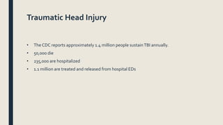
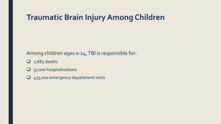
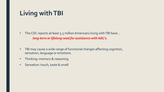
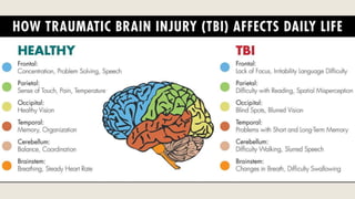
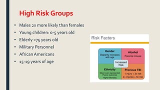
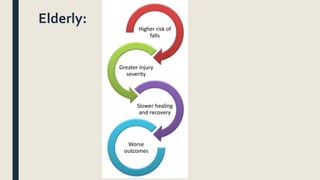
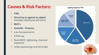
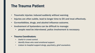
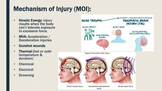
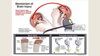
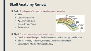
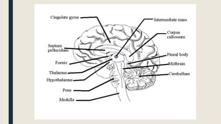
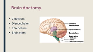
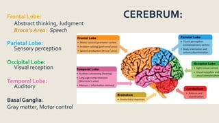
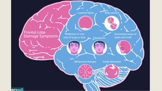
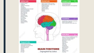
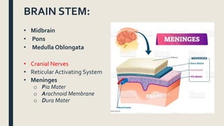
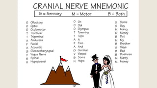
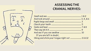
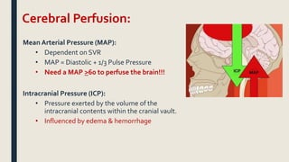
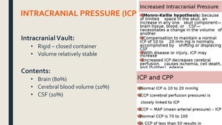
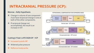
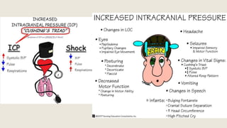
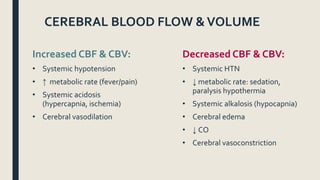
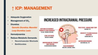
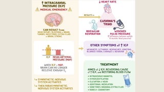
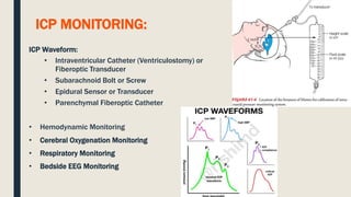
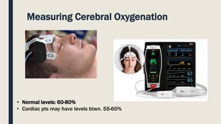
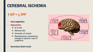
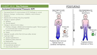
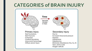
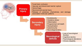
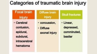
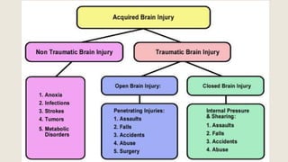
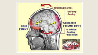
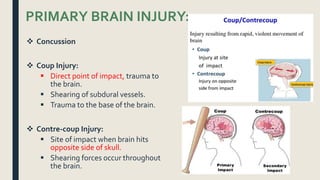
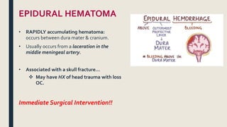
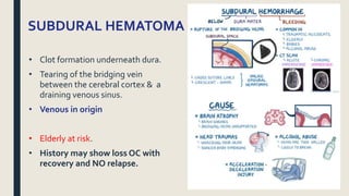
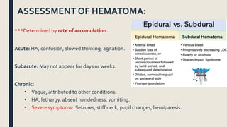
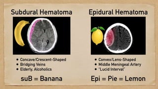
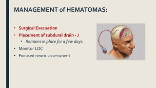
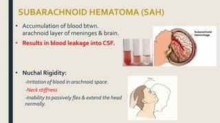
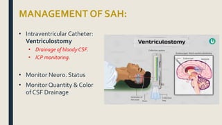
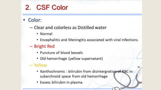
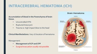
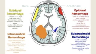
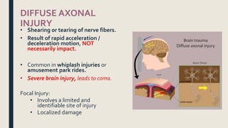
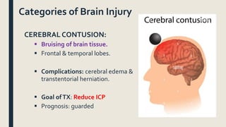
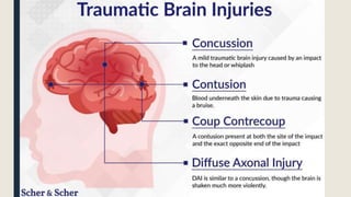
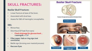
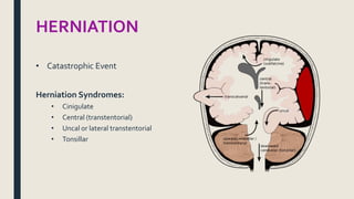
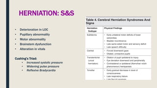
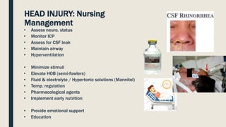
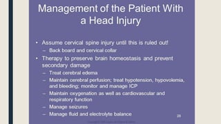
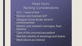
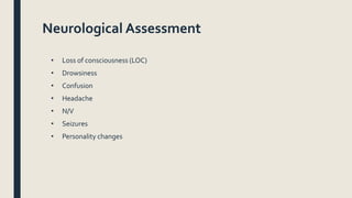
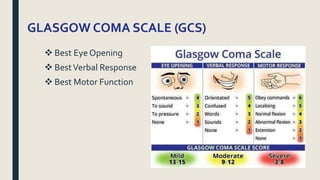
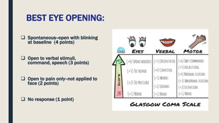
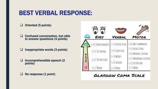
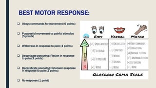
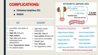
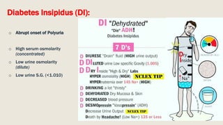
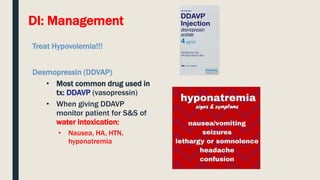
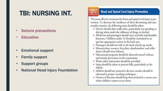
This document discusses traumatic head injuries, including causes, risk factors, pathophysiology, assessment, complications, and nursing management. It describes different types of brain injuries such as concussions, hematomas, herniations, and diffuse axonal injury. Key points include identifying high risk groups for TBI, assessing neurological status using the Glasgow Coma Scale, monitoring for increased intracranial pressure, and treating complications like diabetes insipidus and SIADH. Nursing focuses on maintaining cerebral perfusion, minimizing stimuli, and addressing psychosocial needs through education and support.
A description of brain trauma focusing on psychiatric complications
Types of TBI, epidemiology, aetiology, evaluation, investigations,
It also explores basal skull fractures.
The neuropsychiatric sequelae are described including diffuse axonal injuries, hydrocephalus, neurotransmitter changes, specific mental illness (depression, mania, PTSD, substance abuse, sleep, and psychosis)
Stroke or Cerebrovascular incident, is defined as an abrupt onset of a neurological deficit that is attributable to a focal vascular cause.
The clinical manifestations of stroke are highly variable because of the complex anatomy of the brain
Head injury refers to any injury to the scalp, skull or brain. Common causes include motor vehicle accidents, falls, and assaults. The brain may experience bruising, bleeding, or swelling which increases intracranial pressure. Nurses monitor patients closely for changes in vital signs, pupil size/reactivity, and neurological status that indicate increased pressure. Treatment involves controlling bleeding, maintaining oxygenation and circulation, preventing infection, and monitoring for complications.
This document discusses types of stroke, risk factors, statistics, recurrent strokes, ABCs of stroke support, stroke scales, interventions, drug therapy including fibrinolytics, CT scans, and ischemic versus hemorrhagic strokes. It covers the main types of stroke as hemorrhagic (15% of cases) and ischemic (85% of cases due to blood clots), risk factors like atrial fibrillation, statistics on prevalence and recurrence rates, and the stroke chain of survival steps for rapid diagnosis and treatment.
CVA (cerebrovascular accident), also known as stroke, and TIA (transient ischemic attack) are disruptions in blood flow to the brain. A CVA is caused by ischemia or hemorrhage in the brain and results in cell death, while a TIA's disruption is temporary without cell death. Risk factors include atherosclerosis, hypertension, cardiac issues, and diabetes. Symptoms depend on the location and size of the affected area but may include paralysis, impaired speech/vision, and sensory changes. Treatment focuses on prevention by controlling risk factors and potentially using blood thinners. Nursing care revolves around monitoring for complications and maximizing recovery of functions.
The document describes a 62-year-old man named Robert who fell from a ladder while picking mangoes and was found unconscious. He was brought to the emergency department conscious but confused, complaining of left arm pain. His medical history includes hypertension. A physical exam found signs of head injury including a hematoma and neurological deficits on the right side of his body. The summary provides key details about the patient's condition and situation.
This is a short presentation at Down Town Hospital clinical meeting for DNB Medicine students. It dose not cover the all aspects of stroke care especially Thrombolysis, since it is difficult to practice for Medical specialist, and ischemic stroke is not common in North East India
Acute management of patients with stroke is to stabilize the patient and to complete initial evaluation and assessment, including imaging and laboratory studies, within 60 minutes of patient arrival.
Critical decisions focus on the need for intubation, blood pressure control, and determination of risk/benefit for thrombolytic intervention.
1. A 70-year-old woman collapsed at home and was found confused by her daughter. EMS determined she may have had a stroke and transported her to the hospital within 30 minutes of the collapse.
2. At the hospital, she was found to have high blood pressure, left-sided weakness, and a CT scan showed a blood clot in her carotid artery causing a right hemisphere stroke.
3. Her risk factors included a history of untreated hypertension and a previous transient ischemic attack. She was diagnosed with an ischemic stroke likely due to atherosclerosis.
This document discusses the diagnosis and management of acute stroke. It begins by outlining the objectives of reviewing stroke etiology, identifying stroke location/type based on exam, and acute management of ischemic and hemorrhagic strokes. It then provides statistics on stroke prevalence and costs. The document reviews tools for stroke assessment like the NIH Stroke Scale and discusses etiologies of different stroke types. It includes cases demonstrating physical exam findings and determining stroke location. Management topics covered include airway protection, imaging, and treatment algorithms.
Hemorrhagic stroke accounts for 10-15% of all strokes and is associated with higher mortality than ischemic stroke. Patients often present with headache, altered mental status, seizures, nausea/vomiting, or hypertension. The bleeding occurs directly into the brain from damaged arteries, and mortality is high, with 40-80% dying within 30 days. Risk factors include age, hypertension, amyloidosis, coagulopathies, anticoagulation, cocaine abuse, and genetic conditions. Treatment focuses on stabilizing vital signs, controlling blood pressure and seizures, and reducing intracranial pressure if elevated.
The document discusses cerebrovascular accidents (strokes) including definitions, blood supply to the brain, conditions caused by occlusion of different arteries, classification of strokes, diagnostic studies, and management of strokes. It covers topics such as transient ischemic attacks, the circle of Willis, effects of reduced cerebral blood flow, intravenous thrombolysis with tPA, complications of strokes, and risk factors. Rehabilitation goals and emerging therapies are also mentioned.
This document discusses supportive management strategies for patients experiencing acute stroke. It covers positioning, monitoring and treatment of cerebral edema, management of seizures, blood pressure control, glucose control, potential cardiac issues, the role of hypothermia and neuroprotective agents, prevention of venous thromboembolism, and monitoring for infections. Key recommendations include maintaining normothermia, blood sugars between 140-180 mg/dL, treating cerebral edema with osmotic therapies like mannitol if indicated, and early mobilization to prevent complications like DVT.
Acute management of Stroke By Dr Sanjay jaiswal Neurologist sept2012Sanjay Jaiswal
The document discusses early management of ischemic stroke. It defines stroke as a sudden neurological deficit of vascular origin lasting more than 24 hours. It emphasizes that "time is brain" and every minute of untreated stroke causes the loss of 1.9 million neurons. It outlines risk factors, signs and symptoms of different types of stroke, and the definition of transient ischemic attack. Current acute treatments for ischemic stroke including thrombolysis within 3-4.5 hours and aspirin within 48 hours are discussed.
The document discusses ischemic stroke, including its epidemiology, classification, risk factors, and etiopathogenesis. Some key points:
- Stroke occurs every 5 seconds worldwide and is a leading cause of death and disability globally. Incidence and prevalence varies significantly between countries and regions.
- Strokes are classified based on their underlying cause (ischemic vs hemorrhagic) and further subtyped based on etiology (large vessel atherosclerosis, cardioembolism, small vessel disease, etc).
- Major risk factors for ischemic stroke include hypertension, atrial fibrillation, diabetes, smoking, obesity, high cholesterol, lack of physical activity, and a family history of stroke.
Stroke is a medical condition where blood supply to part of the brain is decreased, causing loss of brain function. It is a leading cause of death and disability. There are two main types - ischemic (caused by clots) and hemorrhagic (caused by bleeding). Risk factors include hypertension, heart disease, smoking, diabetes and obesity. Prevention involves controlling risk factors through lifestyle changes and medications. Anyone experiencing symptoms like weakness or numbness on one side of the body should seek immediate medical attention.
1. The document provides information about stroke, including its definition, risk factors, pathophysiology, early warning signs, and primary impairments. It notes that stroke is caused by either blockage or rupture of blood vessels in the brain.
2. High blood pressure, diabetes, heart disease, smoking, age, race, family history, and prior stroke or TIA are identified as major risk factors. Ischemic and hemorrhagic strokes are described in terms of pathophysiology.
3. Early warning signs include sudden numbness, confusion, vision problems, and difficulty walking or balancing. Primary impairments involve sensation, motor function, coordination, reflexes, and speech/language.
Stroke is a medical emergency caused by interrupted or reduced blood flow to the brain. The main types are ischemic (caused by blockage) and hemorrhagic (caused by bleeding). Risk factors include high blood pressure, atrial fibrillation, diabetes, and smoking. Symptoms depend on the affected brain region and may include paralysis, confusion, and speech problems. Diagnosis involves brain imaging and blood tests. Treatment focuses on restoring blood flow and preventing complications and recurrence through medication and lifestyle changes. Outcomes vary depending on the severity and location of brain damage.
This document provides an overview of radiological imaging in the management of stroke. It discusses:
1) Various imaging modalities used including unenhanced CT, CT angiography, MRI, and their benefits. Diffusion weighted MRI can detect acute ischemia within 30 minutes.
2) Examples of imaging findings for different stroke types like ischemic and hemorrhagic strokes. Ischemic strokes appear as bright lesions on DWI MRI.
3) Surgical interventions for acute stroke management include decompressive hemicraniectomy to reduce intracranial pressure for large hemispheric infarcts, and external ventricular drainage for intraventricular hemorrhage and hydrocephalus.
Dr. Syed Muhammad Ali Shah provides an overview of ischemic stroke. Key points include:
- Stroke is defined as rapid onset of neurological deficit lasting over 24 hours caused by a vascular issue.
- Risk factors include atrial fibrillation, hypertension, smoking, obesity, and high cholesterol.
- Diagnosis involves investigations like CT scans and MRI. Treatment depends on the cause but may include thrombolysis within 4.5 hours, aspirin, rehabilitation, and preventing future strokes through controlling risk factors.
- Future advances include endovascular therapies to remove clots and research on neuroprotection strategies. Prevention through lifestyle changes and medications can reduce stroke risk.
This document provides definitions and information about evaluating and managing patients presenting with stroke. It defines stroke, transient ischemic attack (TIA), and their subtypes. It discusses the epidemiology of stroke and risk factors. Evaluation of a stroke patient involves a neurological exam and considering differential diagnoses. Investigations aim to confirm the vascular nature and type of lesion. Management focuses on treating the underlying cause, preventing complications, and rehabilitation.
Hemorrhagic vs. Ischemic Stroke Prognosis_TPostrelTina Postrel
This document summarizes the prognosis and rehabilitation principles for ischemic and hemorrhagic strokes. It covers classifications of strokes, early and late prognosis for different types, risk factors affecting prognosis, evidence from studies on mortality and functional recovery, and rehabilitation guidelines. The key points are that hemorrhagic strokes generally have poorer early prognosis but better late prognosis compared to ischemic strokes, which have lower initial mortality but slower long-term functional recovery.
This document provides an overview of ischaemic stroke, including its definition, risk factors, pathophysiology, clinical presentation, diagnosis and management. Key points include:
- Ischaemic stroke accounts for 80% of strokes and results from focal brain infarction due to obstruction of cerebral blood flow.
- Major risk factors include hypertension, atrial fibrillation, diabetes, hyperlipidemia and previous stroke or TIA.
- Clinical syndromes depend on the location of brain infarction and can include motor/sensory deficits, aphasia and visual field cuts.
- Diagnosis involves neuroimaging such as CT, MRI and vascular imaging to identify the cause.
- Acute
Strokes are caused by vascular injuries that reduce blood flow to the brain and can cause neurological impairment. Strokes are classified based on their etiology and pathology. Common causes include thrombosis, embolism, and hypoperfusion. The initial evaluation of a patient experiencing an acute ischemic stroke involves assessing their history, performing an examination, and obtaining brain imaging to determine eligibility for thrombolysis or thrombectomy. Specific management focuses on reperfusing ischemic brain tissue using interventions like thrombolysis with rtPA or endovascular thrombectomy within established time windows.
This document provides information about neurological nursing related to increased intracranial pressure and head injuries. It discusses the anatomy and physiology of the brain, types of head injuries including primary and secondary injuries, complications such as hematomas, and management of increased ICP. Nursing assessments, diagnoses, and interventions are also reviewed, including monitoring for signs of increased ICP, maintaining airway and breathing, preventing injury, and using the Glasgow Coma Scale to assess level of consciousness.
The document discusses head injuries and traumatic brain injuries (TBI). It covers causes of TBI like falls and motor vehicle accidents. It then discusses the primary and secondary injuries that can occur from a TBI. It explains increased intracranial pressure and outlines treatments to control pressure like osmotic diuretics, CSF drainage, and fluid restriction. The document also covers assessing and diagnosing different types of brain injuries through imaging and examinations.
This document provides an overview of the management of head injuries. It defines head injury as damage to the head from impact and classifies injuries as closed or open, diffuse or focal. The pathophysiology section explains how small increases in intracranial volume can raise pressure dramatically. Presentation may include altered consciousness, bleeding, seizures or vomiting. Investigations include CT scans to detect fractures or bleeds. Treatment focuses on preventing secondary injuries like hypoxia, controlling pressure, and maintaining perfusion and nutrition. Follow-up is needed as some patients with mild injuries may later develop complications.
This document provides an outline and introduction to the management of head injuries. It discusses the relevant anatomy of the head, definitions of head and brain injuries, epidemiology, causes, classifications based on severity, location and type of injury. It also covers the pathogenesis of head injuries and specific entities like skull fractures, concussions, hemorrhages. The assessment and treatment of head injured patients is outlined along with conclusions.
A head injury can range from mild to severe and is caused by blunt force trauma or penetrating injuries to the skull and brain. Symptoms of a serious head injury include loss of consciousness, persistent headaches, vomiting, or abnormal behavior. Treatment depends on severity but may include monitoring for deterioration, supporting circulation and lowering intracranial pressure by evacuating hematomas or reducing brain swelling.
Intracranial bleeding encompasses all bleeds that may occur within the cranial cavity including Epidural, Subdural, Sub arachnoid, intraparenchymal and Intraventricular haemorrhages. all are discussed in these slides and relevant references are provided for detailed information.
It is important to note that medicine is not learnt online but through series of organised events under specialised supervision in recognised institutions of learning.
1. The document discusses the initial management of traumatic brain injury, including decompressive craniectomy to reduce ICP, hyperosmolar therapy with mannitol or HTS, CSF drainage with EVD, and sedation to control refractory ICP. Early enteral nutrition is recommended if no contraindications.
2. Spinal trauma management involves cervical spine clearance following the Canadian C-spine Rule or Nexus criteria. Imaging includes 3-view c-spine series and thoracolumbar films. Neurological exam uses the ASIA chart.
3. Common neurosurgical emergencies are discussed like raised ICP, stroke, seizures, and brainstem compression from posterior fossa
Head injuries can range from minor scalp lacerations to serious brain injuries. They are a leading cause of death from trauma. The risk is highest among males aged 15-24. Mechanisms include blunt and penetrating injuries. Injuries may involve the scalp, skull, or brain. Scalp injuries include lacerations and hematomas. Skull fractures can be linear, depressed, or involve the skull base. Brain injuries include concussions, diffuse axonal injury, contusions, hematomas, and lacerations. Diagnosis involves assessing Glasgow Coma Scale and using imaging like CT scans. Treatment depends on the type and severity of injury.
This document discusses head injuries, including their epidemiology, pathophysiology, types, and management. Head injuries are a major public health problem worldwide and are mostly caused by road traffic accidents and assaults. The main types of head injuries discussed are cerebral contusions, diffuse axonal injury, cerebral edema, traumatic intracranial hematomas such as extradural, subdural, subarachnoid and intracerebral hemorrhages, and concussions. Initial management focuses on preventing secondary brain damage through measures such as neurological observation, immobilization, intubation if needed, and transport to a dedicated neurological facility for patients with more severe injuries.
Lecture 7 management of head injury patientsTibebe Birhanu
1. Head injuries result in over 1.6 million emergency department visits annually in the US, costing an estimated $100 billion per year. Primary brain injuries include focal injuries like epidural hematomas and diffuse injuries like axonal shearing.
2. Secondary brain injury occurs when the initial injury is exacerbated by processes like edema, ischemia, or herniation. The goal of treatment is to prevent secondary injury by controlling intracranial pressure and maintaining adequate cerebral perfusion and oxygenation.
3. Key aspects of management include airway control, treating hypotension, monitoring the Glasgow Coma Scale, controlling intracranial pressure through interventions like hyperventilation and mannitol if pressure is elevated
TRAUMATIC BRAIN INJURY and anesthetic management.pptxssuser579a28
Traumatic brain injury (TBI) can be caused by a forceful impact or penetration of the skull. There are two main types - penetrating injuries which break the skull, and closed or blunt injuries which cause the brain to move inside the skull. TBI can lead to hematomas, contusions, diffuse axonal injury or intracranial hemorrhage depending on the location and nature of the injury in the brain. The primary injury occurs at the time of impact, while secondary injuries may develop like brain swelling, low oxygen levels, and increased pressure inside the skull in the following hours or days. Emergency treatment focuses on stabilizing the patient's breathing, blood circulation, neck immobilization and rapid evaluation for life-threatening
Head injury types, clinical manifestations, diagnosis and managementVibha Amblihalli
Head injuries can range from minor scalp lacerations to major brain trauma. Common causes include motor vehicle accidents, falls, and sports injuries. Diagnosis involves CT or MRI imaging to identify fractures and intracranial bleeding. Treatment depends on injury severity but may include reducing intracranial pressure, surgical evacuation of hematomas, and preventing complications like seizures. Outcomes range from full recovery to permanent disability or death depending on the nature and extent of brain damage.
1) Head injuries are a common cause of disability and death in children and can range from mild to severe. Increased intracranial pressure following head trauma can lead to secondary brain injury.
2) The goal in managing increased intracranial pressure is to maintain adequate cerebral perfusion pressure through treatments aimed at lowering intracranial pressure such as hyperventilation, mannitol, and sedation/analgesia while supporting mean arterial pressure.
3) Intracranial pressure monitoring can help guide management and detect events that may require intervention to prevent herniation and optimize cerebral oxygen delivery.
This document discusses head injuries, including types, causes, symptoms, diagnosis and management. The main types of head injuries are scalp lacerations, skull fractures, minor head trauma including concussion, and major head trauma such as cerebral contusions and lacerations. Diagnosis involves CT or MRI imaging. Management consists of supportive measures to control ICP, surgical evacuation of hematomas when indicated, and medical therapy depending on the specific head injury. Preventive measures include using safety equipment and reducing accidents.
Head injuries can range from minor scalp lacerations to major brain trauma. Common causes include motor vehicle accidents, falls, assaults, and sports injuries. Death from head injuries can occur immediately after injury, within 2 hours, or up to 3 weeks later. Diagnosis involves CT or MRI imaging. Treatment depends on the type and severity of injury but generally includes supportive care, measures to reduce intracranial pressure, and possibly surgery. Outcomes range from full recovery to permanent neurological deficits or death.
The document discusses head trauma and traumatic brain injury (TBI). It defines TBI as structural or physiological disruption of the brain from head injury. TBI can be closed or open head injuries and is classified by severity using the Glasgow Coma Scale. Common causes of TBI include falls, motor vehicle accidents, contact sports, and assaults. Types of primary brain injuries include intracranial hemorrhages, coup-contrecoup injuries, cerebral lacerations, and cerebral contusions. Secondary brain injuries can result from changes like increased intracranial pressure that affect oxygenation and blood pressure. Management of TBI involves treating primary and secondary injuries, including measures to control increased intracranial pressure.
Burns can cause devastating physical and emotional effects. Classification is based on depth of skin damage from 1st to 4th degree. Severe burns increase risks of infections, loss of mobility, and multi-organ damage. Treatment involves stabilizing fluid and electrolyte shifts, administering IV fluids based on burn size, and preventing complications like shock, hypothermia, and infections. Patients with severe or complex burns may require referral to a specialized burn center.
This document provides information on abdominal trauma, including the clinical presentation, diagnosis, and management of various intra-abdominal injuries that can result from blunt or penetrating trauma. It discusses injuries to organs like the liver, spleen, bowel, pancreas, kidneys, and pelvic structures. Key points covered include the mechanisms of injury, signs and symptoms of each type of injury, appropriate diagnostic tests, and treatment approaches, including both conservative management and indications for surgery. Nursing considerations like monitoring, fluid management, and patient education are also reviewed.
Meningitis is an inflammation of the membranes and cerebrospinal fluid surrounding the brain and spinal cord. It can be caused by either bacteria (septic) or viruses (aseptic). Bacterial meningitis is more severe and its symptoms include headache, fever, stiff neck, nausea, vomiting, and decreased level of consciousness. It is diagnosed through cultures of blood, spinal fluid and imaging tests and treated quickly with antibiotics that cross the blood brain barrier like ceftriaxone. Nursing care focuses on isolation precautions, monitoring for increased intracranial pressure, managing symptoms, and preventing complications through interventions like keeping the head elevated. Vaccines can help prevent common types of bacterial meningitis.
This document provides information on musculoskeletal trauma. It begins with epidemiology statistics on musculoskeletal injuries and discusses the anatomy and physiology of bones, joints, tendons, ligaments and neurovascular structures. It then covers mechanisms of injury, clinical manifestations, emergency management, complications, and nursing management of various musculoskeletal traumas including fractures, dislocations, soft tissue injuries, and pelvic fractures. Specific topics covered in depth include fat embolism, hemorrhage, osteomyelitis, avascular necrosis, crush injuries, compartment syndrome, and rhabdomyolysis.
This document provides information about rape and sexual assault, including definitions, statistics, and the psychological and physical effects on victims. It describes the three phases of Rape Trauma Syndrome (acute, outward adjustment, and renormalization), common reactions in each phase, and long-term coping strategies. The role of Sexual Assault Nurse Examiners is outlined, including conducting a sensitive exam, collecting forensic evidence, and providing treatment and referrals. The goal is to reduce further trauma to victims and support their recovery process.
This document provides information on eye trauma, including causes, assessments, interventions, and nursing care. Key points include:
- Eye injuries are often preventable through safety measures. Common causes include accidents, assaults, and work or sports injuries.
- Assessment of eye trauma involves evaluating visual acuity, eye movements, pupil response, and checking for foreign bodies or orbital fractures. History should include injury details.
- Interventions depend on injury type but may include wound irrigation, patching, antibiotics, surgery for foreign bodies or fractures, and corticosteroids to reduce inflammation. Goals are to prevent infection and further damage.
- Nursing care focuses on monitoring for complications, educating on activity restrictions, and
This document discusses seizure disorders and epilepsy. It begins by differentiating between types of seizures, such as partial and generalized seizures. Diagnostic tests for seizures are outlined, along with the assessment and management of seizures. Various anti-seizure medications are discussed, along with goals of drug therapy and patient education. Nursing priorities for patients with seizures include maintaining a patent airway, providing oxygenation, obtaining vascular access, using seizure precautions, administering appropriate medications, and assessing therapeutic drug levels.
The document discusses spinal cord injuries, including the anatomy of the spinal cord and vertebral column, common mechanisms of injury, types of spinal cord injuries, assessment findings, emergency management, pharmacological interventions like methylprednisolone, and stabilization techniques such as cervical traction. Nursing considerations are also addressed, including monitoring for complications and implementing appropriate nursing diagnoses and interventions.
This document provides information on trauma care including mechanisms of injury, assessments of trauma patients, nursing interventions, and considerations in trauma patients. It discusses the primary and secondary survey process for trauma patients and identifying life-threatening injuries. Nursing priorities include establishing and maintaining a patent airway, ensuring adequate breathing and circulation, providing pain management and comfort measures. Special considerations are discussed for aging trauma patients, those with alcohol or drug use, and supporting family coping after a traumatic event.
This document discusses trauma care and nursing considerations for trauma patients. It covers mechanisms of injury, primary and secondary assessments, common injuries, interventions for airway, breathing and circulation issues, head-to-toe exams, and complications. Key nursing priorities include life-saving interventions, prevention of further injury, managing pain and withdrawal symptoms, and facilitating family support and coping. The elderly are at higher risk for serious injuries from falls and have worse outcomes. Alcohol and drug use are contributing factors in many traumatic injuries.
This document provides an overview of gastrointestinal disorders, including anatomy and physiology of the GI tract, physical assessment techniques, common GI conditions like peptic ulcer disease, and nursing interventions. It describes the functions of the GI organs and accessories like the liver and pancreas. Diagnostic tests and procedures like endoscopy are outlined. Complications of tube feedings and acute GI bleeding from sources like stress ulcers are reviewed.
This document provides information on acute kidney injury (AKI) for nursing students. It begins with learning objectives about the renal system, causes and stages of AKI, and the nurse's role in management. It then reviews anatomy and physiology of the kidneys, normal function, causes of AKI including pre-renal, intrinsic and post-renal, stages of AKI, assessment findings, and nursing interventions for each stage. The goal is for students to understand AKI, recognize patients at risk, implement preventive measures, and provide evidence-based care to optimize outcomes.
This document provides an overview of shock and its pathophysiology. It defines shock as a clinical syndrome resulting from inadequate tissue perfusion due to alterations in circulation. The stages of shock are described as compensatory, progressive, and irreversible. Compensatory mechanisms aimed at maintaining homeostasis in response to shock are discussed for various body systems. Nursing interventions for shock focus on treating its underlying cause, restoring circulating volume and hemodynamics through fluid resuscitation and vasoactive drugs, and minimizing oxygen consumption.
This document discusses endocrine emergencies including disorders of the pancreas, pituitary, adrenal and thyroid glands. It covers the pathophysiology, manifestations, and treatment of diabetic ketoacidosis (DKA) and hyperglycemic hyperosmolar state (HHS), as well as hypoglycemia, Addison's disease, and adrenal crisis. The key aspects of assessment and management are fluid resuscitation, electrolyte replacement, insulin therapy, and treatment of acidosis for DKA and HHS patients. Nursing diagnoses related to ineffective breathing, gas exchange, fluid deficits, and therapeutic management are also reviewed.
Benign breast disorders are common noncancerous conditions that cause pain and discomfort. The most common are fibrocystic breasts and fibroadenomas. Fibrocystic changes are lumpy, tender breasts that occur before menstruation due to hormone fluctuations. Fibroadenomas are solid, rubbery breast nodules usually occurring in women ages 15-35. Mastitis is a breast infection common in lactating women. Mammography and ultrasound are used to diagnose breast conditions. Women should perform monthly breast self-exams to monitor for any changes.
This chapter discusses disorders of the gastrointestinal system in children. It covers various congenital abnormalities that can develop during prenatal formation of the GI tract, including cleft lip/palate, esophageal atresia, and Hirschsprung's disease. Common motility disorders like GERD and constipation are described. Infectious disorders such as ulcers, appendicitis, and inflammatory bowel disease are addressed. The chapter also reviews obstructive issues, malabsorption, hepatic disorders, and postoperative care following GI surgery. Nursing assessment and management are focused on fluid balance, nutrition, pain control, and addressing parental needs.
Basavarajeeyam is a Sreshta Sangraha grantha (Compiled book ), written by Neelkanta kotturu Basavaraja Virachita. It contains 25 Prakaranas, First 24 Chapters related to Rogas& 25th to Rasadravyas.
- Video recording of this lecture in English language: https://youtu.be/kqbnxVAZs-0
- Video recording of this lecture in Arabic language: https://youtu.be/SINlygW1Mpc
- Link to download the book free: https://nephrotube.blogspot.com/p/nephrotube-nephrology-books.html
- Link to NephroTube website: www.NephroTube.com
- Link to NephroTube social media accounts: https://nephrotube.blogspot.com/p/join-nephrotube-on-social-media.html
8 Surprising Reasons To Meditate 40 Minutes A Day That Can Change Your Life.pptxHolistified Wellness
We’re talking about Vedic Meditation, a form of meditation that has been around for at least 5,000 years. Back then, the people who lived in the Indus Valley, now known as India and Pakistan, practised meditation as a fundamental part of daily life. This knowledge that has given us yoga and Ayurveda, was known as Veda, hence the name Vedic. And though there are some written records, the practice has been passed down verbally from generation to generation.
TEST BANK For Basic and Clinical Pharmacology, 14th Edition by Bertram G. Kat...rightmanforbloodline
TEST BANK For Basic and Clinical Pharmacology, 14th Edition by Bertram G. Katzung, Verified Chapters 1 - 66, Complete Newest Version.
Rasamanikya is a excellent preparation in the field of Rasashastra, it is used in various Kushtha Roga, Shwasa, Vicharchika, Bhagandara, Vatarakta, and Phiranga Roga. In this article Preparation& Comparative analytical profile for both Formulationon i.e Rasamanikya prepared by Kushmanda swarasa & Churnodhaka Shodita Haratala. The study aims to provide insights into the comparative efficacy and analytical aspects of these formulations for enhanced therapeutic outcomes.
TEST BANK For Community Health Nursing A Canadian Perspective, 5th Edition by...Donc Test
TEST BANK For Community Health Nursing A Canadian Perspective, 5th Edition by Stamler, Verified Chapters 1 - 33, Complete Newest Version Community Health Nursing A Canadian Perspective, 5th Edition by Stamler, Verified Chapters 1 - 33, Complete Newest Version Community Health Nursing A Canadian Perspective, 5th Edition by Stamler Community Health Nursing A Canadian Perspective, 5th Edition TEST BANK by Stamler Test Bank For Community Health Nursing A Canadian Perspective, 5th Edition Pdf Chapters Download Test Bank For Community Health Nursing A Canadian Perspective, 5th Edition Pdf Download Stuvia Test Bank For Community Health Nursing A Canadian Perspective, 5th Edition Study Guide Test Bank For Community Health Nursing A Canadian Perspective, 5th Edition Ebook Download Stuvia Test Bank For Community Health Nursing A Canadian Perspective, 5th Edition Questions and Answers Quizlet Test Bank For Community Health Nursing A Canadian Perspective, 5th Edition Studocu Test Bank For Community Health Nursing A Canadian Perspective, 5th Edition Quizlet Test Bank For Community Health Nursing A Canadian Perspective, 5th Edition Stuvia Community Health Nursing A Canadian Perspective, 5th Edition Pdf Chapters Download Community Health Nursing A Canadian Perspective, 5th Edition Pdf Download Course Hero Community Health Nursing A Canadian Perspective, 5th Edition Answers Quizlet Community Health Nursing A Canadian Perspective, 5th Edition Ebook Download Course hero Community Health Nursing A Canadian Perspective, 5th Edition Questions and Answers Community Health Nursing A Canadian Perspective, 5th Edition Studocu Community Health Nursing A Canadian Perspective, 5th Edition Quizlet Community Health Nursing A Canadian Perspective, 5th Edition Stuvia Community Health Nursing A Canadian Perspective, 5th Edition Test Bank Pdf Chapters Download Community Health Nursing A Canadian Perspective, 5th Edition Test Bank Pdf Download Stuvia Community Health Nursing A Canadian Perspective, 5th Edition Test Bank Study Guide Questions and Answers Community Health Nursing A Canadian Perspective, 5th Edition Test Bank Ebook Download Stuvia Community Health Nursing A Canadian Perspective, 5th Edition Test Bank Questions Quizlet Community Health Nursing A Canadian Perspective, 5th Edition Test Bank Studocu Community Health Nursing A Canadian Perspective, 5th Edition Test Bank Quizlet Community Health Nursing A Canadian Perspective, 5th Edition Test Bank Stuvia
TEST BANK For An Introduction to Brain and Behavior, 7th Edition by Bryan Kol...rightmanforbloodline
TEST BANK For An Introduction to Brain and Behavior, 7th Edition by Bryan Kolb, Ian Q. Whishaw, Verified Chapters 1 - 16, Complete Newest Versio
TEST BANK For An Introduction to Brain and Behavior, 7th Edition by Bryan Kolb, Ian Q. Whishaw, Verified Chapters 1 - 16, Complete Newest Version
Osteoporosis - Definition , Evaluation and Management .pdfJim Jacob Roy
Osteoporosis is an increasing cause of morbidity among the elderly.
In this document , a brief outline of osteoporosis is given , including the risk factors of osteoporosis fractures , the indications for testing bone mineral density and the management of osteoporosis
Local Advanced Lung Cancer: Artificial Intelligence, Synergetics, Complex Sys...Oleg Kshivets
Overall life span (LS) was 1671.7±1721.6 days and cumulative 5YS reached 62.4%, 10 years – 50.4%, 20 years – 44.6%. 94 LCP lived more than 5 years without cancer (LS=2958.6±1723.6 days), 22 – more than 10 years (LS=5571±1841.8 days). 67 LCP died because of LC (LS=471.9±344 days). AT significantly improved 5YS (68% vs. 53.7%) (P=0.028 by log-rank test). Cox modeling displayed that 5YS of LCP significantly depended on: N0-N12, T3-4, blood cell circuit, cell ratio factors (ratio between cancer cells-CC and blood cells subpopulations), LC cell dynamics, recalcification time, heparin tolerance, prothrombin index, protein, AT, procedure type (P=0.000-0.031). Neural networks, genetic algorithm selection and bootstrap simulation revealed relationships between 5YS and N0-12 (rank=1), thrombocytes/CC (rank=2), segmented neutrophils/CC (3), eosinophils/CC (4), erythrocytes/CC (5), healthy cells/CC (6), lymphocytes/CC (7), stick neutrophils/CC (8), leucocytes/CC (9), monocytes/CC (10). Correct prediction of 5YS was 100% by neural networks computing (error=0.000; area under ROC curve=1.0).
Histololgy of Female Reproductive System.pptxAyeshaZaid1
Dive into an in-depth exploration of the histological structure of female reproductive system with this comprehensive lecture. Presented by Dr. Ayesha Irfan, Assistant Professor of Anatomy, this presentation covers the Gross anatomy and functional histology of the female reproductive organs. Ideal for students, educators, and anyone interested in medical science, this lecture provides clear explanations, detailed diagrams, and valuable insights into female reproductive system. Enhance your knowledge and understanding of this essential aspect of human biology.
Does Over-Masturbation Contribute to Chronic Prostatitis.pptxwalterHu5
In some case, your chronic prostatitis may be related to over-masturbation. Generally, natural medicine Diuretic and Anti-inflammatory Pill can help mee get a cure.
2. OBJECTIVES
■ Identify mechanisms of injury associated with brain trauma.
■ Describe the pathophysiologic changes as a basis for signs and symptoms.
■ Discuss the nursing assessment of patients with brain injuries.
■ Identify appropriate nursing diagnosis and expected outcomes for the brain injured
patient.
■ Plan appropriate interventions for patients with brain injuries.
■ Evaluate the effectiveness of nursing interventions for patients with brain injuries.
3. Traumatic Head Injury
• The CDC reports approximately 1.4 million people sustainTBI annually.
• 50,000 die
• 235,000 are hospitalized
• 1.1 million are treated and released from hospital EDs
4. Traumatic Brain Injury Among Children
Among children ages 0-14,TBI is responsible for:
2,685 deaths
37,000 hospitalizations
435,000 emergency department visits
5. Living withTBI
• The CDC reports at least 5.3 millionAmericans living withTBI have…
long term or lifelong need for assistance with ADL’s.
• TBI may cause a wide range of functional changes affecting cognition,
sensation, language or emotions.
• Thinking: memory & reasoning
• Sensation: touch, taste & smell
6.
7. High Risk Groups
• Males 2x more likely than females
• Young children: 0-5 years old
• Elderly >75 years old
• Military Personnel
• African Americans
• 15-19 years of age
8. Which one is most likely to sustain aTBI
leading to hospitalization and/or death?
10. Causes & Risk Factors:
• Falls
• Struck by or against an object
(includes intentional self-harm)
• MVC’s
• Assaults – firearms
• Low Socioeconomic
• ETOH use
• Rural (MVC, lightening, chemical
exposure)
• Urban (poisonings and homicide)
11. Pre-Hospital Care of theTrauma Patient
• TRIAGE: involves correct
identification of the injuries-
nature and extent.
• Treatment utilizing ATLS
• Levels ofTrauma Care
• Transport to the nearest available
Level I trauma center.
12. The Trauma Patient
• Traumatic injuries: induced suddenly without warning.
• Injuries are often subtle, lead to longer time to DX and treat effectively.
• Co-morbidities, drugs, and alcohol influence outcome.
• Involvement of bystanders can be difficult to manage:
• people need be interviewed, police involvement is necessary.
• Trauma Coordinators:
• Useful in crowd control.
• Identify those who need emotional support.
• Liaison to hospital support-clergy, psychiatry, grief counselors.
13. Mechanism of Injury (MOI):
• Kinetic Energy: injury
results when the body
can’t tolerate exposure
to excessive force.
• MVA: Acceleration /
Deceleration injuries
• Gunshot wounds
• Thermal (hot or cold-
temperature &
duration)
• Chemical
• Electrical
• Drowning
14.
15.
16. Skull Anatomy Review
Scalp: five layers of tissue, protective cover, vascular
• Skin
• ConnectiveTissue
• Aponeurotic Galea
• Loose AreolarTissue
• Pericranium
Skull: Formed by cranium and facial bones.
• Cranium: double layer of solid bone surrounds a spongy middle layer.
• Bones: Frontal,Temporal, Parietal, Occipital and Mastoid
• Vasculature: Middle Meningeal Artery
22. BRAIN STEM:
• Midbrain
• Pons
• Medulla Oblongata
• Cranial Nerves
• Reticular Activating System
• Meninges
o Pia Mater
o Arachnoid Membrane
o Dura Mater
25. Cerebrospinal Fluid
• Clear, colorless fluid
• SG of 1.007
• Produced in the ventricles circulates
around the brain & spinal cord.
• Provides cushion & protection.
Ventricles:
• Center of the brain
• Secretes CSF by filtering blood
• Forms part the blood brain barrier
(BBB)
26. Metabolism & Perfusion: Cerebral Blood
Flow
• High metabolic rate
• Consumes 20% of the body’s oxygen!!
• Largest user of glucose
• Unable to store nutrients
Blood supplied by vertebral and internal
carotid arteries:
Vertebral - supplies posterior brain,
cerebellum, & brain stem
Carotid Arteries: supply the cerebrum
27. Cerebral Perfusion:
Cerebral Blood Flow (CBF)
Dependent upon CPP
Flow requires a pressure gradient.
Cerebral Perfusion Pressure (CPP)
Pressure moving the blood
through the cranium
Autoregulation allows BP changes
to maintain CPP
CPP = MAP - ICP
28.
29. Mean Arterial Pressure (MAP):
• Dependent on SVR
• MAP = Diastolic + 1/3 Pulse Pressure
• Need a MAP >60 to perfuse the brain!!!
Intracranial Pressure (ICP):
• Pressure exerted by the volume of the
intracranial contents within the cranial vault.
• Influenced by edema & hemorrhage
Cerebral Perfusion:
31. Monroe – Kelly Hypothesis:
Change in volume of one component
must have reciprocal change in one or
both of the other components.
If reciprocal change not
accomplished, the result is an
increase in ICP.
Cushing’sTriad: LATE SIGN OF ↑ ICP
Increased systolic BP
Widened pulse pressure
Reflexive bradycardia
INTRACRANIAL PRESSURE (ICP):
45. PRIMARY BRAIN INJURY:
Concussion
Coup Injury:
Direct point of impact, trauma to
the brain.
Shearing of subdural vessels.
Trauma to the base of the brain.
Contre-coup Injury:
Site of impact when brain hits
opposite side of skull.
Shearing forces occur throughout
the brain.
46. EPIDURAL HEMATOMA
• RAPIDLY accumulating hematoma:
occurs between dura mater & cranium.
• Usually occurs from a laceration in the
middle meningeal artery.
• Associated with a skull fracture…
May have HX of head trauma with loss
OC.
Immediate Surgical Intervention!!
47. SUBDURAL HEMATOMA
• Clot formation underneath dura.
• Tearing of the bridging vein
between the cerebral cortex & a
draining venous sinus.
• Venous in origin
• Elderly at risk.
• History may show loss OC with
recovery and NO relapse.
48. ASSESSMENT OF HEMATOMA:
***Determined by rate of accumulation.
Acute: HA, confusion, slowed thinking, agitation.
Subacute: May not appear for days or weeks.
Chronic:
• Vague, attributed to other conditions.
• HA, lethargy, absent mindedness, vomiting.
• Severe symptoms: Seizures, stiff neck, pupil changes, hemiparesis.
49.
50. MANAGEMENT of HEMATOMAS:
• Surgical Evacuation
• Placement of subdural drain - J
• Remains in place for a few days.
• Monitor LOC
• Focused neuro. assessment
51. SUBARACHNOID HEMATOMA (SAH)
• Accumulation of blood btwn.
arachnoid layer of meninges & brain.
• Results in blood leakage into CSF.
• Nuchal Rigidity:
-Irritation of blood in arachnoid space.
-Neck stiffness
-Inability to passively flex & extend the head
normally.
52. MANAGEMENT OF SAH:
• Intraventricular Catheter:
Ventriculostomy
• Drainage of bloody CSF.
• ICP monitoring.
• Monitor Neuro. Status
• Monitor Quantity & Color
of CSF Drainage
53.
54. INTRACEREBRAL HEMATOMA (ICH)
Accumulation of blood in the Parenchyma of brain
tissue.
• Uncontrolled HTN
• Ruptured Aneurysm
• Trauma w. high impact blow to the head
Clinical Manifestations: Vary r/t location of hematoma
Management:
• Management of ICP and CPP
• Surgical evacuation usually not possible
55.
56. DIFFUSE AXONAL
INJURY
• Shearing or tearing of nerve fibers.
• Result of rapid acceleration /
deceleration motion, NOT
necessarily impact.
• Common in whiplash injuries or
amusement park rides.
• Severe brain injury, leads to coma.
Focal Injury:
• Involves a limited and
identifiable site of injury
• Localized damage
59. SKULL FRACTURES:
Linear Skull Fracture: most common!
• May be a simple non-displaced fracture
• May involve nicking of underlying blood
vessels.
Depressed Skull Fracture:
• Bone fragments embedded in brain tissue
• Brain tissue compression
• Dural laceration
• Associated with scalp laceration.
• Requires debridement and surgery
60. Comminuted (open) Skull Fracture:
• Open fracture with communication
btwn. brain & environment.
• Dura is interrupted
• Risk of infection
• Egg shell Fracture
• Result of a fall or blunt force.
SKULL FRACTURES:
61. Basilar Skull Fracture:
• Linear fracture at base of the brain.
• Associated with dural tear.
• Assess for S&S of meningitis / encephalitis.
S&S:
• Rhinorrhea
• Otorrhea (CSF leak from ears)
• Check drainage for glucose level…
>200mg/dL = CSF
• Filter paper: halo or ring sign (not
conclusive)
• Battle sign (bruising over mastoid process)
• Raccoon Eyes
SKULL FRACTURES:
71. GLASGOW COMA SCALE (GCS)
Best Eye Opening
BestVerbal Response
Best Motor Function
72. BEST EYE OPENING:
Spontaneous--open with blinking
at baseline (4 points)
Open to verbal stimuli,
command, speech (3 points)
Open to pain only--not applied to
face (2 points)
No response (1 point)
73. BEST VERBAL RESPONSE:
Oriented (5 points)
Confused conversation, but able
to answer questions (4 points)
Inappropriate words (3 points)
Incomprehensible speech (2
points)
No response (1 point)
74. BEST MOTOR RESPONSE:
Obeys commands for movement (6 points)
Purposeful movement to painful stimulus
(5 points)
Withdraws in response to pain (4 points)
Decorticate posturing: Flexion in response
to pain (3 points)
Decerebrate posturing: Extension response
in response to pain (2 points)
No response (1 point)
76. o Abrupt onset of Polyuria
o High serum osmolarity
(concentrated)
o Low urine osmolarity
(dilute)
o Low urine S.G. (<1.010)
Diabetes Insipidus (DI):
77. DI: Management
Treat Hypovolemia!!!
Desmopressin (DDVAP)
• Most common drug used in
tx: DDAVP (vasopressin)
• When giving DDAVP
monitor patient for S&S of
water intoxication:
• Nausea, HA, HTN,
hyponatremia
78. • Excessive ADH production
• FVO: Increase of total body
water…
- Dilutional hyponatremia
HA, seizures!
- Hypo-osmolarity
- Concentrated urine
o Low serum osmolarity (dilute)
o High urine osmolarity
(concentrated)
o High urine S.G. (<1.030)
SIADH: