European project PROMET - Deep Real-Time Optoacoustic Imaging of Breast Tumors
•
0 likes•153 views

Recommended

Recommended
More Related Content
What's hot
What's hot (20)
Similar to European project PROMET - Deep Real-Time Optoacoustic Imaging of Breast Tumors
Similar to European project PROMET - Deep Real-Time Optoacoustic Imaging of Breast Tumors (20)
Recently uploaded
Recently uploaded (20)
European project PROMET - Deep Real-Time Optoacoustic Imaging of Breast Tumors
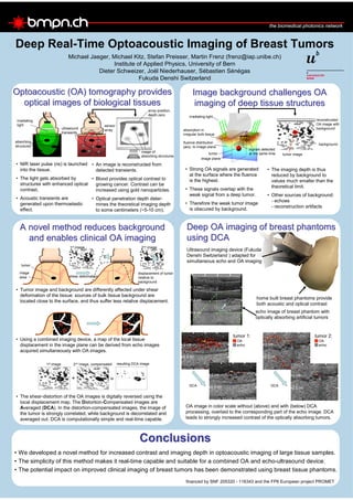
- 1. Deep Real-Time Optoacoustic Imaging of Breast Tumors Michael Jaeger, Michael Kitz, Stefan Preisser, Martin Frenz (frenz@iap.unibe.ch) Institute of Applied Physics, University of Bern Dieter Schweizer, Joël Niederhauser, Sébastien Sénégas Fukuda Denshi Switzerland OptoacousticOptoacoustic (OA) tomography provides(OA) tomography provides optical images of biological tissuesoptical images of biological tissues • NIR laser pulse (ns) is launched into the tissue. • The light gets absorbed by structures with enhanced optical contrast. • Acoustic transients are generated upon thermoelastic effect. • An image is reconstructed from detected transients. • Blood provides optical contrast to growing cancer. Contrast can be increased using gold nanoparticles. • Optical penetration depth deter- mines the theoretical imaging depth to some centimeters (~5-10 cm). Image background challenges OAImage background challenges OA imaging of deep tissue structuresimaging of deep tissue structures A novel method reduces backgroundA novel method reduces background and enables clinical OA imagingand enables clinical OA imaging ConclusionsConclusions • Strong OA signals are generated at the surface where the fluence is the highest. • These signals overlap with the weak signal from a deep tumor. • Therefore the weak tumor image is obscured by background. • The imaging depth is thus reduced by background to values much smaller than the theoretical limit. • Other sources of background: - echoes - reconstruction artifacts Deep OA imaging of breast phantomsDeep OA imaging of breast phantoms using DCAusing DCA home built breast phantoms provide both acoustic and optical contrast Ultrasound imaging device (Fukuda Denshi Switzerland ) adapted for simultaneous echo and OA imaging echo image of breast phantom with optically absorbing artificial tumors OA image in color scale without (above) and with (below) DCA processing, overlaid to the corresponding part of the echo image. DCA leads to strongly increased contrast of the optically absorbing tumors. • Tumor image and background are differently affected under shear deformation of the tissue: sources of bulk tissue background are located close to the surface, and thus suffer less relative displacement. • Using a combined imaging device, a map of the local tissue displacement in the image plane can be derived from echo images acquired simultaneously with OA images. • The shear-distortion of the OA images is digitally reversed using the local displacement map. The Distortion-Compensated images are Averaged (DCA). In the distortion-compensated images, the image of the tumor is strongly correlated, while background is decorrelated and averaged out. DCA is computationally simple and real-time capable. tumor image area displacement of tumor relative to background shear deformation 1st image 2nd image + = 1st image 2nd image, compensated resulting DCA image • We developed a novel method for increased contrast and imaging depth in optoacoustic imaging of large tissue samples. • The simplicity of this method makes it real-time capable and suitable for a combined OA and echo-ultrasound device. • The potential impact on improved clinical imaging of breast tumors has been demonstrated using breast tissue phantoms. 1 cm 2 cm 3 cm tumor 1 array position, depth zero image of absorbing structures absorbing structures irradiating light ultrasound transients sensor array image plane irradiating light absorption in irregular bulk tissue signals detected at the same time tumor image background tumor reconstructed OA image with background fluence distribution perp. to image plane financed by SNF 205320 - 116343 and the FP6 European project PROMET 2 cm 2.5 cm 2 cm 2.5 cm 2 cm 2.5 cm 2 cm 2.5 cmDCA DCA tumor 1: OA echo tumor 2: OA echo