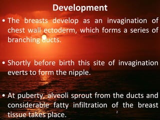
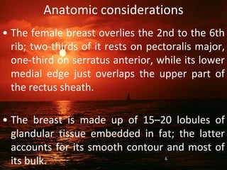
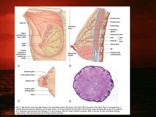
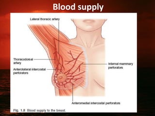
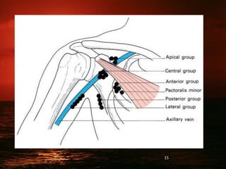
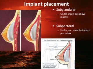
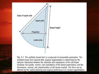
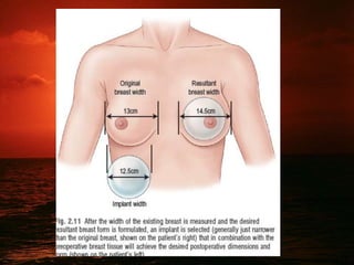
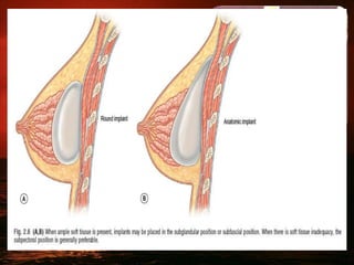
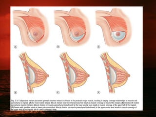
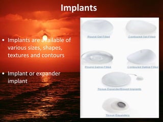
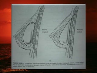
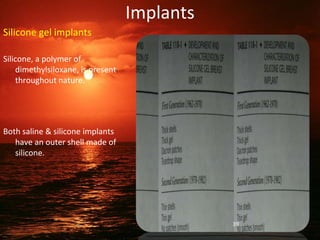
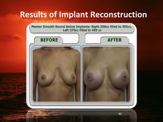
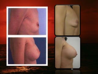
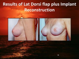
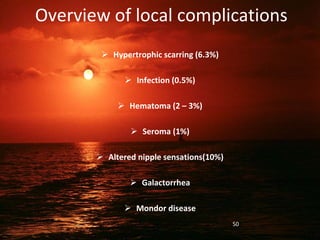
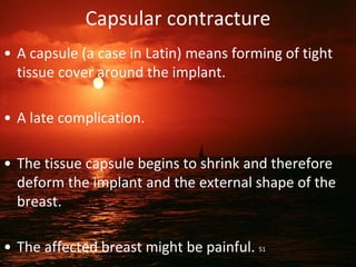
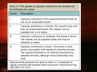
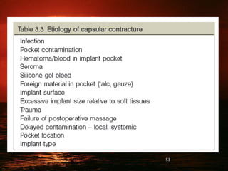
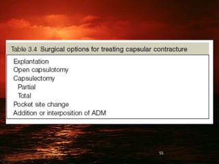
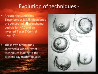
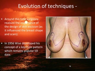
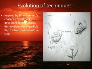
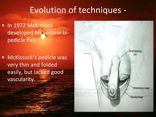
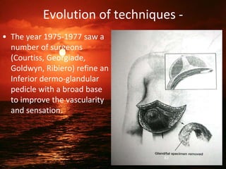
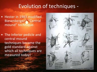
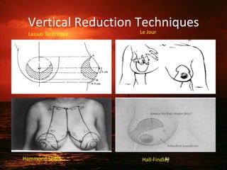
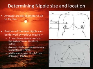
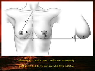
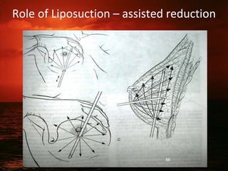
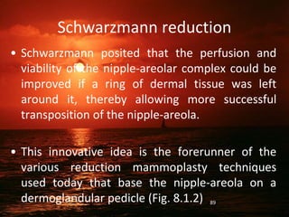
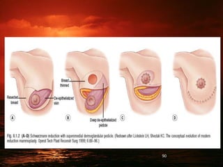
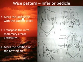
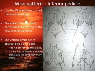
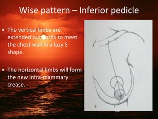
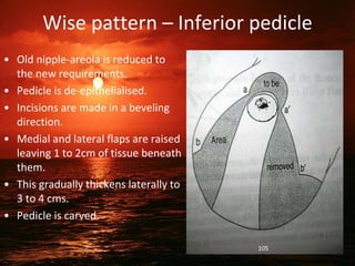
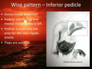
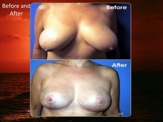
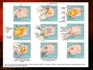
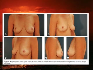
Augmentation
&
Reduction
Mammoplasty
Dr. Md Sohaib Akhtar
PG Department Burns, Plastic &
Reconstructive Surgery
J.N.M.C.
Development
• The breasts develop as an invagination of
chest wall ectoderm, which forms a series of
branching ducts.
• Shortly before birth this site of invagination
everts to form the nipple.
• At puberty, alveoli sprout from the ducts and
considerable fatty infiltration of the breast
tissue takes place.
2
• With pregnancy there is tremendous
development of the alveoli which, in
lactation, secrete the fatty droplets of
milk.
• At the menopause the gland tissue
atrophies.
3
4
5
Anatomic considerations
• The female breast overlies the 2nd to the 6th
rib; two-thirds of it rests on pectoralis major,
one-third on serratus anterior, while its lower
medial edge just overlaps the upper part of
the rectus sheath.
• The breast is made up of 15–20 lobules of
glandular tissue embedded in fat; the latter
accounts for its smooth contour and most of
its bulk. 6
• These lobules are separated by fibrous septa
running from the subcutaneous tissues to the
fascia of the chest wall (the ligaments of
Cooper).
• Each lobule drains by its lactiferous duct on to
the nipple, which is surrounded by the
pigmented areola.
• This area is lubricated by the areolar glands of
Montgomery; these are large, modified
sebaceous glands. 7
8
Blood supply
9
Sensory supply
• Sensation is provided chiefly by branches of the 4th
(& sometimes the 5th) anterolateral intercostal
nerve.
• To a lesser extent, anteromedial branches of the 4th
& 5th intercostal nerves also contributes to
sensation of the breast.
• Inframammary & periareolar incisions are more
likely to result in injury to these intercostal nerves,
10
Breast Anatomy
11
Lymphatic drainage
12
13
The axillary lymph nodes (some 20–30 in number) drain not only the lymphatics of the
breast, but also those of the pectoral region, upper abdominal wall and the upper
limb, and are arranged in five groups:
1. anterior—lying deep to pectoralis major along the lower border of pectoralis
minor;
2. posterior—along the subscapular vessels;
3. lateral—along the axillary vein;
4. central—in the axillary fat;
5. apical (through which all the other axillary nodes drain) —immediately behind
the clavicle at the apex of the axilla above pectoralis minor and along the medial
side of the axillary vein.
14
15
Augmentation
Mammoplasty
16
• No procedure in plastic surgery has been the
subject of greater scrutiny & controversy, both
scientific & political, than breast augmentation.
• More than 2 million American women, or 1% of
adult female population , have breast implants.
• Breast augmentation is the 2nd most commonly
performed cosmetic surgical procedure in US,
after suction assisted lipectomy.
17
• Women b/w 19-34 yrs are the largest
consumers of cosmetic breast implants(50%),
followed by women ages 35 to 50 yrs(42%).
• The history of breast augmentation reflects the
search for the ideal implantable material,
commencing with 19th century attempts to
transplant lipomas into breast defects,
followed by the modern era of polymer-based
devices. 18
Government regulation
• In 1976, the United States Congress passed the
Federal Safe Devices Act, which empowered the
FDA to regulate medical devices.
• Specific concerns of carcinogenicity, autoimmune
diseases, product failure, & impaired
mammographic evaluation led to a moratorium on
use of all silicone gel implants by the FDA in 1992.
19
• Since then saline implants have been available
without restriction, & were officially approved
by the FDA in 2000.
• Since the moratorium on silicone breast
implants, several studies have proved that
these devices are safe & do not cause
connective tissue disorders, malignancy, or
risk to breast feeding infants.
20
Breast augmentation
Technically known as augmentation mammoplasty, is a surgical procedure to enhance
the size and shape of a woman's breast for a number of reasons:
• To enhance the body contour of a woman who, for personal reasons,
feels her breast size is too small.
• To restore breast volume lost due to weight loss or following
pregnancy
• To achieve better symmetry when breasts are moderately
disproportionate in size and shape
• To improve the shape of breasts that are sagging or have lost
firmness, often used with a breast lift procedure
• To provide the foundation of a breast contour when a breast has
been removed or disfigured by surgery to treat breast cancer
• To improve breast appearance or create the appearance of a breast
that is missing or disfigured due to trauma, heredity, or congenital
abnormalities. 21
22
Surgical technique
• Four separate incisional sites:-
1. Inframammary
2. Periareolar
3. Axillary &
4. Transumblical
have been used for placement of breast prostheses.
Each location has its advantages & disadvantages,
depending on the individual surgeon’s experience & the
positioning of the implant in either a subglandular,
subpectoral, or the more recently described “dual-plane”
position. 23
24
• Inframammary crease incisions are preferred for patients
with a well developed crease that conceals the scar.
• In general, the periareolar incision provides excellent
access to all portions of the breast. Exposure of the
Inframammary ligament is excellent with this incision.
• Transection of breast ductal tissues is required, before the
pectoralis major muscle is identified.
• If subpectoral augmentation is chosen, the pectoralis major
muscle is either divided along the obliquity of its fibers or
the lateral border of muscle is elevated. 25
• The periareolar incision tends to heal with minimal
scarring.
• The periareolar incision facilitates revisional
procedures because it allows easier access to all
portions of the breast for capsulorrhaphy or
capsulectomy.
• In addition, the incision may be incorporated into
future periareolar mastopexy designs for ptosis as
the patient ages. 26
Endoscopic applications in augmentation
mammoplasty
Transaxillary approach is available for many years.
• Observed difficulty in attaining precise positioning
of the implant in the lower pole of the breast.
• Advantages:- absence of scar on breast surface, an
avoidance of breast ductal transection, & a low
probability of sensory nerve injury.
• Disadvantage:- trauma to intercostobrachial nerve.
27
• ENDOSCOPIC ASSISTANCE has enhanced
Transaxillary approach by allowing a more
accurate placement of the breast implant
inferiorly & improved control of inframammary
crease.
• Meticulous pre-op marking is extremely
important.
• Implant exchange, capsulorrhaphy, &
capsulotomy to treat implant-related
complications also can be performed with this
technique. 28
29
Transumblical augmentation mammoplasty
• Minimal scarring.
• Using a single infraumblical incision, this technique
uses endoscopic visualization to create a long,
subcutaneous tunnel up to the inferior border of
the breast.
• Difficult technique, hasn’t gained much popularity.
30
Implant placement
• Subglandular
– Under breast but above
muscle
• Subpectoral
– Under pec. major but above
pec. minor
31
32
33
34
35
Implants
• Implants are available of
various sizes, shapes,
textures and contours
• Implant or expander
implant
36
37
Implants
Saline implants
• Since silicone gel implants were banned by the FDA in
1992, saline implants have been available for both
cosmetic & reconstructive surgery.
• An elastomeric solid shell of silicone surrounding a hollow
interior is the principle design of current models.
• Implant survival, excluding trauma 97.9% - 99.5% after 10
years.
38
Implants
Silicone gel implants
Silicone, a polymer of
dimethylsiloxane, is present
throughout nature.
Both saline & silicone implants
have an outer shell made of
silicone.
39
Results of Implant Reconstruction
40
41
Autologous augmentation
• Autologous augmentation eliminates the risk of
implant deflation, contracture, infection, exposure,
& ultimately, exchange or removal.
• Not gained wide popularity because of increased
operative complexity, donor site complications &
scarring, prolonged recovery, & risk of flap failure.
42
• Potential candidates --patients unable to have a
breast implant, patients after explanation of
implants because of complications, or women
desiring either abdominal or gluteal contouring
in addition to breast augmentation.
• De-epithelialized pedicled transverse rectus
abdominis musculocutaneous (TRAM) flaps
have been used for breast reconstruction
following abdominoplasty. 43
Autologous augmentation (contd.)
• Local perforator flaps from the lateral chest wall also can
be used to augment the breasts, which is particularly
advantageous when combined with contour surgery in the
bariatric patient.
• Free perforator flaps have been described as potential
donors for autologous augmentation.
• Specifically, deep inferior epigastric perforator (DIEP),
superior gluteal artery perforator (S-GAP), & superficial
inferior epigastric artery (SIEA) perforator flaps have been
used to augment the breast in patients who desired
simultaneous abdominal or gluteal excision.
44
Pedicled TRAM Flap
TRAM = Transverse Rectus Abdominis Myocutaneous Flap 45
Results of TRAM reconstruction
46
Latissimus Dorsi Pedicled Flap
Commonly used to cover an underlying implant
47
Results of Lat Dorsi flap plus Implant
Reconstruction
48
Complications
49
Overview of local complications
 Hypertrophic scarring (6.3%)
 Infection (0.5%)
 Hematoma (2 – 3%)
 Seroma (1%)
 Altered nipple sensations(10%)
 Galactorrhea
 Mondor disease
50
Capsular contracture
• A capsule (a case in Latin) means forming of tight
tissue cover around the implant.
• A late complication.
• The tissue capsule begins to shrink and therefore
deform the implant and the external shape of the
breast.
• The affected breast might be painful. 51
52
53
54
55
Capsular contracture – Treatment
• Baker grade III or IV capsular contracture often
require treatment.
• Closed capsulotomy can improve capsular
contracture, even to Baker grade I capsular
contracture.
• Open capsulotomy or capsulectomy is the
treatment of choice for symptomatic capsular
contracture.
• Although, capsulectomy has a lower recurrence
rate, it is a more difficult procedure & associated
with more complications. 56
• A more recent method of correcting capsular
contracture involves repositioning the implant
in a DUAL PLANE.
• In this technique, capsulectomy is performed
followed by elevation of the inferior border of
the pectoralis major muscle.
• Approximately the superior 2/3rd of the
implant is placed submuscularly, while the
inferior 1/3rd is located in a subglandular
position.
57
58
Rupture of the implant
• The ruptured implant is very often diagnosed by
ultrasound, mammography or even other
examination.
• Implants which are filled with cohesive gel, even
after the rupture of the capsule of an implant
preserves its original shape & does not leak out.
59
• Recommended to revise the implant if there is
a suspicion on the rupture.
• In implants filled with saline the rupture or
leakage do not pose any medical risk.
•
• It is better in this case to replace this implant
rather within a month
60
61
Autoimmune & connective tissues
disorders
• Numerous case reports in 1980s suggested a
linkage b/w silicone implants & autoimmune &
connective tissues disorders.
• Includes scleroderma , rheumatoid arthritis, SLE ,
Sjogren syndrome, dermatomyositis ,polymyositis,
& polymyalgia rheumatica.
62
Silicone effects on pregnancy ,
lactation , & breast–fed children
• No scientific evidence that silicone is a mutagen or
teratogen.
• Implants do not interfere with lactation or increase
the amount of silicone in breast milk.
• Clinical studies suggests silicone implants are safe
for pregnancy , lactation & breast feeding.
63
Carcinogenesis
• Studies concluded that a lower incidence of Ca.Breast was found in
augmented patients than in nonimplanted control subjects.
• Possible explanations :-
1. Augmented women have less volume in which to develop
cancer compared to nonaugmented women.
2. Silicone may mediate a biologic protective effect against
breast cancer.
3. Also, augmented women are more aware of their breast
composition or more compliant with breast screening
than nonaugmented women
64
Shift of the implant
An example of wrong inserted implant under the muscle
after the surgery – a double fold under the breast
65
Aesthetic complications
• Implant malposition & an unfavorable shape are the 2nd
most common cause, after capsular contracture , of patient
dissatisfaction following augmentation mammoplasty.
• Correction of these types of problems usually involves a
combination of techniques, including removal of implant ,
capsulotomy, capsulectomy, alteration of the
inframammary crease, selection of a different location or
different shape & size implant.
• Residual ptosis is best treated by recognition of the
relationship of the nipple to the inframammary crease &
placement of implant in a subglandular position.
66
• The double bubble deformity results from disruption or
distortion of the inframammary crease.
• Commonly noted after attempted correction of a
constricted, tubular breast , but it might also occur after
subpectoral placement with elevation of the crease or after
disruption of the inframammary ligament & inferior
displacement of the prosthesis.
• Surgical correction involves varying combinations of
inframammary crease reconstruction , pectoralis major
muscle release, & replacement with a smaller implant.
67
68
• Rippling is almost always a complication of saline
implants, primarily affecting the upper pole .
• Risk factors for rippling include underfilling &
textured implants. Textured implants have a
greater likelihood of causing traction on skin
because of their increased adherent properties.
o Prophylactic treatment include using a smooth
implant, subpectoral placement , & overfilling of
implant.
69
1st line Tt :- conversion to subpectoral placement
if previously subglandular, overfilling the
implant, changing textured to a smooth implant,
or converting a saline to a gel implant.
2nd line Tt :- capsulorrhaphy, or partial
capsulectomy, placing external bolsters, or using
Allogenic acellular dermal grafts( AlloDerm ) b/w
implant & skin.
3rd line Tt :- transfer of healthy , well-
vascularised tissue to cover the implant in severe
, refractory cases.
70
Psychological issues
• Patient satisfaction with both saline & silicone
breast implants is very high, ranging from 93% -
97%.
• Women interested in augmentation mammoplasty,
compared to physically similar women, are more
concerned about their appearance & feel that
larger breasts are the ideal breast size.
• Like other cosmetic surgery patients, these women
have a greater prevalence of psychiatric disease
71
Imaging of breast implants
• Three modalities – mammography, sonography, & MRI
• MRI is the most accurate & expensive study with a
sensitivity greater than 95% for a ruptured implant. Shell
folding ( “linguine sign” ) on MRI indicates a rupture
implant .
• Sonographic signs of implant rupture include the
“snowstorm” appearance of free silicone in the breast
tissue or the “stepladder” sign of linear echoes.
• MRI is the radiographic study of choice to determine
implant rupture in patients with Baker III or IV contracture.
72
Medicolegal considerations
• Surgeons must navigate between product liability &
informed consent laws that hold them accountable
for a wide array of health issues, proven & alleged.
• Implant failure or complications create a burden to
the patient & thus a potential stimulus for
litigation.
73
Reduction
Mammoplasty
74
Why need breast reduction ?
• Very large breasts -
– Shoulder pain.
– Cervical pain.
– Upper thoracic pain.
– Severe embarrassment.
– Recurrent infections.
• Inability to exercise.
• Difficulty in breathing.
• Finding clothing which fit.
• Grooving of shoulder strap
areas.
75
Evolution of techniques -
• 1925 – Passot described
nipple transposition into
a button-hole incision
higher on the breast
mound.
76
Evolution of techniques -
• Around the same time
Biesenberger (1928) developed
the concept of a parenchymal
pedicle for NAC and an
inverted T scar (“Central
mound”).
• These two techniques
spawned a wide array of
techniques leading to the
present day mammaplsties.
77
Evolution of techniques -
• Around this time surgeons
realised the importance of
the design of skin excision (as
it influenced the breast shape
and scars).
• In 1956 Wise developed his
concept of a key-hole pattern
which remains popular till
date.
78
Evolution of techniques -
• Inspired by Schwarzmann’s
concepts, Strombeck
developed his horizontal,
dermo-glandular bi-pedicle
flap for transposition of the
NAC.
79
Evolution of techniques -
• Pitanguy in 1962
developed the superior
pedicle technique which is
one of the favorite
techniques till date.
• Skoog soon followed in
1963 with his Supero-
lateral pedicle technique.
80
Evolution of techniques -
• In 1972 McKissock
developed his vertical bi-
pedicle flap.
• McKissock’s pedicle was
very thin and folded
easily, but lacked good
vascularity.
81
Evolution of techniques -
• The year 1975-1977 saw a
number of surgeons
(Courtiss, Georgiade,
Goldwyn, Ribiero) refine an
Inferior dermo-glandular
pedicle with a broad base
to improve the vascularity
and sensation.
82
Evolution of techniques -
• Hester in 1983 modified
Biesenberger’s “Central
mound” technique.
• The Inferior pedicle and
central mound
techniques became the
gold standard against
which all techniques are
measured today!
83
Vertical Reduction Techniques
Lassus Technique Le Jour
Hammond SPAIR Hall-Findlay
84
Determining Nipple size and location
• Average areolar diameter is 38
to 45 mm.
• Position of the new nipple can
be derived by various means –
– 21 cms below sternal notch on
the mid-clavicular line (Penn,
1955).
– Average nipple to infra-mammary
fold distance = 7cms.
– Mid-humeral point plus 2-3 cms
(Pitanguy, 1967).
85
Measurements required prior to reduction mammoplasty.
a=19–21 cm; b=9–11 cm; c=4–5 cm; d=5–8 cm; e=0–2 cm
a
b c c
d
e b
86
TIMING OF SURGERY
When breast growth is complete
In exceptional cases it can be done in a
young or old person
Role of Liposuction – assisted reduction
88
Schwarzmann reduction
• Schwarzmann posited that the perfusion and
viability of the nipple-areolar complex could be
improved if a ring of dermal tissue was left
around it, thereby allowing more successful
transposition of the nipple-areola.
• This innovative idea is the forerunner of the
various reduction mammoplasty techniques
used today that base the nipple-areola on a
dermoglandular pedicle (Fig. 8.1.2) 89
90
Biesenberger Reduction
• Another vanguard event in the history of
breast reduction came with Biesenberger, who
was the first to develop a reproducible
parenchymal pedicle-based technique with a
“cut as you go” skin resection pattern (Fig.
8.1.3)
• Results in an inverted-T scar and relies heavily
on wide sub cutaneous undermining with
folding of the breast pedicle
• High rates of skin and nipple necrosis
91
92
Strombeck horizontal bipedicle
technique
• Strombeck demonstrated a horizontal
bipedicled flap to maintain the nipple-areolar
complex in 1960, thereby capitalizing on the
work of Aufricht and Bames (medial and lateral
perforators for perfusion, as well as
innervation) and Schwarzmann
(dermoglandular pedicle) (Fig. 8.1.4).
93
94
McKissock vertical bipedicled
dermoglandular flap
• The superolateral dermoglandular pedicle was
described by Skoog in 1963, whereas Pitanguy
and Weiner described superiorly-based der-
moglandular pedicle some 10 years later in
1973 (Fig. 8.1.5).
95
96
97
INFERIOR PEDICLE TECHNIQUE
• In 1977, Courtiss and Goldwyn, and
Georgiade, among others, made advances in
inferior pedicle tech- niques (Fig. 8.1.6).2
98
99
• Five years prior, McKissock modi- fied
Strombeck’s horizontal bipedicle technique
into a vertical bipedicle technique.
• This required thinning of the superior and
inferior portions of the bipedicled flap to
allow folding, and resultingly does not have as
much perfusion as other techniques (Fig.
8.1.7).
100
101
Wise pattern – Inferior pedicle
• Mark the landmarks
with the patient erect.
• Transpose the infra-
mammary crease
anteriorly.
• Mark the position of
the new nipple.
102
Wise pattern – Inferior pedicle
• Use the Wise pattern to mark
the key-hole design.
• The apex of the key-hole
corresponds to the top of the
new areolar diameter.
• The vertical limbs are of
approx. 8 to 9 cms each
– 4 to 4.5 cm for the areola and,
– 4.5 cm for the distance b/w the
areola and the infra-mammary
crease.
103
Wise pattern – Inferior pedicle
• The vertical limbs are
extended outwards to meet
the chest wall in a lazy S
shape.
• The horizontal limbs will form
the new infra-mammary
crease.
104
Wise pattern – Inferior pedicle
• Old nipple-areola is reduced to
the new requirements.
• Pedicle is de-epithelialised.
• Incisions are made in a beveling
direction.
• Medial and lateral flaps are raised
leaving 1 to 2cm of tissue beneath
them.
• This gradually thickens laterally to
3 to 4 cms.
• Pedicle is carved.
105
Wise pattern – Inferior pedicle
• Excess tissue is excised.
• Pedicle with lateral and
medial mesenteries is left.
• Pedicle is inset into the
area for the new nipple-
areola.
• Flaps are sutured.
106
Before and
After
107
• The result of the previous decades’
developments in breast reduction surgery –
Wise pattern skin resection with multiple
pedicle options – was labeled in the 1980s as
the “gold standard” because the results
obtained were reliable, reproducible, and
useful for breasts of nearly all shapes and
sizes
108
• Modifications and additions were still being
made to the cadre of pedicle options for the
Wise pattern skin resection technique of
breast reduction in the 1980s, such as
Hester’s central mound technique (Fig. 8.1.8).
109
110
• But the years of experience andfollow-up
after these reduction procedures demon
strated that the reduced breasts tended to
bottom out and lose upper pole fullness (Fig.
8.1.9
111
112
• This problem actually had been
anticipated in previous decades.
• Orlando and Guthrie, originators of the
superomedial pedicle, had devised their
approach in an effort to augment the
upper pole and thereby reduce
bottoming out (Fig. 8.1.10)
113
114
CONCLUSON
• Despite the many recent advances in breast
reduction surgery, the inverted-T scar technique
remains a comfortable and predictable
technique for the surgeon who performs breast
surgery.
• Although there is appropriate increasing interest
in short scar or vertical scar techniques, the
inverted-T option has proven reliable and safe,
which may be as important to the patient as the
length of the scar in the inframammary fold.
115
www.cosmetic-plastic-
surgery.com
116
THANK YOU
117

1.Augmentation & reduction mammoplasty (2).pptx

  • 1.
    Augmentation & Reduction Mammoplasty Dr. Md SohaibAkhtar PG Department Burns, Plastic & Reconstructive Surgery J.N.M.C.
  • 2.
    Development • The breastsdevelop as an invagination of chest wall ectoderm, which forms a series of branching ducts. • Shortly before birth this site of invagination everts to form the nipple. • At puberty, alveoli sprout from the ducts and considerable fatty infiltration of the breast tissue takes place. 2
  • 3.
    • With pregnancythere is tremendous development of the alveoli which, in lactation, secrete the fatty droplets of milk. • At the menopause the gland tissue atrophies. 3
  • 4.
  • 5.
  • 6.
    Anatomic considerations • Thefemale breast overlies the 2nd to the 6th rib; two-thirds of it rests on pectoralis major, one-third on serratus anterior, while its lower medial edge just overlaps the upper part of the rectus sheath. • The breast is made up of 15–20 lobules of glandular tissue embedded in fat; the latter accounts for its smooth contour and most of its bulk. 6
  • 7.
    • These lobulesare separated by fibrous septa running from the subcutaneous tissues to the fascia of the chest wall (the ligaments of Cooper). • Each lobule drains by its lactiferous duct on to the nipple, which is surrounded by the pigmented areola. • This area is lubricated by the areolar glands of Montgomery; these are large, modified sebaceous glands. 7
  • 8.
  • 9.
  • 10.
    Sensory supply • Sensationis provided chiefly by branches of the 4th (& sometimes the 5th) anterolateral intercostal nerve. • To a lesser extent, anteromedial branches of the 4th & 5th intercostal nerves also contributes to sensation of the breast. • Inframammary & periareolar incisions are more likely to result in injury to these intercostal nerves, 10
  • 11.
  • 12.
  • 13.
  • 14.
    The axillary lymphnodes (some 20–30 in number) drain not only the lymphatics of the breast, but also those of the pectoral region, upper abdominal wall and the upper limb, and are arranged in five groups: 1. anterior—lying deep to pectoralis major along the lower border of pectoralis minor; 2. posterior—along the subscapular vessels; 3. lateral—along the axillary vein; 4. central—in the axillary fat; 5. apical (through which all the other axillary nodes drain) —immediately behind the clavicle at the apex of the axilla above pectoralis minor and along the medial side of the axillary vein. 14
  • 15.
  • 16.
  • 17.
    • No procedurein plastic surgery has been the subject of greater scrutiny & controversy, both scientific & political, than breast augmentation. • More than 2 million American women, or 1% of adult female population , have breast implants. • Breast augmentation is the 2nd most commonly performed cosmetic surgical procedure in US, after suction assisted lipectomy. 17
  • 18.
    • Women b/w19-34 yrs are the largest consumers of cosmetic breast implants(50%), followed by women ages 35 to 50 yrs(42%). • The history of breast augmentation reflects the search for the ideal implantable material, commencing with 19th century attempts to transplant lipomas into breast defects, followed by the modern era of polymer-based devices. 18
  • 19.
    Government regulation • In1976, the United States Congress passed the Federal Safe Devices Act, which empowered the FDA to regulate medical devices. • Specific concerns of carcinogenicity, autoimmune diseases, product failure, & impaired mammographic evaluation led to a moratorium on use of all silicone gel implants by the FDA in 1992. 19
  • 20.
    • Since thensaline implants have been available without restriction, & were officially approved by the FDA in 2000. • Since the moratorium on silicone breast implants, several studies have proved that these devices are safe & do not cause connective tissue disorders, malignancy, or risk to breast feeding infants. 20
  • 21.
    Breast augmentation Technically knownas augmentation mammoplasty, is a surgical procedure to enhance the size and shape of a woman's breast for a number of reasons: • To enhance the body contour of a woman who, for personal reasons, feels her breast size is too small. • To restore breast volume lost due to weight loss or following pregnancy • To achieve better symmetry when breasts are moderately disproportionate in size and shape • To improve the shape of breasts that are sagging or have lost firmness, often used with a breast lift procedure • To provide the foundation of a breast contour when a breast has been removed or disfigured by surgery to treat breast cancer • To improve breast appearance or create the appearance of a breast that is missing or disfigured due to trauma, heredity, or congenital abnormalities. 21
  • 22.
  • 23.
    Surgical technique • Fourseparate incisional sites:- 1. Inframammary 2. Periareolar 3. Axillary & 4. Transumblical have been used for placement of breast prostheses. Each location has its advantages & disadvantages, depending on the individual surgeon’s experience & the positioning of the implant in either a subglandular, subpectoral, or the more recently described “dual-plane” position. 23
  • 24.
  • 25.
    • Inframammary creaseincisions are preferred for patients with a well developed crease that conceals the scar. • In general, the periareolar incision provides excellent access to all portions of the breast. Exposure of the Inframammary ligament is excellent with this incision. • Transection of breast ductal tissues is required, before the pectoralis major muscle is identified. • If subpectoral augmentation is chosen, the pectoralis major muscle is either divided along the obliquity of its fibers or the lateral border of muscle is elevated. 25
  • 26.
    • The periareolarincision tends to heal with minimal scarring. • The periareolar incision facilitates revisional procedures because it allows easier access to all portions of the breast for capsulorrhaphy or capsulectomy. • In addition, the incision may be incorporated into future periareolar mastopexy designs for ptosis as the patient ages. 26
  • 27.
    Endoscopic applications inaugmentation mammoplasty Transaxillary approach is available for many years. • Observed difficulty in attaining precise positioning of the implant in the lower pole of the breast. • Advantages:- absence of scar on breast surface, an avoidance of breast ductal transection, & a low probability of sensory nerve injury. • Disadvantage:- trauma to intercostobrachial nerve. 27
  • 28.
    • ENDOSCOPIC ASSISTANCEhas enhanced Transaxillary approach by allowing a more accurate placement of the breast implant inferiorly & improved control of inframammary crease. • Meticulous pre-op marking is extremely important. • Implant exchange, capsulorrhaphy, & capsulotomy to treat implant-related complications also can be performed with this technique. 28
  • 29.
  • 30.
    Transumblical augmentation mammoplasty •Minimal scarring. • Using a single infraumblical incision, this technique uses endoscopic visualization to create a long, subcutaneous tunnel up to the inferior border of the breast. • Difficult technique, hasn’t gained much popularity. 30
  • 31.
    Implant placement • Subglandular –Under breast but above muscle • Subpectoral – Under pec. major but above pec. minor 31
  • 32.
  • 33.
  • 34.
  • 35.
  • 36.
    Implants • Implants areavailable of various sizes, shapes, textures and contours • Implant or expander implant 36
  • 37.
  • 38.
    Implants Saline implants • Sincesilicone gel implants were banned by the FDA in 1992, saline implants have been available for both cosmetic & reconstructive surgery. • An elastomeric solid shell of silicone surrounding a hollow interior is the principle design of current models. • Implant survival, excluding trauma 97.9% - 99.5% after 10 years. 38
  • 39.
    Implants Silicone gel implants Silicone,a polymer of dimethylsiloxane, is present throughout nature. Both saline & silicone implants have an outer shell made of silicone. 39
  • 40.
    Results of ImplantReconstruction 40
  • 41.
  • 42.
    Autologous augmentation • Autologousaugmentation eliminates the risk of implant deflation, contracture, infection, exposure, & ultimately, exchange or removal. • Not gained wide popularity because of increased operative complexity, donor site complications & scarring, prolonged recovery, & risk of flap failure. 42
  • 43.
    • Potential candidates--patients unable to have a breast implant, patients after explanation of implants because of complications, or women desiring either abdominal or gluteal contouring in addition to breast augmentation. • De-epithelialized pedicled transverse rectus abdominis musculocutaneous (TRAM) flaps have been used for breast reconstruction following abdominoplasty. 43
  • 44.
    Autologous augmentation (contd.) •Local perforator flaps from the lateral chest wall also can be used to augment the breasts, which is particularly advantageous when combined with contour surgery in the bariatric patient. • Free perforator flaps have been described as potential donors for autologous augmentation. • Specifically, deep inferior epigastric perforator (DIEP), superior gluteal artery perforator (S-GAP), & superficial inferior epigastric artery (SIEA) perforator flaps have been used to augment the breast in patients who desired simultaneous abdominal or gluteal excision. 44
  • 45.
    Pedicled TRAM Flap TRAM= Transverse Rectus Abdominis Myocutaneous Flap 45
  • 46.
    Results of TRAMreconstruction 46
  • 47.
    Latissimus Dorsi PedicledFlap Commonly used to cover an underlying implant 47
  • 48.
    Results of LatDorsi flap plus Implant Reconstruction 48
  • 49.
  • 50.
    Overview of localcomplications  Hypertrophic scarring (6.3%)  Infection (0.5%)  Hematoma (2 – 3%)  Seroma (1%)  Altered nipple sensations(10%)  Galactorrhea  Mondor disease 50
  • 51.
    Capsular contracture • Acapsule (a case in Latin) means forming of tight tissue cover around the implant. • A late complication. • The tissue capsule begins to shrink and therefore deform the implant and the external shape of the breast. • The affected breast might be painful. 51
  • 52.
  • 53.
  • 54.
  • 55.
  • 56.
    Capsular contracture –Treatment • Baker grade III or IV capsular contracture often require treatment. • Closed capsulotomy can improve capsular contracture, even to Baker grade I capsular contracture. • Open capsulotomy or capsulectomy is the treatment of choice for symptomatic capsular contracture. • Although, capsulectomy has a lower recurrence rate, it is a more difficult procedure & associated with more complications. 56
  • 57.
    • A morerecent method of correcting capsular contracture involves repositioning the implant in a DUAL PLANE. • In this technique, capsulectomy is performed followed by elevation of the inferior border of the pectoralis major muscle. • Approximately the superior 2/3rd of the implant is placed submuscularly, while the inferior 1/3rd is located in a subglandular position. 57
  • 58.
  • 59.
    Rupture of theimplant • The ruptured implant is very often diagnosed by ultrasound, mammography or even other examination. • Implants which are filled with cohesive gel, even after the rupture of the capsule of an implant preserves its original shape & does not leak out. 59
  • 60.
    • Recommended torevise the implant if there is a suspicion on the rupture. • In implants filled with saline the rupture or leakage do not pose any medical risk. • • It is better in this case to replace this implant rather within a month 60
  • 61.
  • 62.
    Autoimmune & connectivetissues disorders • Numerous case reports in 1980s suggested a linkage b/w silicone implants & autoimmune & connective tissues disorders. • Includes scleroderma , rheumatoid arthritis, SLE , Sjogren syndrome, dermatomyositis ,polymyositis, & polymyalgia rheumatica. 62
  • 63.
    Silicone effects onpregnancy , lactation , & breast–fed children • No scientific evidence that silicone is a mutagen or teratogen. • Implants do not interfere with lactation or increase the amount of silicone in breast milk. • Clinical studies suggests silicone implants are safe for pregnancy , lactation & breast feeding. 63
  • 64.
    Carcinogenesis • Studies concludedthat a lower incidence of Ca.Breast was found in augmented patients than in nonimplanted control subjects. • Possible explanations :- 1. Augmented women have less volume in which to develop cancer compared to nonaugmented women. 2. Silicone may mediate a biologic protective effect against breast cancer. 3. Also, augmented women are more aware of their breast composition or more compliant with breast screening than nonaugmented women 64
  • 65.
    Shift of theimplant An example of wrong inserted implant under the muscle after the surgery – a double fold under the breast 65
  • 66.
    Aesthetic complications • Implantmalposition & an unfavorable shape are the 2nd most common cause, after capsular contracture , of patient dissatisfaction following augmentation mammoplasty. • Correction of these types of problems usually involves a combination of techniques, including removal of implant , capsulotomy, capsulectomy, alteration of the inframammary crease, selection of a different location or different shape & size implant. • Residual ptosis is best treated by recognition of the relationship of the nipple to the inframammary crease & placement of implant in a subglandular position. 66
  • 67.
    • The doublebubble deformity results from disruption or distortion of the inframammary crease. • Commonly noted after attempted correction of a constricted, tubular breast , but it might also occur after subpectoral placement with elevation of the crease or after disruption of the inframammary ligament & inferior displacement of the prosthesis. • Surgical correction involves varying combinations of inframammary crease reconstruction , pectoralis major muscle release, & replacement with a smaller implant. 67
  • 68.
  • 69.
    • Rippling isalmost always a complication of saline implants, primarily affecting the upper pole . • Risk factors for rippling include underfilling & textured implants. Textured implants have a greater likelihood of causing traction on skin because of their increased adherent properties. o Prophylactic treatment include using a smooth implant, subpectoral placement , & overfilling of implant. 69
  • 70.
    1st line Tt:- conversion to subpectoral placement if previously subglandular, overfilling the implant, changing textured to a smooth implant, or converting a saline to a gel implant. 2nd line Tt :- capsulorrhaphy, or partial capsulectomy, placing external bolsters, or using Allogenic acellular dermal grafts( AlloDerm ) b/w implant & skin. 3rd line Tt :- transfer of healthy , well- vascularised tissue to cover the implant in severe , refractory cases. 70
  • 71.
    Psychological issues • Patientsatisfaction with both saline & silicone breast implants is very high, ranging from 93% - 97%. • Women interested in augmentation mammoplasty, compared to physically similar women, are more concerned about their appearance & feel that larger breasts are the ideal breast size. • Like other cosmetic surgery patients, these women have a greater prevalence of psychiatric disease 71
  • 72.
    Imaging of breastimplants • Three modalities – mammography, sonography, & MRI • MRI is the most accurate & expensive study with a sensitivity greater than 95% for a ruptured implant. Shell folding ( “linguine sign” ) on MRI indicates a rupture implant . • Sonographic signs of implant rupture include the “snowstorm” appearance of free silicone in the breast tissue or the “stepladder” sign of linear echoes. • MRI is the radiographic study of choice to determine implant rupture in patients with Baker III or IV contracture. 72
  • 73.
    Medicolegal considerations • Surgeonsmust navigate between product liability & informed consent laws that hold them accountable for a wide array of health issues, proven & alleged. • Implant failure or complications create a burden to the patient & thus a potential stimulus for litigation. 73
  • 74.
  • 75.
    Why need breastreduction ? • Very large breasts - – Shoulder pain. – Cervical pain. – Upper thoracic pain. – Severe embarrassment. – Recurrent infections. • Inability to exercise. • Difficulty in breathing. • Finding clothing which fit. • Grooving of shoulder strap areas. 75
  • 76.
    Evolution of techniques- • 1925 – Passot described nipple transposition into a button-hole incision higher on the breast mound. 76
  • 77.
    Evolution of techniques- • Around the same time Biesenberger (1928) developed the concept of a parenchymal pedicle for NAC and an inverted T scar (“Central mound”). • These two techniques spawned a wide array of techniques leading to the present day mammaplsties. 77
  • 78.
    Evolution of techniques- • Around this time surgeons realised the importance of the design of skin excision (as it influenced the breast shape and scars). • In 1956 Wise developed his concept of a key-hole pattern which remains popular till date. 78
  • 79.
    Evolution of techniques- • Inspired by Schwarzmann’s concepts, Strombeck developed his horizontal, dermo-glandular bi-pedicle flap for transposition of the NAC. 79
  • 80.
    Evolution of techniques- • Pitanguy in 1962 developed the superior pedicle technique which is one of the favorite techniques till date. • Skoog soon followed in 1963 with his Supero- lateral pedicle technique. 80
  • 81.
    Evolution of techniques- • In 1972 McKissock developed his vertical bi- pedicle flap. • McKissock’s pedicle was very thin and folded easily, but lacked good vascularity. 81
  • 82.
    Evolution of techniques- • The year 1975-1977 saw a number of surgeons (Courtiss, Georgiade, Goldwyn, Ribiero) refine an Inferior dermo-glandular pedicle with a broad base to improve the vascularity and sensation. 82
  • 83.
    Evolution of techniques- • Hester in 1983 modified Biesenberger’s “Central mound” technique. • The Inferior pedicle and central mound techniques became the gold standard against which all techniques are measured today! 83
  • 84.
    Vertical Reduction Techniques LassusTechnique Le Jour Hammond SPAIR Hall-Findlay 84
  • 85.
    Determining Nipple sizeand location • Average areolar diameter is 38 to 45 mm. • Position of the new nipple can be derived by various means – – 21 cms below sternal notch on the mid-clavicular line (Penn, 1955). – Average nipple to infra-mammary fold distance = 7cms. – Mid-humeral point plus 2-3 cms (Pitanguy, 1967). 85
  • 86.
    Measurements required priorto reduction mammoplasty. a=19–21 cm; b=9–11 cm; c=4–5 cm; d=5–8 cm; e=0–2 cm a b c c d e b 86
  • 87.
    TIMING OF SURGERY Whenbreast growth is complete In exceptional cases it can be done in a young or old person
  • 88.
    Role of Liposuction– assisted reduction 88
  • 89.
    Schwarzmann reduction • Schwarzmannposited that the perfusion and viability of the nipple-areolar complex could be improved if a ring of dermal tissue was left around it, thereby allowing more successful transposition of the nipple-areola. • This innovative idea is the forerunner of the various reduction mammoplasty techniques used today that base the nipple-areola on a dermoglandular pedicle (Fig. 8.1.2) 89
  • 90.
  • 91.
    Biesenberger Reduction • Anothervanguard event in the history of breast reduction came with Biesenberger, who was the first to develop a reproducible parenchymal pedicle-based technique with a “cut as you go” skin resection pattern (Fig. 8.1.3) • Results in an inverted-T scar and relies heavily on wide sub cutaneous undermining with folding of the breast pedicle • High rates of skin and nipple necrosis 91
  • 92.
  • 93.
    Strombeck horizontal bipedicle technique •Strombeck demonstrated a horizontal bipedicled flap to maintain the nipple-areolar complex in 1960, thereby capitalizing on the work of Aufricht and Bames (medial and lateral perforators for perfusion, as well as innervation) and Schwarzmann (dermoglandular pedicle) (Fig. 8.1.4). 93
  • 94.
  • 95.
    McKissock vertical bipedicled dermoglandularflap • The superolateral dermoglandular pedicle was described by Skoog in 1963, whereas Pitanguy and Weiner described superiorly-based der- moglandular pedicle some 10 years later in 1973 (Fig. 8.1.5). 95
  • 96.
  • 97.
  • 98.
    INFERIOR PEDICLE TECHNIQUE •In 1977, Courtiss and Goldwyn, and Georgiade, among others, made advances in inferior pedicle tech- niques (Fig. 8.1.6).2 98
  • 99.
  • 100.
    • Five yearsprior, McKissock modi- fied Strombeck’s horizontal bipedicle technique into a vertical bipedicle technique. • This required thinning of the superior and inferior portions of the bipedicled flap to allow folding, and resultingly does not have as much perfusion as other techniques (Fig. 8.1.7). 100
  • 101.
  • 102.
    Wise pattern –Inferior pedicle • Mark the landmarks with the patient erect. • Transpose the infra- mammary crease anteriorly. • Mark the position of the new nipple. 102
  • 103.
    Wise pattern –Inferior pedicle • Use the Wise pattern to mark the key-hole design. • The apex of the key-hole corresponds to the top of the new areolar diameter. • The vertical limbs are of approx. 8 to 9 cms each – 4 to 4.5 cm for the areola and, – 4.5 cm for the distance b/w the areola and the infra-mammary crease. 103
  • 104.
    Wise pattern –Inferior pedicle • The vertical limbs are extended outwards to meet the chest wall in a lazy S shape. • The horizontal limbs will form the new infra-mammary crease. 104
  • 105.
    Wise pattern –Inferior pedicle • Old nipple-areola is reduced to the new requirements. • Pedicle is de-epithelialised. • Incisions are made in a beveling direction. • Medial and lateral flaps are raised leaving 1 to 2cm of tissue beneath them. • This gradually thickens laterally to 3 to 4 cms. • Pedicle is carved. 105
  • 106.
    Wise pattern –Inferior pedicle • Excess tissue is excised. • Pedicle with lateral and medial mesenteries is left. • Pedicle is inset into the area for the new nipple- areola. • Flaps are sutured. 106
  • 107.
  • 108.
    • The resultof the previous decades’ developments in breast reduction surgery – Wise pattern skin resection with multiple pedicle options – was labeled in the 1980s as the “gold standard” because the results obtained were reliable, reproducible, and useful for breasts of nearly all shapes and sizes 108
  • 109.
    • Modifications andadditions were still being made to the cadre of pedicle options for the Wise pattern skin resection technique of breast reduction in the 1980s, such as Hester’s central mound technique (Fig. 8.1.8). 109
  • 110.
  • 111.
    • But theyears of experience andfollow-up after these reduction procedures demon strated that the reduced breasts tended to bottom out and lose upper pole fullness (Fig. 8.1.9 111
  • 112.
  • 113.
    • This problemactually had been anticipated in previous decades. • Orlando and Guthrie, originators of the superomedial pedicle, had devised their approach in an effort to augment the upper pole and thereby reduce bottoming out (Fig. 8.1.10) 113
  • 114.
  • 115.
    CONCLUSON • Despite themany recent advances in breast reduction surgery, the inverted-T scar technique remains a comfortable and predictable technique for the surgeon who performs breast surgery. • Although there is appropriate increasing interest in short scar or vertical scar techniques, the inverted-T option has proven reliable and safe, which may be as important to the patient as the length of the scar in the inframammary fold. 115
  • 116.
  • 117.