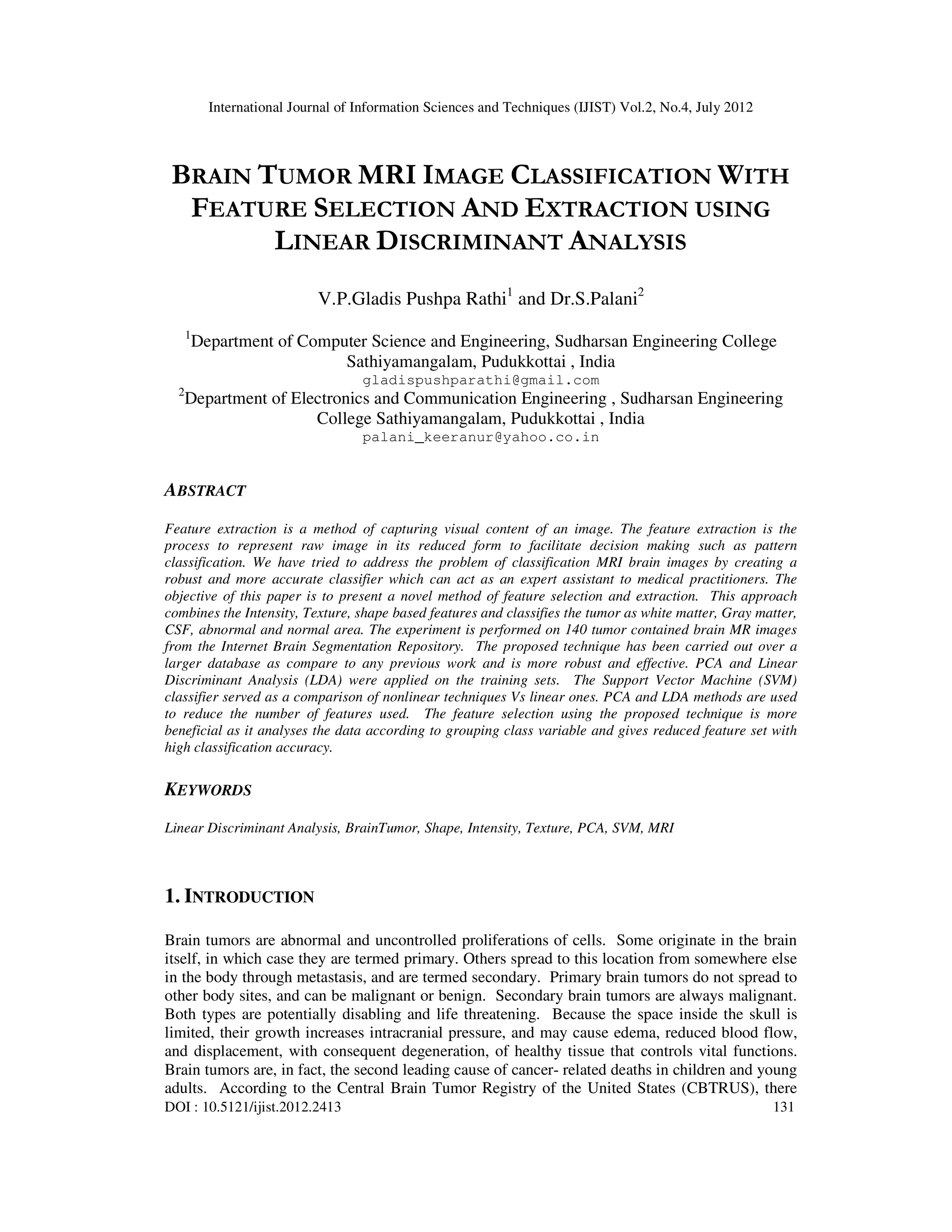
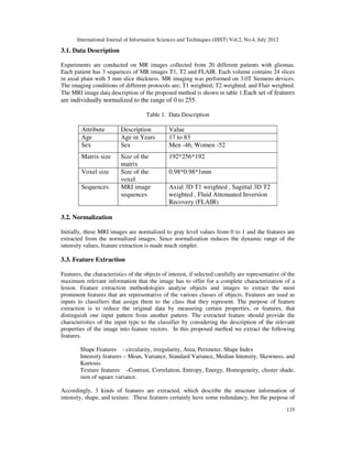
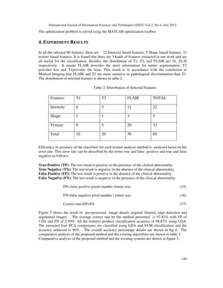
This document presents a novel approach for brain tumor classification in MRI images using feature selection and extraction. It extracts intensity, texture, and shape-based features from MRI images and applies principal component analysis (PCA) and linear discriminant analysis (LDA) for dimensionality reduction. Support vector machines (SVM) are then used to classify tumors as white matter, gray matter, CSF, abnormal or normal tissue. The technique is tested on 140 brain MRI images and achieves high classification accuracy compared to previous methods.
This document reviews various automated techniques that have been developed for brain tumor detection. It summarizes research done by several researchers on methods like sequential floating forward selection, color coding schemes using brain atlases, neural networks, region growing segmentation combined with area calculation, symmetry analysis of tumor areas in MRI images, and combining clustering and classification algorithms. The paper concludes that image segmentation plays an important role in medical applications like tumor diagnosis and that more robust techniques are needed for high accuracy and reliability.
A Survey On Brain Tumor Detection TechniquesIRJET Journal
This document summarizes various techniques that have been proposed for detecting brain tumors from MRI scanned images. It discusses how features can be extracted from images using pixel intensity to detect tumor location. Techniques mentioned include preprocessing images, segmentation, and using classifiers like support vector machines. MATLAB software is often used to implement these techniques and detect tumors. The document reviews several papers on topics like region growing segmentation methods, discrete wavelet transforms combined with neural networks, and challenges in brain tumor detection and identification.
MRI Image Segmentation Using Gradient Based Watershed Transform In Level Set ...IJERA Editor
This document summarizes a research paper on segmenting MRI brain images using a gradient-based watershed transform within a level set method. The paper begins with an introduction on the importance of accurate brain image segmentation for medical diagnosis. It then reviews existing segmentation methods and their limitations. The proposed method uses a two-level gradient watershed transform combined with morphological operations within a level set framework to segment brain images. Experimental results showed this approach achieved better segmentation accuracy than traditional methods.
IRJET- A Study on Brain Tumor Detection Algorithms for MRI ImagesIRJET Journal
This document discusses algorithms for detecting brain tumors in MRI images. It begins with an abstract that outlines the key stages of brain tumor detection using image processing techniques: pre-processing, segmentation, feature extraction, and classification. It then reviews several existing techniques for brain tumor segmentation and classification, noting their advantages and limitations. Specifically, it examines algorithms using Bayesian techniques, neural networks, clustering, and deep learning. The document proposes using a Spearman algorithm for segmentation combined with a convolutional neural network classifier to overcome limitations of other methods and provide more accurate tumor detection.
3D Segmentation of Brain Tumor ImagingIJAEMSJORNAL
A brain tumor is a collection of anomalous cells that grow in or around the brain. Brain tumors affect the humans badly, it can disrupt proper brain function and be life-threatening. In this project, we have proposed a system to detect, segment, and classify the tumors present in the brain. Once the brain tumor is identified at the very beginning, proper treatments can be done and it may be cured.
IRJET- Analysis of Brain Tumor Classification by using Multiple Clustering Al...IRJET Journal
This document analyzes and compares multiple clustering algorithms for brain tumor classification using MRI and PET images. It first discusses using Gray Level Co-occurrence Matrix (GLCM) to extract texture features from the images. It then analyzes the performance of k-means clustering, fuzzy c-means, Gustafson-Kessel algorithm, and density-based spectral clustering for tumor detection. The Gustafson-Kessel algorithm was found to be the most efficient based on performance.
PREDICTION OF MALIGNANCY IN SUSPECTED THYROID TUMOUR PATIENTS BY THREE DIFFER...cscpconf
This document compares three classification methods - artificial neural networks, decision trees, and logistic regression - for predicting malignancy in thyroid tumor patients using a clinical dataset. It describes each method and applies them to a dataset of 259 thyroid tumor patients. The artificial neural network achieved 98% accuracy on the training set and 92% on the validation set. The decision tree method used 150 cases to build a model and achieved 86% accuracy. Logistic regression analysis resulted in 88% accuracy. The artificial neural network was able to accurately predict malignancy and identified important attributes like multiple nodules and family cancer history.
IRJET- Brain Tumor Detection using Convolutional Neural NetworkIRJET Journal
This document summarizes research on using convolutional neural networks (CNNs) to detect brain tumors from MRI images. It begins with an abstract describing how earlier tumor detection was done manually by doctors, which took more time and was sometimes inaccurate. CNN models provide quicker and more precise results. The document then reviews several existing techniques for brain tumor segmentation and classification, noting their advantages and limitations. It proposes using a CNN-based classifier to overcome these limitations by comparing trained and test data to get the best results. Key steps in tumor detection using image processing techniques are described as image pre-processing, segmentation, feature extraction, and classification.
A MODIFIED MAXIMUM RELEVANCE MINIMUM REDUNDANCY FEATURE SELECTION METHOD BASE...gerogepatton
Parkinson’s disease is a complex chronic neurodegenerative disorder of the central nervous system. One of the common symptoms for the Parkinson’s disease subjects, is vocal performance degradation. Patients usually advised to follow personalized rehabilitative treatment sessions with speech experts. Recent research trends aim to investigate the potential of using sustained vowel phonations for replicating the speech experts’ assessments of Parkinson’s disease subjects’ voices. With the purpose of improving the accuracy and efficiency of Parkinson’s disease treatment, this article proposes a two-stage diagnosis model to evaluate an LSVT dataset. Firstly, we propose a modified minimum Redundancy-Maximum Relevance (mRMR) feature selection approach, based on Cuckoo Search and Tabu Search to reduce the features numbers. Secondly, we apply simple random sampling technique to dataset to increase the samples of the minority class. Promisingly, the developed approach obtained a classification Accuracy rate of 95% with 24 features by 10-fold CV method.
A novel framework for efficient identification of brain cancer region from br...IJECEIAES
Diagnosis of brain cancer using existing imaging techniques, e.g., Magnetic Resonance Imaging (MRI) is shrouded with various degrees of challenges. At present, there are very few significant research models focusing on introducing some novel and unique solutions towards such problems of detection. Moreover, existing techniques are found to have lesser accuracy as compared to other detection schemes. Therefore, the proposed paper presents a framework that introduces a series of simple and computationally cost-effective techniques that have assisted in leveraging the accuracy level to a very higher degree. The proposed framework takes the input image and subjects it to non-conventional segmentation mechanism followed by optimizing the performance using directed acyclic graph, Bayesian Network, and neural network. The study outcome of the proposed system shows the significantly higher degree of accuracy in detection performance as compared to frequently existing approaches.
Brain Tumor is basically the unusual growth of some new cells found in the brain. This can happen in any area of the brain. Tumor are categorized by finding the origin of the cell which has tumor and if the cells are cancerous or not. Segmentation process is carried out to find if brain tumor exists or not, then the response of the patient to the tests performed is collected, different therapy sessions and also by creating models which has tumor growth in it. This one is different from the other types of tumor. Anyone can suffer from this disease. Primary tumors are basically Benign or Malignant. Here, we propose CNN Convolutional Neural Network based approach for improving accuracy. It also have capacity to detect certain features without any interaction from human beings. With the help of this model it classifies whether the MRI brain scan has tumor or not. There are other different algorithms, but this paper shows that CNN gives more accuracy than the rest. This model gives validation accuracy between 77 85 . gives more precise and accurate results. CNN also let us to train large data sets and cross validate results, hence the most easy and reliable model to use. Anagha Jayakumar | Mehtab Mehdi "Brain Tumor Detection using Neural Network" Published in International Journal of Trend in Scientific Research and Development (ijtsrd), ISSN: 2456-6470, Volume-5 | Issue-1 , December 2020, URL: https://www.ijtsrd.com/papers/ijtsrd38105.pdf Paper URL : https://www.ijtsrd.com/computer-science/other/38105/brain-tumor-detection-using-neural-network/anagha-jayakumar
IRJET- Breast Cancer Detection from Histopathology Images: A ReviewIRJET Journal
This document provides a review of techniques for detecting breast cancer from histopathology images. It discusses how histopathology examines tissue samples under a microscope to study diseases at a microscopic level. Detecting cell nuclei is an important first step, as is identifying mitosis (cell division) and metastasis (cancer spreading). The document reviews several techniques that use convolutional neural networks to automatically analyze histopathology images and detect breast cancer, including techniques for nuclei detection and segmentation. These automatic methods aim to assist pathologists by improving efficiency and reducing human error compared to manual analysis.
IRJET - Machine Learning Applications on Cancer Prognosis and PredictionIRJET Journal
This document discusses machine learning applications for cancer prognosis and prediction using MRI images. It presents a methodology for detecting brain tumors from MRI reports using image segmentation in MATLAB. The key steps include pre-processing MRI images, segmenting the tumor area using algorithms like fuzzy C-means and watershed, extracting features from the tumor region, and classifying tumors as benign or malignant. The proposed system achieved encouraging results for accuracy and precision in automatic brain tumor detection and classification. Future work may involve classifying tumor types and monitoring tumor growth over time using sequential patient images.
IRJET- MRI Brain Image Segmentation using Machine Learning TechniquesIRJET Journal
This document discusses machine learning techniques for segmenting brain MRI images. It presents five machine learning methods - K-means clustering, Fuzzy C-means clustering, Watershed segmentation, Support Vector Machine (SVM) classification, and Convolutional Neural Networks (CNN). The methods are applied to segment brain MRI images into gray matter, white matter and cerebrospinal fluid. Segmented images are compared to a ground truth image to analyze segmentation accuracy of the different methods. Accurate segmentation of brain MRI images is important for medical diagnosis and analysis.
AN EFFICIENT PSO BASED ENSEMBLE CLASSIFICATION MODEL ON HIGH DIMENSIONAL DATA...ijsc
As the size of the biomedical databases are growing day by day, finding an essential features in the disease prediction have become more complex due to high dimensionality and sparsity problems. Also, due to the
availability of a large number of micro-array datasets in the biomedical repositories, it is difficult to analyze, predict and interpret the feature information using the traditional feature selection based classification models. Most of the traditional feature selection based classification algorithms have computational issues such as dimension reduction, uncertainty and class imbalance on microarray datasets. Ensemble classifier is one of the scalable models for extreme learning machine due to its high efficiency, the fast processing speed for real-time applications. The main objective of the feature selection
based ensemble learning models is to classify the high dimensional data with high computational efficiency
and high true positive rate on high dimensional datasets. In this proposed model an optimized Particle swarm optimization (PSO) based Ensemble classification model was developed on high dimensional microarray
datasets. Experimental results proved that the proposed model has high computational efficiency compared to the traditional feature selection based classification models in terms of accuracy , true positive rate and error rate are concerned.
A review deep learning for medical image segmentation using multi modality fu...Aykut DİKER
This paper reviews deep learning approaches for medical image segmentation using multi-modality fusion. It finds that the number of papers on this topic has increased significantly in recent years, as deep learning methods have achieved superior performance over traditional methods. The paper categorizes fusion strategies as early fusion, where modalities are combined before network processing, and late fusion, where each modality is processed separately before fusion. While early fusion is simpler, late fusion can achieve more accurate results by learning complex relationships between modalities. Overall, the paper aims to provide an overview of deep learning fusion methods for multi-modal medical image segmentation.
ENHANCED SYSTEM FOR COMPUTER-AIDED DETECTION OF MRI BRAIN TUMORSsipij
This paper presents a system for detecting and classifying brain tumors in MRI images. Features were extracted from 105 brain images, including mean, standard deviation, and derivative. Two classifiers, SVM and KNN, were tested on the features to classify images as normal or abnormal. The SVM classifier achieved 100% accuracy on the test set, demonstrating the system's ability to successfully separate the two classes.
International Journal of Engineering Research and Applications (IJERA) is an open access online peer reviewed international journal that publishes research and review articles in the fields of Computer Science, Neural Networks, Electrical Engineering, Software Engineering, Information Technology, Mechanical Engineering, Chemical Engineering, Plastic Engineering, Food Technology, Textile Engineering, Nano Technology & science, Power Electronics, Electronics & Communication Engineering, Computational mathematics, Image processing, Civil Engineering, Structural Engineering, Environmental Engineering, VLSI Testing & Low Power VLSI Design etc.
IRJET- Brain Tumor Detection and Classification with Feed Forward Back Propag...IRJET Journal
This document presents a method for detecting and classifying brain tumors in MRI images using a feed forward back propagation neural network. It first preprocesses MRI images by dividing them into blocks and applying Haar transforms for noise removal and edge preservation. Statistical, GLCM, morphological and edge features are then extracted from each block. These features are used to identify abnormal areas. The blocks are then classified as normal or tumor using a feed forward back propagation neural network, which can model nonlinear relationships and is trained to reduce error rates. The method achieves 98% classification accuracy on a benchmark MRI dataset. It results in high accuracy tumor detection with less iterations, reducing computation time compared to previous methods.
IJRET : International Journal of Research in Engineering and Technology is an international peer reviewed, online journal published by eSAT Publishing House for the enhancement of research in various disciplines of Engineering and Technology. The aim and scope of the journal is to provide an academic medium and an important reference for the advancement and dissemination of research results that support high-level learning, teaching and research in the fields of Engineering and Technology. We bring together Scientists, Academician, Field Engineers, Scholars and Students of related fields of Engineering and Technology
Performance analysis of automated brain tumor detection from MR imaging and CT scan using basic image processing techniques based on various hard and soft computing has been performed in our work. Moreover, we applied six traditional classifiers to detect brain tumor in the images. Then we applied CNN for brain tumor detection to include deep learning method in our work. We compared the result of the traditional one having the best accuracy (SVM) with the result of CNN. Furthermore, our work presents a generic method of tumor detection and extraction of its various features.
Development of Computational Tool for Lung Cancer Prediction Using Data MiningEditor IJCATR
The requirement for computerization of detection of lung cancer disease arises ever since recent-techniques which involve
manual-examination of the blood smear as the first step toward diagnosis. This is quite time-consuming, and their accurateness depends
upon the ability of operator's. So, prevention of lung cancer is very essential. This paper has surveyed various techniques used by previous
authors like ANN (Artificial Neural Network), image processing, LDA (Linear Dependent Analysis), SOM (Self Organizing Map) etc.
This document presents an intelligent visualization framework for multi-dimensional data sets. The framework includes pre-processing, feature selection, classification, rule refinement, and visualization phases. In the feature selection phase, principal component analysis and rough sets are used to select important features. Classification is done using rough set rules generation. The rules are then refined using entropy and genetic algorithms. Finally, the refined rules and reducts are visualized using nodes, edges, charts and grids to help experts understand the data. Experimental results on breast cancer and prostate cancer data sets demonstrate the performance of the approach.
A Review on Brain Disorder Segmentation in MR ImagesIJMER
This document reviews various methods for automatically detecting brain tumors from MRI scans using computer-aided systems. It summarizes segmentation and classification approaches that have been used, including thresholding, region growing, genetic algorithms, clustering, and neural networks. The most common techniques are thresholding, region-based segmentation, and support vector machines or neural networks for classification. While these methods have achieved some success, challenges remain in developing systems that can accurately classify tumor types with high performance on diverse datasets. Future work may explore combining discrete and continuous segmentation approaches to improve computational efficiency and detection accuracy.
An Enhanced Feature Selection Method to Predict the Severity in Brain Tumorijtsrd
This document summarizes a research paper that proposes an enhanced feature selection method using rough set theory and particle swarm optimization to predict the severity of brain tumors. The key points are:
1) Traditional feature selection methods have limitations in handling incomplete or uncertain medical image data. Rough set theory can effectively deal with this by selecting prominent feature subsets that have the same discernibility as the original set.
2) A new rough set attribute reduction method is proposed that uses particle swarm optimization as the search method. This approach is compared to other rough set reduction algorithms.
3) Experimental results on a brain tumor dataset show the proposed method generates more efficient reduced subsets and decision rules that achieve higher classification accuracy than other intelligent methods.
Comparing the performance of linear regression versus deep learning on detect...journalBEEI
This document compares the performance of linear regression versus deep learning models for detecting melanoma skin cancer using images. Two machine learning models were developed - one using linear regression for image classification and one using a convolutional neural network (CNN) for object detection. Both models were trained on 600 skin images from a public database and tested on 120 separate images. The testing results showed that the CNN model achieved 70% accuracy compared to 68% for the linear regression model. More importantly, the linear regression model had a 43% false-negative rate, much higher than the CNN's 25% rate. A high false-negative rate could result in delayed treatment and worse health outcomes. Therefore, the document concludes that the CNN model is the best approach for detecting
IRJET- A Novel Segmentation Technique for MRI Brain Tumor ImagesIRJET Journal
This document summarizes several research papers on techniques for segmenting brain tumors in MRI images. It discusses challenges in brain tumor segmentation and describes various approaches that have been proposed, including methods using feature selection, kernel sparse representation, multiple kernel learning (MKL), and post-processing techniques. The document also reviews state-of-the-art segmentation, registration, and modeling methods for brain tumor images and their performance.
Survey of various methods used for integrating machine learning into brain tu...Drjabez
This document surveys various machine learning methods used for integrating machine learning into brain tumor detection and classification from MRI images. It discusses preprocessing techniques like median filtering, Gaussian high pass filtering, and morphology dilation to enhance images. Segmentation techniques covered include thresholding, edge detection, region-based, watershed, Berkeley wavelet transform, K-means clustering, and neural networks. Feature extraction calculates correlation, skewness. Classification algorithms discussed are multi-layer perceptron, naive Bayes, and support vector machines. The document provides an overview of key steps and methods for machine learning-based brain tumor detection and segmentation from MRI images.
BRAIN TUMOR DETECTION USING CNN & ML TECHNIQUESIRJET Journal
1) The document proposes two methods for detecting brain tumors using MRI images - one using traditional machine learning classifiers after segmentation with FCM and feature extraction, and one using a convolutional neural network.
2) For the first method, MRI images undergo preprocessing like skull stripping and noise removal before segmentation with Fuzzy C-Means clustering and morphological operations. Features are then extracted and classified with models like KNN, logistic regression, random forest.
3) For the second method, a 5-layer CNN is used to directly classify tumor images. The CNN includes convolutional, max pooling, flatten, and dense layers to reduce parameters and detect tumors with 92.42% accuracy.
Hybrid model for detection of brain tumor using convolution neural networksCSITiaesprime
The development of aberrant brain cells, some of which may turn cancerous, is known as a brain tumor. Magnetic resonance imaging (MRI) scans are the most common technique for finding brain tumors. Information about the aberrant tissue growth in the brain is discernible from the MRI scans. In numerous research papers, machine learning, and deep learning algorithms are used to detect brain tumors. It takes extremely little time to forecast a brain tumor when these algorithms are applied to MRI pictures, and better accuracy makes it easier to treat patients. The radiologist can make speedy decisions because of this forecast. The proposed work creates a hybrid convolution neural networks (CNN) model using CNN for feature extraction and logistic regression (LR). The pre-trained model visual geometry group 16 (VGG16) is used for the extraction of features. To reduce the complexity and parameters to train we eliminated the last eight layers of VGG16. From this transformed model the features are extracted in the form of a vector array. These features fed into different machine learning classifiers like support vector machine (SVM), naïve bayes (NB), LR, extreme gradient boosting (XGBoost), AdaBoost, and random forest for training and testing. The performance of different classifiers is compared. The CNN-LR hybrid combination outperformed the remaining classifiers. The evaluation measures such as recall, precision, F1-score, and accuracy of the proposed CNN-LR model are 94%, 94%, 94%, and 91% respectively.
MRI Image Segmentation by Using DWT for Detection of Brain Tumorijtsrd
Brain tumor segmentation is one of the critical tasks in the medical image processing. Some early diagnosis of brain tumor helps in improving the treatment and also increases the survival rate of the patients. The manual segmentation for cancer diagnosis of brain tumor and generation of MRI images in clinical routine is difficult and time consuming. The aim of this research paper is to review of MRI based brain tumor segmentation methods for the treatment of cancer like diseases. The magnetic resonance imaging used for detection of tumor and diagnosis of tissue abnormalities. The computerized medical image segmentation helps the doctors in treatment in a simple way with fast decision making. The brain tumor segmentation assessed by computer based surgery, tumor growth, developing tumor growth models and treatment responses. This research focuses on the causes of brain tumor, brain tumor segmentation and its classification, MRI scanning process and different segmentation methodologies. Ishu Rana | Gargi Kalia | Preeti Sondhi ""MRI Image Segmentation by Using DWT for Detection of Brain Tumor"" Published in International Journal of Trend in Scientific Research and Development (ijtsrd), ISSN: 2456-6470, Volume-3 | Issue-4 , June 2019, URL: https://www.ijtsrd.com/papers/ijtsrd25116.pdf
Paper URL: https://www.ijtsrd.com/computer-science/bioinformatics/25116/mri-image-segmentation-by-using-dwt-for-detection-of-brain-tumor/ishu-rana
Brain Tumor Segmentation and Volume Estimation from T1-Contrasted and T2 MRIsCSCJournals
This document presents a novel, fully automatic method for brain tumor segmentation and volume estimation using T1-contrasted and T2 MRI scans. The method involves 5 main steps: 1) preprocessing images using anisotropic diffusion filtering, 2) segmenting tumor regions using k-means clustering, 3) combining segmented regions using logical and morphological operations, 4) applying temporal smoothing for error checking, and 5) measuring the tumor volume. The method was tested on brain volumes from a public dataset and compared to 5 other state-of-the-art algorithms, outperforming them in accuracy and closeness to actual tumor volumes.
This document presents a model to detect and classify brain tumors using watershed algorithm for image segmentation and convolutional neural networks (CNN). The model takes MRI images as input, pre-processes the images by converting them to grayscale and removing noise, then uses watershed algorithm for image segmentation and CNN for tumor classification. The CNN architecture achieves classification of three tumor types. Previous related works that also used deep learning methods for brain tumor detection and classification are discussed. The proposed system methodology involves inputting MRI images, pre-processing, segmentation using watershed algorithm, and classification of tumorous vs non-tumorous cells using CNN.
An Investigation into Brain Tumor Segmentation Techniques IIRindia
A tumor is an anomalous mass in the brain which can be cancerous. Such anomalous growth within this restricted space or inside the covering skull can cause problems. Detecting brain tumors from images of medical modalities like CT scan or MRI involves segmentation (Division into parts) for analysis and can be a challenging task. Accurate segmentation of brain images is very essential for proper diagnosis of tumor and non-tumor areas for clinical analysis. This paper details on segmentation algorithms for brain images, advantages, disadvantages and a comparison of the algorithms.
Automatic brain tumor detection using adaptive region growing with thresholdi...IAESIJAI
Brain cancer affects many people around the world. It's not just limited to the elderly; it is also recognized in children. With the development of image processing, early detection of mental development is possible. Some designers suggest deformable models, histogram averaging, or manual division. Due to constant manual intervention, these cycles can be uncomfortable and tiring. This research introduces a high-level system for the removal of malignant tumors from attractive reverberation images, based on a programmed and rapid distribution strategy for surface extraction and recreation for clinicians. To test the proposed system, acquired tomography images from the Cancer Imaging Archive were used. The results of the study strongly demonstrate that the intended structure is viable in brain tumor detection.
Tumor Detection and Classification of MRI Brain Images using SVM and DNNijtsrd
The brain is one of the most complex organ in the human body that works with billions of cells. A cerebral tumor occurs when there is an uncontrolled division of cells that form an abnormal group of cells around or within the brain. This cell group can affect the normal functioning of brain activity and can destroy healthy cells. Brain tumors are classified as benign or low grade Grade 1 and 2 and malignant tumors or high grade Grade 3 and 4 . The proposed methodology aims to differentiate between normal brain and tumor brain Benign or Melign . The proposed method in this paper is automated framework for differentiate between normal brain and tumor brain. Then our method is used to predict the diseases accurately. Then these methods are used to predict the disease is affected or not by using a comparison method. These methodology are validated by a comprehensive set of comparison against competing and well established image registration methods, by using real medical data sets and classic measures typically employed as a benchmark by the medical imaging community our proposed method is mostly used in medical field. It is used to easily detect the diseases. We demonstrate the accuracy and effectiveness of the preset framework throughout a comprehensive set of qualitative comparisons against several influential state of the art methods on various brain image databases. Sanmathi. R | Sujitha. K | Susmitha. G | Gnanasekaran. S ""Tumor Detection and Classification of MRI Brain Images using SVM and DNN"" Published in International Journal of Trend in Scientific Research and Development (ijtsrd), ISSN: 2456-6470, Volume-4 | Issue-2 , February 2020,
URL: https://www.ijtsrd.com/papers/ijtsrd30192.pdf
Paper Url : https://www.ijtsrd.com/engineering/electronics-and-communication-engineering/30192/tumor-detection-and-classification-of-mri-brain-images-using-svm-and-dnn/sanmathi-r
Automated Intracranial Neoplasm Detection Using Convolutional Neural NetworksIRJET Journal
This document presents a study that uses convolutional neural networks to automatically detect intracranial neoplasms (brain tumors) from MRI scans. The researchers developed a CNN model that achieved 97.87% accuracy in identifying tumors. They used preprocessed MRI images to train and test the model for tumor detection. Convolutional neural networks are a type of deep learning that can provide efficient results for medical image classification tasks like tumor detection compared to traditional methods. The study demonstrates that CNNs are a promising approach for automated brain tumor identification from MRI scans.
Brain tumor classification using artificial neural network on mri imageseSAT Journals
Abstract
In this paper, an attempt has been made to summarize the multi-resolution transformation and the different classifiers useful to
analyze the brain tumor using MRI. X-ray, MRI, Ultrasound etc. are different techniques used to scan brain tumor images.
Radiologist prefers MRI to get detail information about tumor to help him diagnoses. In this paper we have used MRI of brain
tumor for analysis. We have used Digital image processing tool for detection of the tumor. The identification, detection and
classification of brain tumor have been done by extracting features from MRI with the help of wavelet transformation. The MRI of
brain tumor is classified into two categories normal and abnormal brain. In this work Digital image processing has been used as
a tool for getting clear and exact details about tumor in earlier stages. This helps the physicians and practitioners for diagnoses.
Key word – Brain tumor, Wavelet transform, segmentation.
Classification of Abnormalities in Brain MRI Images Using PCA and SVMIJERA Editor
The impact of digital image processing is increasing by the day for its use in the medical and research areas. Medical image classification scheme has been on the increase in order to help physicians and medical practitioners in their evaluation and analysis of diseases. Several classification schemes such as Artificial Neural Network (ANN), Bayes Classification, Support Vector Machine (SVM) and K-Means Nearest Neighbor have been used. In this paper, we evaluate and compared the performance of SVM and PCA by analyzing diseased image of the brain (Alzheimer) and normal (MRI) brain. The results show that Principal Components Analysis outperforms the Support Vector Machine in terms of training time and recognition time.
This document presents a genetic algorithm-based classification method for classifying different types of lung cancer in needle biopsy images. It first segments cell nuclei from biopsy images and extracts color, texture, and shape features from the nuclei. A dictionary learning approach is used to build discriminative subdictionaries for each feature type. In testing, features from an image are classified at the cell level and then fused at the image level via majority voting. The method achieves higher accuracy than using single features or existing classification methods, demonstrating its effectiveness in classifying lung cancer types in biopsy images.
Non negative matrix factorization ofr tuor classificationSahil Prajapati
The PPT aware about you the concept of Non Negative Matrix Factorization and how theses techniques can be used to treat cancer by the use of the coding such as a MATLAB,LABVIEW software to locate the tumor or the cancer part with the different approaches and tachniques.
Go through the PPT to know and how one can improvise my work for better results??
Please help me if one come up with other techniques.
A Survey on Segmentation Techniques Used For Brain Tumor DetectionEditor IJMTER
In recent years Brain tumor is one of the most commonly found causes for death among
children and adults. Early detection of tumor is a must in order to reduce the death rate. For tumor
detection various image techniques can be used. In this paper we mainly concentrate on the images
obtained from MRI scans. In MRI images, the tumor may appear clearly, but for further treatment
the physician need to be a qualified and well experienced person. In order to help the radiologist in
detection computer-aided diagnosis was developed. The generation of a CAD system consists of
several processes and among them segmentation is considered to the most important process. Image
Segmentation is a process of partitioning an image into multiple segments. The main objective of
segmentation is to represent the image into a simplified form so as to increase the efficiency and
accuracy of the system. Therefore the segmentation of brain tumor can be considered as an important
role in the medical image process. Hence in this paper we concentrate on the recently used
segmentation techniques for the detection of tumor using MRI images.
This document discusses a study that proposes a framework for classifying brain tumors using an ensemble of deep features extracted from pre-trained convolutional neural networks (CNNs) and machine learning (ML) classifiers. The framework uses 13 pre-trained CNNs to extract deep features from magnetic resonance (MRI) brain images, which are then evaluated and the top 3 features selected using 9 ML classifiers. The selected features are concatenated to create an ensemble feature, which is classified using ML classifiers. The study evaluates this approach on 3 brain MRI datasets with different numbers of classes to classify tumors. Experimental results show that ensembling deep features can improve performance significantly, and support vector machines generally perform best, especially on larger datasets.
A review on detecting brain tumors using deep learning and magnetic resonanc...IJECEIAES
Early detection and treatment in the medical field offer a critical opportunity to survive people. However, the brain has a significant role in human life as it handles most human body activities. Accurate diagnosis of brain tumors dramatically helps speed up the patient's recovery and the cost of treatment. Magnetic resonance imaging (MRI) is a commonly used technique due to the massive progress of artificial intelligence in medicine, machine learning, and recently, deep learning has shown significant results in detecting brain tumors. This review paper is a comprehensive article suitable as a starting point for researchers to demonstrate essential aspects of using deep learning in diagnosing brain tumors. More specifically, it has been restricted to only detecting brain tumors (binary classification as normal or tumor) using MRI datasets in 2020 and 2021. In addition, the paper presents the frequently used datasets, convolutional neural network architectures (standard and designed), and transfer learning techniques. The crucial limitations of applying the deep learning approach, including a lack of datasets, overfitting, and vanishing gradient problems, are also discussed. Finally, alternative solutions for these limitations are obtained.
Survey on Segmentation Techniques for Spinal Cord ImagesIIRindia
Medical imaging is a technique which is used to expose the interior part of the body, to diagnose the diseases and to treat them as well. Different modalities are used to process the medical images. It helps the human specialists to make diagnosis ailments. In this paper, we surveyed segmentation on the spinal cord images using different techniques such as Data mining, Support vector machine, Neural Networks and Genetic Algorithm which are applied to find the disorders and syndromes affected in the spinal cord system. As a result, we have gained knowledge in an identified disarrays and ailments affected in lumbar vertebra, thoracolumbar vertebra and spinal canal. Finally how the Disc Similarity Index values are generated in each method is also analysed.
Computer Aided System for Detection and Classification of Breast CancerIJITCA Journal
Breast cancer is one of the most important causes of death among all type of cancers for grown-up and
older women, mainly in developed countries, and its rate is rising. Since the cause of this disease is not yet
known, early detection is the best way to decrease the breast cancer mortality. At present, early detection of
breast cancer is attained by means of mammography. An intelligent computer-aided diagnosis system can
be very helpful for radiologist in detecting and diagnosing cancerous cell patterns earlier and faster than
typical screening programs. This paper proposes a computer aided system for automatic detection and
classification of breast cancer in mammogram images. Intuitionistic Fuzzy C-Means clustering technique
has been used to identify the suspicious region or the Region of Interest automatically. Then, the feature
data base is designed using histogram features, Gray Level Concurrence wavelet features and wavelet
energy features. Finally, the feature database is submitted to self-adaptive resource allocation network
classifier for classification of mammogram image as normal, benign or malignant. The proposed system is
verified with 322 mammograms from the Mammographic Image Analysis Society Database. The results
show that the proposed system produces better results.
Similar to BRAIN TUMOR MRIIMAGE CLASSIFICATION WITH FEATURE SELECTION AND EXTRACTION USING LINEAR DISCRIMINANT ANALYSIS (20)
Call for Papers - 5th International Conference on Cloud, Big Data and IoT (CB...ijistjournal
5th International Conference on Cloud, Big Data and IoT (CBIoT 2024) will act as a major forum for the presentation of innovative ideas, approaches, developments, and research projects in the areas of Cloud, Big Data and IoT. It will also serve to facilitate the exchange of information between researchers and industry professionals to discuss the latest issues and advancement in the area of Cloud, Big Data and IoT.
Authors are solicited to contribute to the conference by submitting articles that illustrate research results, projects, surveying works and industrial experiences that describe significant advances in Cloud, Big Data and IoT.
PERFORMANCE ANALYSIS OF PARALLEL IMPLEMENTATION OF ADVANCED ENCRYPTION STANDA...ijistjournal
Cryptography is the study of mathematical techniques related to aspects of information security such as confidentiality, data integrity, entity authentication, and data origin authentication. Most cryptographic algorithms function more efficiently when implemented in hardware than in software running on single processor. However, systems that use hardware implementations have significant drawbacks: they are unable to respond to flaws discovered in the implemented algorithm or to changes in standards. As an alternative, it is possible to implement cryptographic algorithms in software running on multiple processors. However, most of the cryptographic algorithms like DES (Data Encryption Standard) or 3DES have some drawbacks when implemented in software: DES is no longer secure as computers get more powerful while 3DES is relatively sluggish in software. AES (Advanced Encryption Standard), which is rapidly being adopted worldwide, provides a better combination of performance and enhanced network security than DES or 3DES by being computationally more efficient than these earlier standards. Furthermore, by supporting large key sizes of 128, 192, and 256 bits, AES offers higher security against brute-force attacks.
In this paper, AES has been implemented with single processor. Then the result has been compared with parallel implementations of AES with 2 varying different parameters such as key size, number of rounds and extended key size, and show how parallel implementation of the AES offers better performance yet flexible enough for cryptographic algorithms.
Submit Your Research Articles - International Journal of Information Sciences...ijistjournal
The International Journal of Information Science & Techniques (IJIST) focuses on information systems science and technology coercing multitude applications of information systems in business administration, social science, biosciences, and humanities education, library sciences management, depiction of data and structural illustration, big data analytics, information economics in real engineering and scientific problems.
This journal provides a forum that impacts the development of engineering, education, technology management, information theories and application validation. It also acts as a path to exchange novel and innovative ideas about Information systems science and technology.
INFORMATION THEORY BASED ANALYSIS FOR UNDERSTANDING THE REGULATION OF HLA GEN...ijistjournal
Considering information entropy (IE), HLA surface expression (SE) regulation phenomenon is considered as information propagation channel with an amount of distortion. HLA gene SE is considered as sink regulated by the inducible transcription factors (TFs) (source). Previous work with a certain number of bin size, IEs for source and receiver is computed and computation of mutual information characterizes the dependencies of HLA gene SE on some certain TFs in different cells types of hematopoietic system under the condition of leukemia. Though in recent time information theory is utilized for different biological knowledge generation and different rules are available in those specific domains of biomedical areas; however, no such attempt is made regarding gene expression regulation, hence no such rule is available. In this work, IE calculation with varying bin size considering the number of bins is approximately half of the sample size of an attribute also confirms the previous inferences.
Call for Research Articles - 5th International Conference on Artificial Intel...ijistjournal
5th International Conference on Artificial Intelligence and Machine Learning (CAIML 2024) will provide an excellent international forum for sharing knowledge and results in theory, methodology and applications of Artificial Intelligence and Machine Learning. The Conference looks for significant contributions to all major fields of the Artificial Intelligence, Machine Learning in theoretical and practical aspects. The aim of the Conference is to provide a platform to the researchers and practitioners from both academia as well as industry to meet and share cutting-edge development in the field.
Authors are solicited to contribute to the conference by submitting articles that illustrate research results, projects, surveying works and industrial experiences that describe significant advances in the areas of Computer Science, Engineering and Applications.
Online Paper Submission - International Journal of Information Sciences and T...ijistjournal
The International Journal of Information Science & Techniques (IJIST) focuses on information systems science and technology coercing multitude applications of information systems in business administration, social science, biosciences, and humanities education, library sciences management, depiction of data and structural illustration, big data analytics, information economics in real engineering and scientific problems.
This journal provides a forum that impacts the development of engineering, education, technology management, information theories and application validation. It also acts as a path to exchange novel and innovative ideas about Information systems science and technology.
SYSTEM IDENTIFICATION AND MODELING FOR INTERACTING AND NON-INTERACTING TANK S...ijistjournal
System identification from the experimental data plays a vital role for model based controller design. Derivation of process model from first principles is often difficult due to its complexity. The first stage in the development of any control and monitoring system is the identification and modeling of the system. Each model is developed within the context of a specific control problem. Thus, the need for a general system identification framework is warranted. The proposed framework should be able to adapt and emphasize different properties based on the control objective and the nature of the behavior of the system. Therefore, system identification has been a valuable tool in identifying the model of the system based on the input and output data for the design of the controller. The present work is concerned with the identification of transfer function models using statistical model identification, process reaction curve method, ARX model, genetic algorithm and modeling using neural network and fuzzy logic for interacting and non interacting tank process. The identification technique and modeling used is prone to parameter change & disturbance. The proposed methods are used for identifying the mathematical model and intelligent model of interacting and non interacting process from the real time experimental data.
Call for Research Articles - 4th International Conference on NLP & Data Minin...ijistjournal
4th International Conference on NLP & Data Mining (NLDM 2024) will provide an excellent international forum for sharing knowledge and results in theory, methodology and applications of Natural Language Computing and Data Mining.
Authors are solicited to contribute to the conference by submitting articles that illustrate research results, projects, surveying works and industrial experiences that describe significant advances in the following areas, but are not limited to.
Research Article Submission - International Journal of Information Sciences a...ijistjournal
The International Journal of Information Science & Techniques (IJIST) focuses on information systems science and technology coercing multitude applications of information systems in business administration, social science, biosciences, and humanities education, library sciences management, depiction of data and structural illustration, big data analytics, information economics in real engineering and scientific problems.
This journal provides a forum that impacts the development of engineering, education, technology management, information theories and application validation. It also acts as a path to exchange novel and innovative ideas about Information systems science and technology.
Call for Papers - International Journal of Information Sciences and Technique...ijistjournal
The International Journal of Information Science & Techniques (IJIST) focuses on information systems science and technology coercing multitude applications of information systems in business administration, social science, biosciences, and humanities education, library sciences management, depiction of data and structural illustration, big data analytics, information economics in real engineering and scientific problems.
This journal provides a forum that impacts the development of engineering, education, technology management, information theories and application validation. It also acts as a path to exchange novel and innovative ideas about Information systems science and technology.
Implementation of Radon Transformation for Electrical Impedance Tomography (EIT)ijistjournal
Radon Transformation is generally used to construct optical image (like CT image) from the projection data in biomedical imaging. In this paper, the concept of Radon Transformation is implemented to reconstruct Electrical Impedance Topographic Image (conductivity or resistivity distribution) of a circular subject. A parallel resistance model of a subject is proposed for Electrical Impedance Topography(EIT) or Magnetic Induction Tomography(MIT). A circular subject with embedded circular objects is segmented into equal width slices from different angles. For each angle, Conductance and Conductivity of each slice is calculated and stored in an array. A back projection method is used to generate a two-dimensional image from one-dimensional projections. As a back projection method, Inverse Radon Transformation is applied on the calculated conductance and conductivity to reconstruct two dimensional images. These images are compared to the target image. In the time of image reconstruction, different filters are used and these images are compared with each other and target image.
Online Paper Submission - 6th International Conference on Machine Learning & ...ijistjournal
6th International Conference on Machine Learning & Applications (CMLA 2024) will provide an excellent international forum for sharing knowledge and results in theory, methodology and applications of on Machine Learning & Applications.
Authors are solicited to contribute to the conference by submitting articles that illustrate research results, projects, surveying works and industrial experiences that describe significant advances in the following areas, but are not limited to.
Submit Your Research Articles - International Journal of Information Sciences...ijistjournal
The International Journal of Information Science & Techniques (IJIST) focuses on information systems science and technology coercing multitude applications of information systems in business administration, social science, biosciences, and humanities education, library sciences management, depiction of data and structural illustration, big data analytics, information economics in real engineering and scientific problems.
This journal provides a forum that impacts the development of engineering, education, technology management, information theories and application validation. It also acts as a path to exchange novel and innovative ideas about Information systems science and technology.
BER Performance of MPSK and MQAM in 2x2 Almouti MIMO Systemsijistjournal
Almouti published the error performance of the 2x2 space-time transmit diversity scheme using BPSK. One of the key techniques employed for correcting such errors is the Quadrature amplitude modulation (QAM) because of its efficiency in power and bandwidth.. In this paper we explore the error performance of the 2x2 MIMO system using the Almouti space-time codes for higher order PSK and M-ary QAM. MATLAB was used to simulate the system; assuming slow fading Rayleigh channel and additive white Gaussian noise. The simulated performance curves were compared and evaluated with theoretical curves obtained using BER tool on the MATLAB by setting parameters for random generators. The results shows that the technique used do find a place in correcting error rates of QAM system of higher modulation schemes. The model can equally be used not only for the criteria of adaptive modulation but for a platform to design other modulation systems as well.
Online Paper Submission - International Journal of Information Sciences and T...ijistjournal
The International Journal of Information Science & Techniques (IJIST) focuses on information systems science and technology coercing multitude applications of information systems in business administration, social science, biosciences, and humanities education, library sciences management, depiction of data and structural illustration, big data analytics, information economics in real engineering and scientific problems.
This journal provides a forum that impacts the development of engineering, education, technology management, information theories and application validation. It also acts as a path to exchange novel and innovative ideas about Information systems science and technology.
Call for Papers - International Journal of Information Sciences and Technique...ijistjournal
The International Journal of Information Science & Techniques (IJIST) focuses on information systems science and technology coercing multitude applications of information systems in business administration, social science, biosciences, and humanities education, library sciences management, depiction of data and structural illustration, big data analytics, information economics in real engineering and scientific problems.
This journal provides a forum that impacts the development of engineering, education, technology management, information theories and application validation. It also acts as a path to exchange novel and innovative ideas about Information systems science and technology.
International Journal of Information Sciences and Techniques (IJIST)ijistjournal
The International Journal of Information Science & Techniques (IJIST) focuses on information systems science and technology coercing multitude applications of information systems in business administration, social science, biosciences, and humanities education, library sciences management, depiction of data and structural illustration, big data analytics, information economics in real engineering and scientific problems.
This journal provides a forum that impacts the development of engineering, education, technology management, information theories and application validation. It also acts as a path to exchange novel and innovative ideas about Information systems science and technology.
BRAIN TUMOR MRIIMAGE CLASSIFICATION WITH FEATURE SELECTION AND EXTRACTION USI...ijistjournal
Feature extraction is a method of capturing visual content of an image. The feature extraction is the process to represent raw image in its reduced form to facilitate decision making such as pattern classification. We have tried to address the problem of classification MRI brain images by creating a robust and more accurate classifier which can act as an expert assistant to medical practitioners. The objective of this paper is to present a novel method of feature selection and extraction. This approach combines the Intensity, Texture, shape based features and classifies the tumor as white matter, Gray matter, CSF, abnormal and normal area. The experiment is performed on 140 tumor contained brain MR images from the Internet Brain Segmentation Repository. The proposed technique has been carried out over a larger database as compare to any previous work and is more robust and effective. PCA and Linear Discriminant Analysis (LDA) were applied on the training sets. The Support Vector Machine (SVM) classifier served as a comparison of nonlinear techniques Vs linear ones. PCA and LDA methods are used to reduce the number of features used. The feature selection using the proposed technique is more beneficial as it analyses the data according to grouping class variable and gives reduced feature set with high classification accuracy.
Research Article Submission - International Journal of Information Sciences a...ijistjournal
The International Journal of Information Science & Techniques (IJIST) focuses on information systems science and technology coercing multitude applications of information systems in business administration, social science, biosciences, and humanities education, library sciences management, depiction of data and structural illustration, big data analytics, information economics in real engineering and scientific problems.
This journal provides a forum that impacts the development of engineering, education, technology management, information theories and application validation. It also acts as a path to exchange novel and innovative ideas about Information systems science and technology.
A MEDIAN BASED DIRECTIONAL CASCADED WITH MASK FILTER FOR REMOVAL OF RVINijistjournal
In this paper A Median Based Directional Cascaded with Mask (MBDCM) filter has been proposed, which is based on three different sized cascaded filtering windows. The differences between the current pixel and its neighbors aligned with four main directions are considered for impulse detection. A direction index is used for each edge aligned with a given direction. Minimum of these four direction indexes is used for impulse detection under each masking window. Depending on the minimum direction indexes among these three windows new value to substitute the noisy pixel is calculated. Extensive simulations showed that the MBDCM filter provides good performances of suppressing impulses from both gray level and colored benchmarked images corrupted with low noise level as well as for highly dense impulses. MBDCM filter gives better results than MDWCMM filter in suppressing impulses from highly corrupted digital images.
Understanding Inductive Bias in Machine LearningSUTEJAS
This presentation explores the concept of inductive bias in machine learning. It explains how algorithms come with built-in assumptions and preferences that guide the learning process. You'll learn about the different types of inductive bias and how they can impact the performance and generalizability of machine learning models.
The presentation also covers the positive and negative aspects of inductive bias, along with strategies for mitigating potential drawbacks. We'll explore examples of how bias manifests in algorithms like neural networks and decision trees.
By understanding inductive bias, you can gain valuable insights into how machine learning models work and make informed decisions when building and deploying them.
A SYSTEMATIC RISK ASSESSMENT APPROACH FOR SECURING THE SMART IRRIGATION SYSTEMSIJNSA Journal
The smart irrigation system represents an innovative approach to optimize water usage in agricultural and landscaping practices. The integration of cutting-edge technologies, including sensors, actuators, and data analysis, empowers this system to provide accurate monitoring and control of irrigation processes by leveraging real-time environmental conditions. The main objective of a smart irrigation system is to optimize water efficiency, minimize expenses, and foster the adoption of sustainable water management methods. This paper conducts a systematic risk assessment by exploring the key components/assets and their functionalities in the smart irrigation system. The crucial role of sensors in gathering data on soil moisture, weather patterns, and plant well-being is emphasized in this system. These sensors enable intelligent decision-making in irrigation scheduling and water distribution, leading to enhanced water efficiency and sustainable water management practices. Actuators enable automated control of irrigation devices, ensuring precise and targeted water delivery to plants. Additionally, the paper addresses the potential threat and vulnerabilities associated with smart irrigation systems. It discusses limitations of the system, such as power constraints and computational capabilities, and calculates the potential security risks. The paper suggests possible risk treatment methods for effective secure system operation. In conclusion, the paper emphasizes the significant benefits of implementing smart irrigation systems, including improved water conservation, increased crop yield, and reduced environmental impact. Additionally, based on the security analysis conducted, the paper recommends the implementation of countermeasures and security approaches to address vulnerabilities and ensure the integrity and reliability of the system. By incorporating these measures, smart irrigation technology can revolutionize water management practices in agriculture, promoting sustainability, resource efficiency, and safeguarding against potential security threats.
CHINA’S GEO-ECONOMIC OUTREACH IN CENTRAL ASIAN COUNTRIES AND FUTURE PROSPECTjpsjournal1
The rivalry between prominent international actors for dominance over Central Asia's hydrocarbon
reserves and the ancient silk trade route, along with China's diplomatic endeavours in the area, has been
referred to as the "New Great Game." This research centres on the power struggle, considering
geopolitical, geostrategic, and geoeconomic variables. Topics including trade, political hegemony, oil
politics, and conventional and nontraditional security are all explored and explained by the researcher.
Using Mackinder's Heartland, Spykman Rimland, and Hegemonic Stability theories, examines China's role
in Central Asia. This study adheres to the empirical epistemological method and has taken care of
objectivity. This study analyze primary and secondary research documents critically to elaborate role of
china’s geo economic outreach in central Asian countries and its future prospect. China is thriving in trade,
pipeline politics, and winning states, according to this study, thanks to important instruments like the
Shanghai Cooperation Organisation and the Belt and Road Economic Initiative. According to this study,
China is seeing significant success in commerce, pipeline politics, and gaining influence on other
governments. This success may be attributed to the effective utilisation of key tools such as the Shanghai
Cooperation Organisation and the Belt and Road Economic Initiative.
DEEP LEARNING FOR SMART GRID INTRUSION DETECTION: A HYBRID CNN-LSTM-BASED MODELgerogepatton
As digital technology becomes more deeply embedded in power systems, protecting the communication
networks of Smart Grids (SG) has emerged as a critical concern. Distributed Network Protocol 3 (DNP3)
represents a multi-tiered application layer protocol extensively utilized in Supervisory Control and Data
Acquisition (SCADA)-based smart grids to facilitate real-time data gathering and control functionalities.
Robust Intrusion Detection Systems (IDS) are necessary for early threat detection and mitigation because
of the interconnection of these networks, which makes them vulnerable to a variety of cyberattacks. To
solve this issue, this paper develops a hybrid Deep Learning (DL) model specifically designed for intrusion
detection in smart grids. The proposed approach is a combination of the Convolutional Neural Network
(CNN) and the Long-Short-Term Memory algorithms (LSTM). We employed a recent intrusion detection
dataset (DNP3), which focuses on unauthorized commands and Denial of Service (DoS) cyberattacks, to
train and test our model. The results of our experiments show that our CNN-LSTM method is much better
at finding smart grid intrusions than other deep learning algorithms used for classification. In addition,
our proposed approach improves accuracy, precision, recall, and F1 score, achieving a high detection
accuracy rate of 99.50%.
Harnessing WebAssembly for Real-time Stateless Streaming PipelinesChristina Lin
Traditionally, dealing with real-time data pipelines has involved significant overhead, even for straightforward tasks like data transformation or masking. However, in this talk, we’ll venture into the dynamic realm of WebAssembly (WASM) and discover how it can revolutionize the creation of stateless streaming pipelines within a Kafka (Redpanda) broker. These pipelines are adept at managing low-latency, high-data-volume scenarios.
Introduction- e - waste – definition - sources of e-waste– hazardous substances in e-waste - effects of e-waste on environment and human health- need for e-waste management– e-waste handling rules - waste minimization techniques for managing e-waste – recycling of e-waste - disposal treatment methods of e- waste – mechanism of extraction of precious metal from leaching solution-global Scenario of E-waste – E-waste in India- case studies.
KuberTENes Birthday Bash Guadalajara - K8sGPT first impressionsVictor Morales
K8sGPT is a tool that analyzes and diagnoses Kubernetes clusters. This presentation was used to share the requirements and dependencies to deploy K8sGPT in a local environment.
A review on techniques and modelling methodologies used for checking electrom...nooriasukmaningtyas
The proper function of the integrated circuit (IC) in an inhibiting electromagnetic environment has always been a serious concern throughout the decades of revolution in the world of electronics, from disjunct devices to today’s integrated circuit technology, where billions of transistors are combined on a single chip. The automotive industry and smart vehicles in particular, are confronting design issues such as being prone to electromagnetic interference (EMI). Electronic control devices calculate incorrect outputs because of EMI and sensors give misleading values which can prove fatal in case of automotives. In this paper, the authors have non exhaustively tried to review research work concerned with the investigation of EMI in ICs and prediction of this EMI using various modelling methodologies and measurement setups.
BRAIN TUMOR MRIIMAGE CLASSIFICATION WITH FEATURE SELECTION AND EXTRACTION USING LINEAR DISCRIMINANT ANALYSIS
1. International Journal of Information Sciences and Techniques (IJIST) Vol.2, No.4, July 2012
DOI : 10.5121/ijist.2012.2413 131
BRAIN TUMOR MRI IMAGE CLASSIFICATION WITH
FEATURE SELECTION AND EXTRACTION USING
LINEAR DISCRIMINANT ANALYSIS
V.P.Gladis Pushpa Rathi1
and Dr.S.Palani2
1
Department of Computer Science and Engineering, Sudharsan Engineering College
Sathiyamangalam, Pudukkottai , India
gladispushparathi@gmail.com
2
Department of Electronics and Communication Engineering , Sudharsan Engineering
College Sathiyamangalam, Pudukkottai , India
palani_keeranur@yahoo.co.in
ABSTRACT
Feature extraction is a method of capturing visual content of an image. The feature extraction is the
process to represent raw image in its reduced form to facilitate decision making such as pattern
classification. We have tried to address the problem of classification MRI brain images by creating a
robust and more accurate classifier which can act as an expert assistant to medical practitioners. The
objective of this paper is to present a novel method of feature selection and extraction. This approach
combines the Intensity, Texture, shape based features and classifies the tumor as white matter, Gray matter,
CSF, abnormal and normal area. The experiment is performed on 140 tumor contained brain MR images
from the Internet Brain Segmentation Repository. The proposed technique has been carried out over a
larger database as compare to any previous work and is more robust and effective. PCA and Linear
Discriminant Analysis (LDA) were applied on the training sets. The Support Vector Machine (SVM)
classifier served as a comparison of nonlinear techniques Vs linear ones. PCA and LDA methods are used
to reduce the number of features used. The feature selection using the proposed technique is more
beneficial as it analyses the data according to grouping class variable and gives reduced feature set with
high classification accuracy.
KEYWORDS
Linear Discriminant Analysis, BrainTumor, Shape, Intensity, Texture, PCA, SVM, MRI
1. INTRODUCTION
Brain tumors are abnormal and uncontrolled proliferations of cells. Some originate in the brain
itself, in which case they are termed primary. Others spread to this location from somewhere else
in the body through metastasis, and are termed secondary. Primary brain tumors do not spread to
other body sites, and can be malignant or benign. Secondary brain tumors are always malignant.
Both types are potentially disabling and life threatening. Because the space inside the skull is
limited, their growth increases intracranial pressure, and may cause edema, reduced blood flow,
and displacement, with consequent degeneration, of healthy tissue that controls vital functions.
Brain tumors are, in fact, the second leading cause of cancer- related deaths in children and young
adults. According to the Central Brain Tumor Registry of the United States (CBTRUS), there
2. International Journal of Information Sciences and Techniques (IJIST) Vol.2, No.4, July 2012
132
will be 64,530 new cases of primary brain and central nervous system tumors diagnosed by the
end of 2011. Overall more than 600,000 people currently live with the disease. [2]
Early and accurate diagnosis of brain tumor is the key for implementing successful therapy and
treatment planning. However the Diagnosis is a very challenging task due to the large variance
and complexity of tumor characterization in images, such as size, shape, location and intensities
and can only be performed by professional neuro radiologists. In the recent past several research
works have been done for the diagnosis and treatment of brain tumor. The most important
advantage of MR imaging is that it is non-invasive technique.
The use of computer technology in medical decision support is now widespread and pervasive
across a wide range of medical area such as cancer research, gastroenterology, brain tumors etc.
MRI is the viable option now for the study of tumor in soft tissues. The method clearly finds
tumor types, size and location. MRI is a magnetic field which builds up a picture and has no
known side effects related to radiation exposure. It has much higher details in soft tissues.
Researcher had proposed various features for classifying tumor in MRI. The statistical, Intensity,
Symmetry, Texture features etc, which utilize gray value of tumors are used here for classifying
the tumor. However the gray values of MRI tend to change due to over –enhancement or in the
presence of noise.[4]
In image processing, feature extraction is a special form of dimensionality reduction. When the
input data to an algorithm is too large to be processed and it is suspected to be notoriously
redundant (much data, but not much information) then the input data will be transformed into a
reduced representation set of features (also named features vector). Transforming the input data
into the set of features is called feature extraction. If the features extracted are carefully chosen it
is expected that the features set will extract the relevant information from the input data in order
to perform the desired task using this reduced representation instead of the full size input.[3]
This paper presents a novel approach for feature extraction and selection. Feature extraction
involves simplifying the amount of resources required to describe a large set of data accurately.
When performing analysis of complex data, one of the major problems stems from the number of
variables is involved. Analysis with a large number of variables generally requires a large amount
of memory and computation power or a classification algorithm which over fits the training
sample and generalizes poorly to new samples. Feature extraction is a general term for methods
of constructing combinations of the variables to get around these problems while still describing
the data with sufficient accuracy.
Feature selection is the technique of selecting a subset of relevant features for building robust
learning models by removing most irrelevant and redundant features from the data, feature
selection helps improve the performance of learning models by:
• Alleviating the effect of the curse of dimensionality.
• Enhancing generalization capability.
• Speeding up learning process.
• Improving model interpretability.
Feature selection also helps people acquire better understanding about their data by telling them
which are the important features and how they are related with each other. In the proposed
method by using PCA+ LDA, we obtain a combining process for feature reduction. The first
processing step is PCA transformation without dimension reduction, in other words, all the
eigenvalues are kept in a matrix. Then numbers of eigen values, which have highest and effective
values, are computed. The average cumulative sum of the eigenvalues, obtained from PCA, is
3. International Journal of Information Sciences and Techniques (IJIST) Vol.2, No.4, July 2012
133
depicted against the number of eigenvalues. It shows that the sum of two largest eigenvalues has
the value of 99.99 percentages of the whole eigenvalues. This means that the third eigenvalue
will not affect the results. Therefore, we have an action of LDA in second step where feature
matrix dimensionality reduction discounts features from 15 to 2. Limiting the feature vectors by
such a combining process leads to an increase in accuracy rates and a decrease in complexity and
computational time.
This Paper is organized as follows. Section 2 describes the related works .In section 3 we
describe normalization, and feature extraction , selection and comparative analysis of PCA and
LDA In section 4 tumor classification and experimental results are discussed. The conclusions are
given in section 5.
2. RELATED WORKS
For the diagnostic process in pathology, we can discern two main steps. First pathologists observe
tissue and recognize certain histological attributes related to the degree of tumor malignancy. In a
second step interpret their histological findings and come up with a decision related to tumor
grade. In most of the cases, pathologists are unaware of precisely how many attributes have been
considered in their decision but they are able to classify tumors almost instantly and unconscious
of the complexity of the task performed.
Pathologists are capable to verbalize their impression of particular features. For example, they can
call mitosis and apoptosis as “present” or “absent” but they do not know how precisely these
concepts have to be taken into account in the decision process. To this end, although the same set
of features is recognized by different histopathologists, each one is likely to reach to a different
diagnostic output. To confine subjectivity, considerable efforts have been made based on
computer-assisted methods with a considerable high level of accuracy. It proposes data-driven
grading models such as statistical vector machines, artificial neural networks, and decision trees
coupled with image analysis techniques to incorporate quantitative histological features.
However, besides the retention and enhancement of achieved diagnostic accuracies in supporting
medical decision, one of the main objectives, is to enlarge the inter-operability and increase
transparency in decision-making. The latter is major importance in clinical practice, where a
premium is placed on the reasoning and comprehensibility of consulting systems.
A number of approaches have been used to segment and predict the grade and volume of the brain
tumor. EI papageevgious et.al (applied soft computing 2008) in their work proposed a fuzzy
cognitive map (FCM) to find the grade value of tumor. Authors used the soft computing method
of fuzzy cognitive maps to represent and model expert’s knowledge FCM grading model
achieved a diagnostic output accuracy of 90.26% & 93.22 % of brain tumors of low grade and
high grade respectively. They proposed the technique only for Characterization and accurate
determination of grade [1].
Shafab Ibrahim, Noor Elaiza in their work proposed an implementation of evaluation method
known as image mosaicing in evaluating the MRI brain abnormalities segmentation study. 57
mosaic images are formed by cutting various shapes and size of abnormalities and pasting it onto
normal brain tissue. PSO, ANFIS, FCM are used to segment the mosaic images formed.
Statistical analysis method of receiver operating characteristic (ROC) was used to calculate the
accuracy [7].
4. International Journal of Information Sciences and Techniques (IJIST) Vol.2, No.4, July 2012
134
S.Karpagam, S.Gowri, in their work proposed detection of tumor growth by advanced diameter
technique using MRI data. To find the volume of brain tumor they proposed diameter and graph
based methods. The result shows tumor growth and volume [8].
Matthew C.clrk Lawrence et.al proposed a system that automatically segments and lables tumor
in MRI of the human brain. They proposed a system which integrates knowledge based
techniques with multispectral analysis. The results of the system generally correspond well to
ground truth, both on a per state basis and more importantly in tracking total volume during
treatment over time [5].
Carlos A.Patta, Khan IbleKharuddin and Robert, in their work suggested a enhanced
implementation of artificial neural network algorithm to perform segmentation of brain MRI data
learning vector quantization and is used for segmentation. Their result suggests excellent brain
tissue segmentation [6].
In this paper a new and improved method is implemented by combining LDA & PCA for feature
reduction and SVM is used for classification of MRI images. Compared to the previous work
suggested in the literature discussed above high accuracy is achieved for feature selection and
extraction.
3. PROPOSED METHOD
The architecture of our system is illustrated in Figure 1.The major components of our system are
Brain tumor Database, Normalisation, Feature selection, Feature extraction and Classification.
Figure 1. Architecture of proposed method
5. International Journal of Information Sciences and Techniques (IJIST) Vol.2, No.4, July 2012
135
3.1. Data Description
Experiments are conducted on MR images collected from 20 different patients with gliomas.
Each patient has 3 sequences of MR images T1, T2 and FLAIR. Each volume contains 24 slices
in axial plain with 5 mm slice thickness. MR imaging was performed on 3.0T Siemens devices.
The imaging conditions of different protocols are; T1 weighted, T2 weighted, and Flair weighted.
The MRI image data description of the proposed method is shown in table 1.Each set of features
are individually normalized to the range of 0 to 255.
Table 1. Data Description
Attribute Description Value
Age Age in Years 17 to 83
Sex Sex Men -46, Women -52
Matrix size Size of the
matrix
192*256*192
Voxel size Size of the
voxel
0.98*0.98*1mm
Sequences MRI image
sequences
Axial 3D T1 weighted , Sagittal 3D T2
weighted , Fluid Attenuated Inversion
Recovery (FLAIR)
3.2. Normalization
Initially, these MRI images are normalized to gray level values from 0 to 1 and the features are
extracted from the normalized images. Since normalization reduces the dynamic range of the
intensity values, feature extraction is made much simpler.
3.3. Feature Extraction
Features, the characteristics of the objects of interest, if selected carefully are representative of the
maximum relevant information that the image has to offer for a complete characterization of a
lesion. Feature extraction methodologies analyse objects and images to extract the most
prominent features that are representative of the various classes of objects. Features are used as
inputs to classifiers that assign them to the class that they represent. The purpose of feature
extraction is to reduce the original data by measuring certain properties, or features, that
distinguish one input pattern from another pattern. The extracted feature should provide the
characteristics of the input type to the classifier by considering the description of the relevant
properties of the image into feature vectors. In this proposed method we extract the following
features.
Shape Features - circularity, irregularity, Area, Perimeter, Shape Index
Intensity features – Mean, Variance, Standard Variance, Median Intensity, Skewness, and
Kurtosis
Texture features –Contrast, Correlation, Entropy, Energy, Homogeneity, cluster shade,
sum of square variance.
Accordingly, 3 kinds of features are extracted, which describe the structure information of
intensity, shape, and texture. These features certainly have some redundancy, but the purpose of
6. International Journal of Information Sciences and Techniques (IJIST) Vol.2, No.4, July 2012
136
this step is to find the potential by useful features. In the next step the feature selection will be
performed to reduce the redundancy.
3.4. Feature Selection
Feature selection (also known as subset selection) is a process commonly used in machine
learning, wherein a subset of the features available from the data is selected for application of a
learning algorithm. The best subset contains the least number of dimensions that contributes to
high accuracy; we discard the remaining, unimportant dimensions.
3.4.1. Forward Selection
This selection process starts with no variables and adds them one by one, at each step
adding the one that decreases the error the most, until any further addition does not
significantly decrease the error. We use a simple ranking based feature selection criterion,
a two –tailed t-test, which measures the significance of a difference of means between
two distributions, and therefore evaluates the discriminative power of each individual
feature in separating two classes.
Figure 2. Steps for forward selection
7. International Journal of Information Sciences and Techniques (IJIST) Vol.2, No.4, July 2012
137
The features are assumed to come from normal distributions with unknown, but equal
variances. Since the correlation among features has been completely ignored in this
feature ranking method, redundant features can be inevitably selected, which ultimately
affects the classification results. Therefore, we use this feature ranking method to select
the more discriminative feature, e.g.by applying a cut-off ratio (p value<0.1), and then
apply a feature subset selection method on the reduced feature space, as detailed below.
Figure 2 shows the procedure for forward selection
3.4.2. Backward Selection
This selection process starts with all the variables and removes them one by one, at each step
removing the one that decreases the error the most (or increases it only slightly), until any further
removal increases the error significantly. To reduce over fitting, the error referred to above is the
error on a validation set that is distinct from the training set. The support vector machine
recursive feature elimination algorithm is applied to find a subset of features that optimizes the
performance of the classifier. This algorithm determines the ranking of the features based on a
backward sequential selection method that remove one feature at a time. At each time, the
removed feature makes the variation of SVM based leave-one-out error bound smallest, compared
to removing other features
3.5. Classification
There are many possible techniques for classification of data. Principal Component Analysis
(PCA) and Linear Discriminant Analysis (LDA) are the two commonly used techniques for data
classification and dimensionality reduction. Linear Discriminant Analysis easily handles the case
where the within-class frequencies are unequal and their performance has been examined on
randomly generated test data. This method maximizes the ratio of between-class variance to the
within-class variance in any particular data set thereby guaranteeing maximal separability. The
use of Linear Discriminant Analysis for data classification is applied to classification problem in
speech recognition We decided to implement an algorithm for LDA in hopes of providing better
classification compared to Principal Components Analysis. The prime difference between LDA
and PCA is that PCA does more of feature classification and LDA does data classification. In
PCA, the shape and location of the original data sets change when transformed to a different
space whereas LDA doesn’t change the location but only tries to provide more class separability
and draw a decision region between the given classes. The classification process is divided into
the training phase and the testing phase. In the training phase known data are given. In the testing
phase, unknown data are given and the classification is performed using the classifier after
training. The accuracy of the classification depends on the efficiency of the training.
3.5.1. Principal Component Analysis
Principal components are the projection of the original features onto the eigenvectors and
correspond to the largest eigenvalues of the covariance matrix of the original feature set.
Principle components provide linear representation of the original data using the least
number of components with the mean squared error minimized
PCA can be used to approximate the original data with lower dimensional feature vectors. The
basic approach is to compute the eigenvectors of the covariance matrix of the original data, and
approximate it by a linear combination of the leading eigenvectors. By using PCA procedure, the
test image can be identified by first, projecting the image onto the eigen space to obtain the
8. International Journal of Information Sciences and Techniques (IJIST) Vol.2, No.4, July 2012
138
corresponding set of weights, and then comparing with the set of weights of the faces in the
training set.
The problem of low-dimensional feature representation can be stated as follows:
Let X=(x1 , x 2, x 3, x 4…… x i …… x n) represent the n×N data matrix, where each xi is a face
vector of dimension n, concatenated from a p×q face image. Here n represents the total number
of pixels(p,q) in the face image and N is the number of face images in the training set . The PCA
can be considered as a linear transformation from the original image vector to a projection feature
vector, i.e
Y = WT
X (1)
where Y is the m×N feature vector matrix, m is the dimension of the feature vector, and
transformation matrix W is an n×m transformation matrix whose columns are the eigenvectors
corresponding to the m largest eigen values computed using equation(2)
λei= Sei (2)
where ei and λ are eigenvectors and eigen values of the matrix respectively. Here the total
scatter matrix S and the mean image of all samples are defined as
s=ΣN
i=1 (xi-µ) (xi-µ)T ,
µ=1/N ΣN
i=1 xi (3)
after applying the linear transformation WT
the scatter of the transformed feature vectors
{ y1,y2,…..yN} is WT
SW. In PCA , the projection Wopt is chosen to maximize the determinant
of the total scatter matrix of the projected samples, i.e.,
Wopt =arg MAX –w
| W T
SW | = [w1, w2 …..wm ] (4)
where { w,i=1,2,….m} is the set of n-dimensional eignvectors of S corresponding to the m
largest eigen values. In other words, the input vector (face) in an n-dimensional space is reduced
to a feature vector in an m- dimensional subspace. We can see that the dimension of the reduced
feature vector m is much less than the dimension of the input faces vector n.
3.5.2. Linear Discriminant Analysis
LDA methods are used in statistics, pattern recognition, and machine learning to find a linear
combination of features. LDA attempts to express 1ess one dependent variable as a linear
combination of other features or measurements. LDA is also closely related to PCA and factor
analysis in that they both look for linear combination of variables which best explain the data.
LDA explicitly attempts to model the difference between the classes of data. PCA on the other
hand does not take into account of any difference in class, and factor analysis builds the feature.
Combination is based on differences rather than similarities. LDA searches for those vectors in
the underlying space that best discriminable among classes. More formally given a number of
independent features relative to which the data is described, LDA creates a linear combination of
those which yields the largest mean differences between the desired classes. We define two
measures: 1) one is called within- class scatter matrix as given by
Sw= ( )( )T
j
i
c
j
Nj
i
j
i x
x j
1 1
j µ
µ −
−
∑∑
= =
(5)
where xi
j
is the ith
sample of class j, µj is the mean of class j, c is the number of classes, and µj is
the number of samples in class j and 2)between class scatter matrix
9. International Journal of Information Sciences and Techniques (IJIST) Vol.2, No.4, July 2012
139
Sb= ( )( )T
c
j
µ
µ
µ
µ j
1
j −
−
∑
=
(6)
where µ represents the mean of all classes.
2.5.2. Support Vector Machine
Support vector machines are a state of the art pattern recognition technique grown up from
statistical learning theory. The basic idea of applying SVMs for solving classification problems
can be stated briefly as follows: a) Transform the input space to higher dimension feature space
through a non-linear mapping function and b) Construct the separating hyperplane with maximum
distance from the closest points of the training set.
In the case of linear separable data, the SVM tries to find among all hyper planes that minimize
the training error, the one that separates the training data with maximum distance from their
closest points
0
=
+
• b
x
w (7)
with w and b are weight and bias parameters respectively.
In order to define the maximal margin hyperplane (MMH) the following constrains must be
fulfilled:
Minimize ( ) 1
||
||
2
1 2
≥
+
• b
x
w
withy
w i
i (8)
This is a classic nonlinear optimization problem with inequality constraints. It can be solved by
the karush-kuhn-Tucker (KKT) theorem by introducing Lagrange multipliers
maximize j
T
i
j
i
j
l
j
i
i
l
i
i x
x
a
a
y
y
a ∑
∑ =
=
−
1
,
1 2
1
(9)
subject to 0
0
1
≥
=
∑
=
i
i
l
i
i anda
y
a (10)
The solution of w is:
w= i
i
l
i
i x
y
a
∑
=1
(11)
The only nonzero solutions define those training data (usually a small percentage of the initial
data set) that are necessary to form the MMH and are called support vectors. The optimal hyper
plane theory is generalized for non-linear overlapping data by the transformation of the input
vectors into a higher dimensional feature space through a mapping function
( ) [ ] f
T
n
n
n
i R
x
a
x
a
x
a
x
z
R
x ∈
Φ
Φ
Φ
=
→
∈ )
(
),.....,
(
),
( 2
2
1
1 (12)
The KKT conditions transform to
Maximize )
(
2
1
1
,
1
j
i
j
i
j
l
j
i
i
l
i
i x
x
K
a
a
y
y
a ∑
∑ =
=
− (13)
Subject to 0
0
1
≥
=
∑
=
i
l
i
i
i anda
y
a (14)
10. International Journal of Information Sciences and Techniques (IJIST) Vol.2, No.4, July 2012
140
The optimization problem is solved using the MATLAB optimization toolbox
4. EXPERIMENT RESULTS
In all the selected 60 features, there are 22 Intensity based features, 5 Shape based features, 33
texture based features. It is found that there are 3 kinds of features extracted in our work and are
all useful for the classification. Besides, the distribution of T1, T2, and FLAIR are 10, 20,30
respectively. It means FLAIR provides the most information for tumor segmentation, T2
provides less and T1provides the least. This result is in accordance with the conclusion in
Medical Imaging that FLAIR and T2 are more sensitive in pathological discrimination than T1.
The distribution of selected features is shown in table 2.
Table 2: Distribution of Selected Features
Features T1 T2 FLAIR TOTAL
Intensity 6 5 11 22
Shape 1 1 3 5
Texture 8 5 20 33
Total 10 20 30 60
Efficiency or accuracy of the classifiers for each texture analysis method is analysed based on the
error rate. This error rate can be described by the terms true and false positive and true and false
negative as follows:
True Positive (TP): The test result is positive in the presence of the clinical abnormality.
True Negative (TN): The test result is negative in the absence of the clinical abnormality.
False Positive (FP): The test result is positive in the absence of the clinical abnormality.
False Negative (FN): The test result is negative in the presence of the clinical abnormality
FP= false positive pixels number /tumor size (15)
FN=false negative pixel number / tumor size (16)
Correct rate=FP+FN (17)
Figure 3 shows the result of pre-processed image details original, blurred, edge detection and
segmented images . The average correct rate by the method presented is 97.82% with FP of
1.0% and FN of 2.50%. All the features produce classification accuracy of 98.87% using LDA.
The extracted four PCA components are classified using LDA and SVM classification and the
accuracy achieved is 96%. . The overall accuracy percentage details are shown in fig 4. The
comparative analysis of the proposed method and the existing algorithms are shown in table 3.
Comparative analysis of the proposed method and the existing systems are shown in figure 5.
11. International Journal of Information Sciences and Techniques (IJIST) Vol.2, No.4, July 2012
141
(a) (b)
(c ) (d)
(e )
Fig: 3 Pre-processing results a) Original image b) blurred image c) edge detection d)
Segmentation e) normalization and all process
12. International Journal of Information Sciences and Techniques (IJIST) Vol.2, No.4, July 2012
142
Fig: 4 overall accuracy performance of the proposed method
Table 3.Comparative analysis
Classification accuracy FP FN Correct rate With FS Without FS
Proposed method 1.00% 2.50% 97.82% 98.87% 98.77%
KNN 2.75% 7.51% 93.50% 98.48% 95.47%
Fuzzy connectedness 2.95% 5.02% 92.04% 98.35% 97.47%
AdaBoost 3.15% 6.07% 90.05% 98.74% 98.55%
Fig: 5 Comparative analysis of existing algorithms and the proposed method
In this proposed system we used SVM for classification . Here we use two steps for
classification one is SVM without continuous training another one is SVM with
continuous training. The corresponding outputs are shown in figure 6 and figure 7 .Using
13. International Journal of Information Sciences and Techniques (IJIST) Vol.2, No.4, July 2012
143
this process we can easily identify the classification process and the accuracy. Continuous
training gives more identification of the similar properties.
Fig 6 SVM without continuous training: Random weights, Random data and Sepeable
data
14. International Journal of Information Sciences and Techniques (IJIST) Vol.2, No.4, July 2012
144
(a) (b)
( C ) ( d)
Fig 7. SVM with continuous training: Random weights, Random data and Seperable data
5. CONCLUSIONS
Brain Tumor MRI image Classification with feature selection and extraction have been carried
out in the past with limited successs. The method suggested in this paper for the above work
includes the steps, Image collection, Normalization, Intensity, shape and Texture feature
extraction, feature selection and classification. In this method the shape, Intensity and Texture
features are extracted and used for classification. Vital features are selected using LDA. The
results are compared with PCA dimension reduction techniques. The number of features selected
or features extracted by PCA and the classification accuracy by SVM is 98.87%. In this method
we train the system by both continuous and without continuous data. So we minimize the error
rate as well as increase the classification accuracy. Thus the proposed method performs better
15. International Journal of Information Sciences and Techniques (IJIST) Vol.2, No.4, July 2012
145
than the existing works. It is expected that the information of new imaging technique fMRI and
the Image MOMENTS when added into the scheme will give more accurate results.
ACKNOWLEDGEMENTS
The work done by V.P.Gladis Pushpa Rathi, Dr. S.Palani is supported by Sudharsan Engineering
College Sathiyamangalam, Pudukkottai, India
REFERENCES
[1] E.I papageorgiou, P.P Spyridonos, “ Brain Tumor characterization using the soft computing
technique of fuzzy cognitive maps”Applied soft computing 2008, vol 8,pp 820-828
[2] www.CEwebsource.com
[3] M.Egmont –Petersen, D.de ridder ,” Image processing with neural networks- a
[4] V.P.GladisPushparathi, S.Palani, “Linear Discriminant analysis for brain tumor
classification using Feature Selection”,Int. J. Communication and Engineering , vol 5, issue
4 , pp 1179-1185
[5] Matthew C.Clark, Lawrence O.Hall ,” Automatic tumor segmentation using Knowledge
based techniques,”IEEE transactions on medical imaging, vol 17, No 2, April 1998
[6] Carlos A Parra, Khan Iftekharuddin ,” Automated brain data segmentation and Pattern
recognition using ANN” Proc. CIRAS, 2003
[7] Shafaf Ibrahim, Noor Elaiza Abdul Khalid,” Image Mosaicing for evalustion of MRI Brain
Tissue abnormalities segmentation study”,Int.J.Biology and Biomedical Engineering, issue
4, volume 5, 2011, pp 181-189
[8] S.Karpagam, S.Gowri,”Detection of tumor growth by advanced diameter technique using
MRI data”, Proc.The world congress of Engineering 2011 Vol I WEC 2011, London, U.K
[9] V.P.GladisPushparathi, S.Palani, “A novel approach for feature extraction and Selection on
MRI images for brain tumor classification “,Proc, CCSEA, SEA,CLOUD, DKMP, CS & IT-
CSCP 2012,NewDelhi, pp. 225–234,
[10] Dr.S.Palani, V.P.GladisPushpaRathi,” Detection and characterization of braintumor using
segmentation based on HSOM , Wavelet packet feature spaces and ANN”,
Ieeeexplore.ieee.org/xpl/freeabstract.jsp/ anumber=5942097(2010)
[11] HuiLiu, YunfongZhang, CaigingZhana,”intelligent computing for the analysis of brain
MRI” Journal of Computational information systems “, 7:5(2011) pp1472-1478
[12] H.S. Zadech, H.S. Windham,” Optimal linear transformation for MRI feature extraction”,
IEEE Trans. Med. Imaging 15 (1996) 749-767.
[13] K.M. Iftekharuddin, “On techniques in fractal analysis and their applications in brian MRI”,
in: T.L. Cornelius (Ed.), Medical imaging systems: technology and applications, Analysis
and Computational Methods, vol. 1, World Scientific Publications, 2005, ISBN 981-256-
993-6.
[14] J.C. Bezdek, L.O. Hall, L.P. Clarke,” Review of MR image segmentation techniques using
pattern recognition”, Med. Phys. 20 (4)(1993)1033-1048.
[15] S.Shan, W.A.Sandhom, M.H Gramat,” A new approach to brain tumor Diagnosis using
fuzzy logic based genetic programming”, Proc. 25th Annual International Conference of the
IEEE EMBS Mexico,17-21,2003
16. International Journal of Information Sciences and Techniques (IJIST) Vol.2, No.4, July 2012
146
Authors
V.P.GladisPushpaRathi graduated from Cape Institute of Technow doing her Ph.D in
AnnaUniversity,Trichy. She is a faculty member of the department of Computer Science
and Engineering, Sudharsan Engineering College. She has 5 years of teaching
experience. Her field of Interest includes Digital Image Processing, Soft computing, and
Datamining.
Dr.S Palani graduated from P.S.G. College of Technology, Coimbatore, did his post
graduate studies at IIT Kharagpur and the doctoral degree from Regional Engineering
College Trichy. He is a faculty member of the department of Electrical and Electronics
Engineering, Sudharsan Engineering College, Pudukkottai, India. He has more than 40
years of teaching experience. His field of Interest includes Control Systems, Electrical
Engineering and Digital signal Processing.