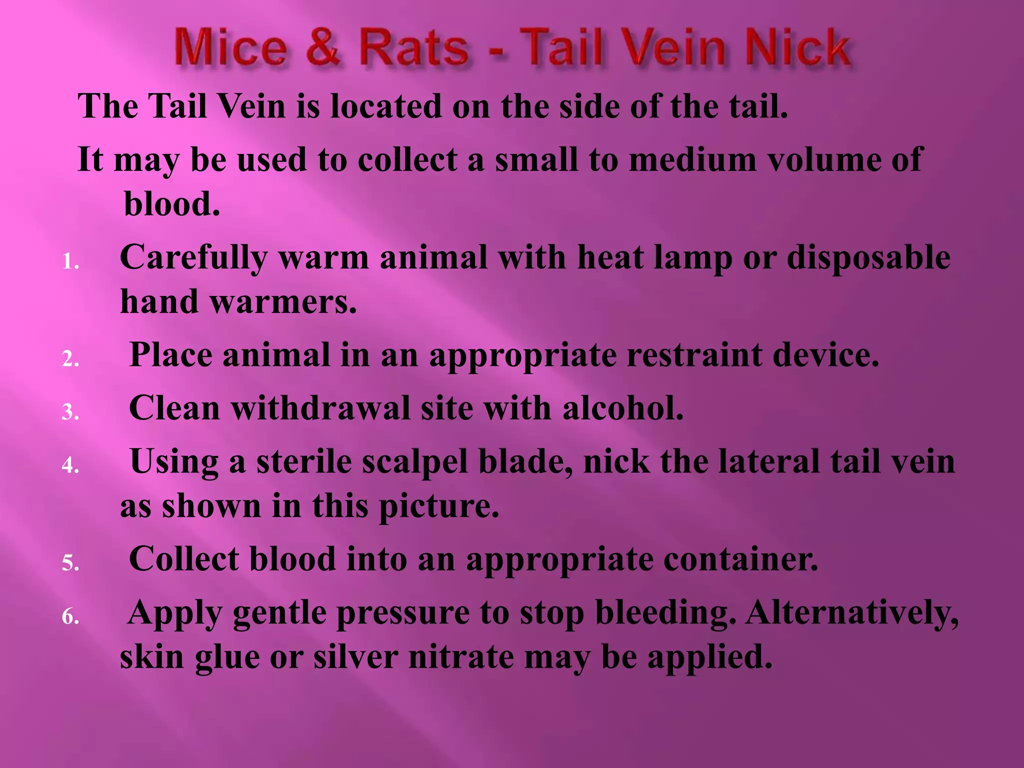
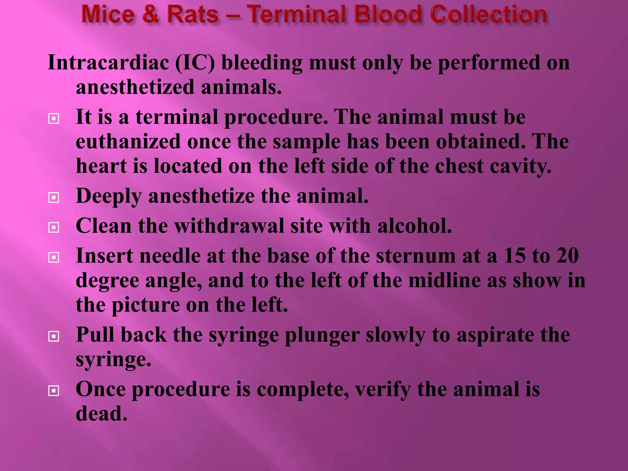
2. As with any procedure, training is critically
important.
The comfort and level of skill of an investigator
with a procedure as well as the sample volume
and frequency of sampling should be considered
when choosing a method.
3.
4.
5.
6.
7. The Mandibular Vein is located under the jawbone of
the mouse.
This technique can be used for collecting large volumes
of blood.
Restrain the animal by grasping the skin along its
back with your left hand (if right handed).
Clean the withdrawal site with alcohol.
Locate the hairless area at the angle of the mandible
(see middle picture). This is the landmark for the
puncture site.
Insert needle or lancet.
Collect blood into an appropriate container.
Release the animal, and apply light pressure to stop
bleeding.
8.
9. The Saphenous Vein is located along the lower portion of the hind
leg.
This technique is used for collecting small volumes of blood.
Unless the rat is anesthetized, this technique may require two
people.
For mice, restrain in a restraint tube. Place the animal head first
into the tube, and restrain the hind leg against the tube edge.
Pluck or clip the hair on the leg.
Clean withdrawal site with alcohol, and apply a small amount of
petroleum based ointment.
Apply gentle pressure to the medial surface of the leg (as shown in
picture). The vessel should become visible as blood pools.
Prick the vessel with a needle or lancet, and collect blood into an
appropriate container.
Release animal, and apply pressure to stop bleeding.
10.
11. The Orbital Sinus method may be used to collect
large volumes of blood.
Appropriate anesthesia must be used for this
procedure. Animals may receive topical
anesthetic (i.e., tetracaine ophthalmic drops) or
general anesthesia.
12. 1. Restrain the animal by grasping the skin along its
back with your left hand (if right-handed).
2. Apply anesthetic drops to eye. Use caution not to
touch the eye with the applicator.
3. Allow drop to take affect and then wipe away any
excess fluid.
4. Place a hematocrit tube at the medial canthus of the
eye (as shown in bottom illustration).
5. With a rotating motion, insert tube through the
conjunctiva membrane.
6. Continue rotating the tube until blood flows.
7. Collect blood into an appropriate container.
8. Release animal, and apply gentle pressure to stop
bleeding
13.
14. The Tail Vein is located on the side of the tail.
It may be used to collect a small to medium volume of
blood.
1. Carefully warm animal with heat lamp or disposable
hand warmers.
2. Place animal in an appropriate restraint device.
3. Clean withdrawal site with alcohol.
4. Using a sterile scalpel blade, nick the lateral tail vein
as shown in this picture.
5. Collect blood into an appropriate container.
6. Apply gentle pressure to stop bleeding. Alternatively,
skin glue or silver nitrate may be applied.
15.
16.
17. Intracardiac (IC) bleeding must only be performed on
anesthetized animals.
It is a terminal procedure. The animal must be
euthanized once the sample has been obtained. The
heart is located on the left side of the chest cavity.
Deeply anesthetize the animal.
Clean the withdrawal site with alcohol.
Insert needle at the base of the sternum at a 15 to 20
degree angle, and to the left of the midline as show in
the picture on the left.
Pull back the syringe plunger slowly to aspirate the
syringe.
Once procedure is complete, verify the animal is
dead.
18.
19. The animal is kept in a restrainer.
The hind foot around ankle is held and medial
dorsal pedal vessel is located on top of the foot.
The foot is cleaned with absolute alcohol and
dorsal pedal vein is punctured with 23G/27G
needle.
Drops of blood that would appear on the skin
surface are collected in a capillary tube and a
little pressure is applied to stop the bleeding .
20.
21. Tarsal vein is identified in one of the hind legs of
large animals. This method is commonly
recommended for guinea pig.
One person has to restrain the animal properly.
Tarsal vein may be visible in blue color.
The surface hairs are removed by applying a suitable
hair remover. A local anesthetic cream is applied on
the collection site.
After 20 to 30 minutes, blood sample is collected
slowly by using 22G needle.
Maximum three samples can be taken per leg and 0.1
to 0.3 ml of blood can be collected per sample.
After the sample collection, gentle pressure is applied
with finger for 2 minutes to stop bleeding
22.
23. This method is commonly adopted for rabbits.
The animal should be placed in a restrainer.
Ear is cleaned with 95% v/v alcohol and local
anesthetic cream is applied on the collection site 10
min prior to sampling.
Size 11 surgical blade is used to cut the marginal ear
vein and blood is collected in a collecting tube.
Otherwise, a 26G needle may be used to collect blood
from animal marginal vein.
After collecting blood, clean sterile cotton is kept on
the collection site and finger pressure is applied to
stop the bleeding
24.
25. Draw blood into vacutainer tube containing ~1.8 mg EDTA per ml blood
Invert vacutainer tubes carefully 10 times to mix blood and anticoagulant and store at
room temperature until centrifugation.
Samples should undergo centrifugation immediately. This should be carried out for a
minimum of 10 minutes at 1000-2000 at room temperature .
This will give three layers: (from top to bottom) plasma, leucocytes (buffy coat),
erythrocytes.
Carefully aspirate the supernatant (plasma) at room temperature and pool in a
centrifuge tube. Take care not to disrupt the cell layer or transfer any cells.
Inspect plasma for turbidity. Turbid samples should be centrifuged and aspirated
again to remove remaining insoluble matter.
Aliquot plasma into cryovials and store at –80 °C.