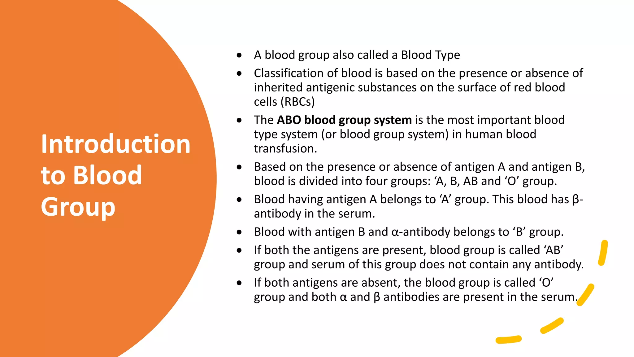
BLOOD BANK
PROTOCOLS
Javed Bhatti
BS Biochemistry
What is Blood
Bank
A blood bank is a center where
blood gathered as a result of blood
donation is stored as preserved for
later use in blood transfusion. The
term ‘Blood Bank’ typically refers
to division of a hospital where the
storage of blood products occurs
and where proper testing is
performed.
Introduction
to Blood
Group
 A blood group also called a Blood Type
 Classification of blood is based on the presence or absence of
inherited antigenic substances on the surface of red blood
cells (RBCs)
 The ABO blood group system is the most important blood
type system (or blood group system) in human blood
transfusion.
 Based on the presence or absence of antigen A and antigen B,
blood is divided into four groups: ‘A, B, AB and ‘O’ group.
 Blood having antigen A belongs to ‘A’ group. This blood has β-
antibody in the serum.
 Blood with antigen B and α-antibody belongs to ‘B’ group.
 If both the antigens are present, blood group is called ‘AB’
group and serum of this group does not contain any antibody.
 If both antigens are absent, the blood group is called ‘O’
group and both α and β antibodies are present in the serum.
Recipient Donor
Type O- O+ B- B+ A- A+ AB- AB+
AB+
AB-
A+
A-
B+
B-
O+
O-
Donor
criteria
• Age: 18-60 Years
• Weight: >55 kg
• Hb: > 12g/dL
• If previously donated, at least 4 months should be elapsed since the
date of previous donation.
• Free from any serious disease condition or pregnancy.
• Free from "Risk Factors".
• Drug addicts
• Hepatitis B & C
• Syphilis
• Malaria
• HIV-AIDS
Routine Tests in
Blood Bank
1. Red Cell Suspension
 Materials:
1. Sample of blood
2. Saline
3. Test tubes 75x12 mm
 Method:
1. Place 0.2 -0.5 ml of blood into the tube (2-3 drops).
2. Fill the tube with the saline.
3. Centrifuge at 200 G for 1-2 minutes until the RBC's are packed.
(G=relative centrifugal force)
4. Decant the supernatant.
5. Tap the tube to resuspend the RBC's in the residual fluid. This
constitutes one wash. Repeat step 2-5 at least twice. The last wash
should always have a clear supernatant with no signs of haemolysis.
6. To make a 5% cell suspension add 1 volume of the packed RBC's to 19
volumes of saline.
7. To make a 3% suspension, add 1 volume packed RBC's to 32 volumes of
saline.
3. Blood Grouping
Slide Method
Take 3 slides and label them with A, B,
D
Put 1 drop of Anti-A on the slide
labeled as A, 1 drop of Anti-B on Slide
B, and 1 drop of Anti- D on slide D
Add 1 drop of blood on slide A, 1 drop
on slide B and 1 drop on slide D
Mix the antisera with blood
Check for agglutination.
Tube Method
Forward
Grouping
Prepare 2-5% suspension of test
sample in normal saline
Set three tubes , label them as A,B, D
Add two drops of anti A , anti-B, anti D
in three different tubes
Add one drop of 2-5% cell suspension
(Ratio of 2:1)
Mix contents well and centrifuge at
1500 rpm for 1 minute
Observe for hemolysis
Gently disperse cell button and check
for agglutination
Conform negative results under
microscope.
Reverse
Grouping:
• Prepare 2-5% suspension of pooled cells
A,B,O
• Label three tubes A cells, B cells and O cells
• Place two drops of serum in each tube
• Add one drop of cell suspension ( A cell to A
tube, B cell to B tube and one drop of O cell
to O tube)
• Centrifuge tubes at 1500 rpm for 1 minute
• Gently disperse for agglutination.
• Check negative results on microscope.
2 vol of test
Serum / plasma +
1 vol of 5%
suspension of
reagent red cells in
respective tubes
Shake & leave at room temp (20-24 C) for 5 min
Centrifuge at 1000 rpm for 1 min
Centrifuge & record the results similarly as for cell grouping
Reverse Grouping
3. Weak D
Testing (Du)
Testing
• Principle and Application:
Some red cells possess the D antigen but it is expressed so weakly that the cells are not
agglutinated directly by anti-D sera. An indirect antiglobulin test is necessary to
identify patients with the Weak D (formerly known as Du )phenotype. Weak D testing is
done on all prenatal patients and candidates for Rh immune globulin. Weak D testing is
also done on Rh negative donors to ensure they are truly D negative. It may or may not
be done routinely on Rh negative candidates for transfusion, depending on the policy
of the transfusing institution. If routine weak D testing is done, weak D positive
patients should receive Rh positive blood.
• Sample
Any sample satisfactory for ABO and Rh testing is acceptable. The weak D is most often
performed directly from the same tube set up for the Rh test, if the patient cells were
a washed 3% suspension.
• Reagents, Equipment, And Supplies
 37oC incubator
 Wash bottle with physiologic saline
 Coombs serum - either polyspecific or anti-IgG
 Coombs control cells
 All reagents, equipment, and supplies used in the Rh TESTING procedure
PROCEDURE
• Prepare a washed, 3% suspension of patient cells, and set up the D and DC (Rh Control) tubes, if not
already done. (SEE ABO/Rh TYPING PROCEDURE)
• Record the D and DC immediate spin results. If the Rh test is negative, continue with step 3.
• Incubate both tubes at 37oC for 15 to 30 minutes.
• Centrifuge and read for agglutination as usual. If the Rh test is negative, continue with step 5.
• Wash both tubes 3-4 times with saline.
• Immediately after the last wash, add one drop Coombs serum to each tube and centrifuge in the
serofuge the time appropriate for the Coombs spin calibration.
• Immediately resuspend gently and examine for agglutination using the lighted agglutination viewer.
• Record results in the appropriate column on the worksheet
• Confirm all negative results by adding one drop Coombs control cells to all tubes showing no
agglutination and centrifuge 15-30 seconds at high speed in the serofuge.
• Gently resuspend and examine for agglutination. Agglutination should be present in this step or the test
is invalid.
INTERPRETATION
• A negative result in the immediate spin phase but
agglutination in the D tube following incubation
(with no agglutination in the DC tube) indicates a
positive test for weak D. Lack of agglutination is a
negative test and the patient is considered truly D
negative. Agglutination in the DC tube invalidates
the test.
• A true weak D should give at least a 2+ positive
result. Weaker results may be due to mixed field
agglutination in an Rh negative individual who
received Rh positive blood, or vice-versa. Obtain a
recent transfusion history on patients who give
inconclusive weak D results.
4. Cross Match
 A pre-requisite for blood transfusion
 Purpose: to avoid reactions of mismatched
transfusion
◦ PROCEDURE:
 In test tube place 2 drops of recipient’s
serum
 Add washed donor red cell suspension
 Mix and incubate at 37degree C for 30 mins
 Centrifuge at 3000 rpm for 1 minute
 Examine for agglutination and hemolysis
◦ INTERPRETATION:
◦ Matched - no agglutination and hemolysis
◦ Mismatched - either agglutination or hemolysis
Blood bank

Blood bank

  • 2.
  • 3.
    What is Blood Bank Ablood bank is a center where blood gathered as a result of blood donation is stored as preserved for later use in blood transfusion. The term ‘Blood Bank’ typically refers to division of a hospital where the storage of blood products occurs and where proper testing is performed.
  • 4.
    Introduction to Blood Group  Ablood group also called a Blood Type  Classification of blood is based on the presence or absence of inherited antigenic substances on the surface of red blood cells (RBCs)  The ABO blood group system is the most important blood type system (or blood group system) in human blood transfusion.  Based on the presence or absence of antigen A and antigen B, blood is divided into four groups: ‘A, B, AB and ‘O’ group.  Blood having antigen A belongs to ‘A’ group. This blood has β- antibody in the serum.  Blood with antigen B and α-antibody belongs to ‘B’ group.  If both the antigens are present, blood group is called ‘AB’ group and serum of this group does not contain any antibody.  If both antigens are absent, the blood group is called ‘O’ group and both α and β antibodies are present in the serum.
  • 6.
    Recipient Donor Type O-O+ B- B+ A- A+ AB- AB+ AB+ AB- A+ A- B+ B- O+ O-
  • 7.
    Donor criteria • Age: 18-60Years • Weight: >55 kg • Hb: > 12g/dL • If previously donated, at least 4 months should be elapsed since the date of previous donation. • Free from any serious disease condition or pregnancy. • Free from "Risk Factors". • Drug addicts • Hepatitis B & C • Syphilis • Malaria • HIV-AIDS
  • 8.
  • 9.
    1. Red CellSuspension  Materials: 1. Sample of blood 2. Saline 3. Test tubes 75x12 mm  Method: 1. Place 0.2 -0.5 ml of blood into the tube (2-3 drops). 2. Fill the tube with the saline. 3. Centrifuge at 200 G for 1-2 minutes until the RBC's are packed. (G=relative centrifugal force) 4. Decant the supernatant. 5. Tap the tube to resuspend the RBC's in the residual fluid. This constitutes one wash. Repeat step 2-5 at least twice. The last wash should always have a clear supernatant with no signs of haemolysis. 6. To make a 5% cell suspension add 1 volume of the packed RBC's to 19 volumes of saline. 7. To make a 3% suspension, add 1 volume packed RBC's to 32 volumes of saline.
  • 10.
  • 11.
    Slide Method Take 3slides and label them with A, B, D Put 1 drop of Anti-A on the slide labeled as A, 1 drop of Anti-B on Slide B, and 1 drop of Anti- D on slide D Add 1 drop of blood on slide A, 1 drop on slide B and 1 drop on slide D Mix the antisera with blood Check for agglutination.
  • 12.
  • 13.
    Forward Grouping Prepare 2-5% suspensionof test sample in normal saline Set three tubes , label them as A,B, D Add two drops of anti A , anti-B, anti D in three different tubes Add one drop of 2-5% cell suspension (Ratio of 2:1) Mix contents well and centrifuge at 1500 rpm for 1 minute Observe for hemolysis Gently disperse cell button and check for agglutination Conform negative results under microscope.
  • 14.
    Reverse Grouping: • Prepare 2-5%suspension of pooled cells A,B,O • Label three tubes A cells, B cells and O cells • Place two drops of serum in each tube • Add one drop of cell suspension ( A cell to A tube, B cell to B tube and one drop of O cell to O tube) • Centrifuge tubes at 1500 rpm for 1 minute • Gently disperse for agglutination. • Check negative results on microscope.
  • 15.
    2 vol oftest Serum / plasma + 1 vol of 5% suspension of reagent red cells in respective tubes Shake & leave at room temp (20-24 C) for 5 min Centrifuge at 1000 rpm for 1 min Centrifuge & record the results similarly as for cell grouping Reverse Grouping
  • 16.
    3. Weak D Testing(Du) Testing • Principle and Application: Some red cells possess the D antigen but it is expressed so weakly that the cells are not agglutinated directly by anti-D sera. An indirect antiglobulin test is necessary to identify patients with the Weak D (formerly known as Du )phenotype. Weak D testing is done on all prenatal patients and candidates for Rh immune globulin. Weak D testing is also done on Rh negative donors to ensure they are truly D negative. It may or may not be done routinely on Rh negative candidates for transfusion, depending on the policy of the transfusing institution. If routine weak D testing is done, weak D positive patients should receive Rh positive blood. • Sample Any sample satisfactory for ABO and Rh testing is acceptable. The weak D is most often performed directly from the same tube set up for the Rh test, if the patient cells were a washed 3% suspension. • Reagents, Equipment, And Supplies  37oC incubator  Wash bottle with physiologic saline  Coombs serum - either polyspecific or anti-IgG  Coombs control cells  All reagents, equipment, and supplies used in the Rh TESTING procedure
  • 17.
    PROCEDURE • Prepare awashed, 3% suspension of patient cells, and set up the D and DC (Rh Control) tubes, if not already done. (SEE ABO/Rh TYPING PROCEDURE) • Record the D and DC immediate spin results. If the Rh test is negative, continue with step 3. • Incubate both tubes at 37oC for 15 to 30 minutes. • Centrifuge and read for agglutination as usual. If the Rh test is negative, continue with step 5. • Wash both tubes 3-4 times with saline. • Immediately after the last wash, add one drop Coombs serum to each tube and centrifuge in the serofuge the time appropriate for the Coombs spin calibration. • Immediately resuspend gently and examine for agglutination using the lighted agglutination viewer. • Record results in the appropriate column on the worksheet • Confirm all negative results by adding one drop Coombs control cells to all tubes showing no agglutination and centrifuge 15-30 seconds at high speed in the serofuge. • Gently resuspend and examine for agglutination. Agglutination should be present in this step or the test is invalid.
  • 18.
    INTERPRETATION • A negativeresult in the immediate spin phase but agglutination in the D tube following incubation (with no agglutination in the DC tube) indicates a positive test for weak D. Lack of agglutination is a negative test and the patient is considered truly D negative. Agglutination in the DC tube invalidates the test. • A true weak D should give at least a 2+ positive result. Weaker results may be due to mixed field agglutination in an Rh negative individual who received Rh positive blood, or vice-versa. Obtain a recent transfusion history on patients who give inconclusive weak D results.
  • 19.
    4. Cross Match A pre-requisite for blood transfusion  Purpose: to avoid reactions of mismatched transfusion ◦ PROCEDURE:  In test tube place 2 drops of recipient’s serum  Add washed donor red cell suspension  Mix and incubate at 37degree C for 30 mins  Centrifuge at 3000 rpm for 1 minute  Examine for agglutination and hemolysis ◦ INTERPRETATION: ◦ Matched - no agglutination and hemolysis ◦ Mismatched - either agglutination or hemolysis