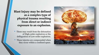
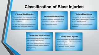
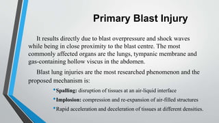
Dr. Md. Zaki Shahriar Sourav
MBBS (DU), FCPS Part 2 Trainee (General Surgery)
HMO, Casualty Block -01
Dhaka Medical College and Hospital
• These may result from the detonation
of High-order explosives or the
deflagration of Low-order explosives.
• These injuries are compounded when
they occur within a confined space.
Blast Injury may be defined
as a complex type of
physical trauma resulting
from direct or indirect
exposure to an explosion.
Epidemiology of blast
Injuries from civilian and terrorist explosions are increasing
worldwide.
A recent review of 3000 victims of terrorist bombings showed
that
• over 13% immediate mortality
• 30% hospitalization rate for survivors
• Hospital mortality of initial survivors was 1.4% for all
patients and 12% for critically injured patients
• One of the most common causes of immediate mortality was
head injury
• Blast injury patients compared to non-blast injury cases have
more severe injuries. On average ISS>25.
• Frequently require surgery or ICU admissions and prolonged
hospital stay and rehabilitation.
Figure: Worldwide trend in terrorist
explosive events from 1999 to 2006
• High Order Explosives: Detonation of these results in energy
release by explosions which is a rapid formation, release and
gases. The energy released with the blast causes significant
of the air through which it passes and the blast wave travels at a
faster than the speed of sound. For example: TNT, RDX etc.
• Low-order explosives: These are materials that give out
energy as a result of rapid burning, called deflagration. Here the
reaction rate is much slower. Although the total energy output is
high explosives, the lower rate results in lesser pressure. For
gunpowder.
The extent and pattern of
injuries from explosions are
multifactorial. They can be
grossly categorised as Agent
Factors, Host Factors and
Environmental Factors. No
two events are identical and
the spectrum of events varies
widely.
Factors affecting the extent of Blast
Injuries
Agent Factor
Size and weight
Material used
Delivery System etc
Host Factors
Age, Gender, BMI
Fitness
Nutrition
Environmental
factors
Open space vs
Confined space
•Structural collapse
•Shielding
•Building and other
debris
Pathophysiology
During an explosion, chemical potential energy is converted to heat, kinetic and sound energy.
Detonation of a High explosive generates gases quicker than the speed of sound. The resulting pulse wave
or overpressure of air pushes outward from the point of onset. This positive pressure accelerating outward
from the explosion is called the “Blast Wind”. It is followed by a partial vacuum which causes air to
flow back in, thus a push-pull effect is created and it can have damaging effects on structures and
humans. These effects may have even more devastating effects in closed areas.
Understanding Blast
Mechanics
Classification of Blast Injuries
• This occurs due to blast overpressure. Most
are marked at the interface between air and
solid or soft tissue.
• e.g. Blast lung, tympanic membrane injury,
intestinal blast injury, globe rupture etc.
• Caused by fragments or materials that are
accelerated as a result of the explosion
• e.g. All penetrating injuries
• It is caused by the gross movement of the
person, object or infrastructure as a result of
the force of the Blast wind.
• e.g. Blunt and crush injuries, traumatic
amputation
• Miscellaneous group of injuries that do not
fall within the other categories.
• e.g. Burns, inhalational injuries, late-onset
respiratory problems, injuries caused by
falling masonry,
• Injuries are caused by the intentional addition
of either biological or radioactive material to
an explosive device.
• e.g. Radiation sickness, Infection
Primary Blast Injury
It results directly due to blast overpressure and shock waves
while being in close proximity to the blast centre. The most
commonly affected organs are the lungs, tympanic membrane and
gas-containing hollow viscus in the abdomen.
Blast lung injuries are the most researched phenomenon and the
proposed mechanism is:
•Spalling: disruption of tissues at an air-liquid interface
•Implosion: compression and re-expansion of air-filled structures
•Rapid acceleration and deceleration of tissues at different densities.
• Tympanic membrane rupture is the most common primary blast injury.
• Patients may be asymptomatic or have a degree of transient hearing loss and
otorrhoea.
• Perforation may be Small (<3mm), Medium (3-5mm) or Large (>5mm)
• Most perforations heal by conservative management; that is analgesia, antibiotics and
good oral hygiene, sterile dressing and avoiding water to ear, over a course of 4
weeks.
• However, large and symptomatic perforations may require surgical intervention.
Primary Blast Lung Injury
Lungs are the 2nd most susceptible to blast injury and are
associated with the highest mortality even among survivors.
Caused by blast wave against the chest wall, not through
oropharynx or trachea.
Pulmonary barotrauma includes:
• Pulmonary contusions
• Haemothorax
• Pneumothorax
• Pneumomediastinum
• Systemic air embolism
Clinical features include apnoea or progressive hypoxia,
bradycardia and hypotension. Delayed or inflammatory response
to intrapulmonary haemorrhage and oedema may also be
present. Signs may be present initially or may manifest last.
Fig: Diagrammatic
representation of Spalling and
Implosion
Image: Pulmonary Contusion
with corresponding histology
slide
Blast Lung Continued
Symptoms:
• Dyspnoea- unable to complete
sentences in a single breath
• Cough
• Haemoptysis
• Retrosternal pain
Signs:
• Tachycardia
• Cyanosis
• Reduced breath sounds
• Coarse crepitations
• Subcutaneous emphysema etc.
• Investigations: Trauma series Xray, Whole
body CT-Scan, Routine/Baseline blood tests
• The diagnosis of blast lung is clinical with
the finding of hypoxia following exposure to
blast. Chest x-rays and CT scans show typical
bat-wing appearances.
• Patient should be managed according to
ATLS protocol. Treatment of blast lung is
mainly supportive. Mechanical ventilation
and Thoracostomy or Pleural decompression
may be necessary.
Below: Chest x-ray showing Bat-wing appearance or
bilateral pulmonary infiltrates.
Right: Chest CT-scans showing Blast Lung features
Abdominal Blast Injuries
Abdominal blast injuries are not common and account for
only about 3% of all incidents.
Intestinal barotrauma is, however, more common with
underwater air blasts.
Clinical features may be delayed. The colon is the organ
most frequently affected and there may be serosal
haemorrhage, or bowel perforation resulting in
pneumoperitoneum or features of peritonitis. Solid organs
are more resistant to primary blasts.
Mesenteric ischaemia from gas embolism may cause
delayed rupture or perforation of large or small intestine.
The patient is to be managed according to ATLS protocol
and surgical intervention where mandated.
Secondary Blast Injuries
These are penetrating or lacerating
injuries caused by fragments/shrapnel
carried off by the force of the blast or
explosion.
This is the most common cause of death
from a blast. Projectiles accelerated by an
explosion can strike at great distances
from the blast centre and the mode of
injury may be similar to that of ballistics.
The source of fragments may be the
device casing, glass, nail or porcelain
intentionally incorporated into a bomb,
nearby building infrastructure and even
bones.
Management of
Secondary Blast Injuries
Imaging: X-ray, CT-scan. (MRI not
advisable)
Management is similar to that of conventional
penetrating trauma.
• Wounds are considered to be grossly
contaminated. Hence, adequate surgical
toileting and debridement.
• Tetanus prophylaxis.
• Antibiotic injection if within the decisive
period.
• Remove fragments immediately if
accessible or in joint spaces, adjacent to
structures with risk of further injury and
bleeding.
• Plan for serial debridement and delayed
primary closure.
Tertiary Blast Injuries
These are a result of the immense force
generated by a blast wind that propels or
throws away personnel, objects or
infrastructure. It can also be due to structural
collapse or falling masonry. Injuries caused are
similar to that of any blunt trauma. Tertiary
blast injuries may result in:
• Head Injuries
• Bone fractures
• Traumatic Amputations
• Crush syndrome
• Compartment syndrome etc
Quaternary Injury: These are
miscellaneous groups of injuries that do not fall
within the other categories.
These include burns, inhalational injuries,
late-onset respiratory problems as well as acute
or chronic psychological disturbances (e.g.
PTSD) etc.
Quinary injuries: These refer to injuries
caused by the intentional addition of either
biological or radioactive material to an
explosive device.
Imaging Methods
The scope of imaging to aid in the diagnosis and treatment of Blast
Injuries includes:
• Plain radiograph: a chest and abdominal radiograph may be very useful
in the acute setting to identify any signs of trauma, foreign body fractures
etc
• Ultrasound: a FAST scan is used in a haemodynamically unstable
patient to identify injuries that require prompt intervention such as a
tension pneumothorax or cardiac tamponade
• CT-scan: it is the most sensitive imaging tool. CT angiography may be
used to identify vascular injuries as well.
• MRI: in the acute setting, MRI may be contraindicated until the presence
of metallic foreign bodies has been excluded.
General Management
Patients should be managed according to ATLS protocol.
Primary Survey and Simultaneous
Resuscitation:
c: Exsanguinating Haemorrhage
A: Airway patency and Cervical Spine
protection
B: Breathing
C: Circulation
D: Neurological disability
E: Exposure in a controlled environment
Secondary Survey:
Allergic History
Past Medical History
History of medication
Last meal
A brief account of the Event from the patient.
Head-to-toe examination.
*Special considerations: Patients with upper
airway burns need to be intubated early despite
full consciousness and haemodynamic stability
due to delayed onset of laryngeal oedema.
Ventilation can be reversed after 48 to 72 hours
after oedema subsides.
Admission Criteria
High-risk patients such as those
with the following:
• Significant burns (>10% TBSA)
• Upper airway burns
• Suspected air embolism
• Radiation
• Shrapnel contamination
• Abnormal vital signs
• Abnormal lung examination
findings
• Clinical or radiological evidence
of pulmonary contusion or
pneumothorax
• Abdominal pain or vomiting
• Penetrating injuries to the thorax,
abdomen, neck or cranial cavity
• Reduced GCS
References
• Bailey and Love’s Short Practice of Surgery; 28th edition.
• Clinical Surgery in general, 4th edition
• Primary Blast Injuries- An updated Concise Review by Daniel Dante, Published in World
Journal of Surgery 2012
• Primary Blast Injury: Update on diagnosis and treatment by Amber E Ritenour published in
Journal of Critical Care Medicine 2008, vol 36.
• Blast Injury Science and Engineering by Anthony Bull
Blast Injury

Blast Injury

  • 1.
    Dr. Md. ZakiShahriar Sourav MBBS (DU), FCPS Part 2 Trainee (General Surgery) HMO, Casualty Block -01 Dhaka Medical College and Hospital
  • 2.
    • These mayresult from the detonation of High-order explosives or the deflagration of Low-order explosives. • These injuries are compounded when they occur within a confined space. Blast Injury may be defined as a complex type of physical trauma resulting from direct or indirect exposure to an explosion.
  • 3.
    Epidemiology of blast Injuriesfrom civilian and terrorist explosions are increasing worldwide. A recent review of 3000 victims of terrorist bombings showed that • over 13% immediate mortality • 30% hospitalization rate for survivors • Hospital mortality of initial survivors was 1.4% for all patients and 12% for critically injured patients • One of the most common causes of immediate mortality was head injury • Blast injury patients compared to non-blast injury cases have more severe injuries. On average ISS>25. • Frequently require surgery or ICU admissions and prolonged hospital stay and rehabilitation.
  • 4.
    Figure: Worldwide trendin terrorist explosive events from 1999 to 2006
  • 5.
    • High OrderExplosives: Detonation of these results in energy release by explosions which is a rapid formation, release and gases. The energy released with the blast causes significant of the air through which it passes and the blast wave travels at a faster than the speed of sound. For example: TNT, RDX etc. • Low-order explosives: These are materials that give out energy as a result of rapid burning, called deflagration. Here the reaction rate is much slower. Although the total energy output is high explosives, the lower rate results in lesser pressure. For gunpowder.
  • 6.
    The extent andpattern of injuries from explosions are multifactorial. They can be grossly categorised as Agent Factors, Host Factors and Environmental Factors. No two events are identical and the spectrum of events varies widely. Factors affecting the extent of Blast Injuries Agent Factor Size and weight Material used Delivery System etc Host Factors Age, Gender, BMI Fitness Nutrition Environmental factors Open space vs Confined space •Structural collapse •Shielding •Building and other debris
  • 7.
    Pathophysiology During an explosion,chemical potential energy is converted to heat, kinetic and sound energy. Detonation of a High explosive generates gases quicker than the speed of sound. The resulting pulse wave or overpressure of air pushes outward from the point of onset. This positive pressure accelerating outward from the explosion is called the “Blast Wind”. It is followed by a partial vacuum which causes air to flow back in, thus a push-pull effect is created and it can have damaging effects on structures and humans. These effects may have even more devastating effects in closed areas.
  • 8.
  • 10.
    Classification of BlastInjuries • This occurs due to blast overpressure. Most are marked at the interface between air and solid or soft tissue. • e.g. Blast lung, tympanic membrane injury, intestinal blast injury, globe rupture etc. • Caused by fragments or materials that are accelerated as a result of the explosion • e.g. All penetrating injuries • It is caused by the gross movement of the person, object or infrastructure as a result of the force of the Blast wind. • e.g. Blunt and crush injuries, traumatic amputation • Miscellaneous group of injuries that do not fall within the other categories. • e.g. Burns, inhalational injuries, late-onset respiratory problems, injuries caused by falling masonry, • Injuries are caused by the intentional addition of either biological or radioactive material to an explosive device. • e.g. Radiation sickness, Infection
  • 12.
    Primary Blast Injury Itresults directly due to blast overpressure and shock waves while being in close proximity to the blast centre. The most commonly affected organs are the lungs, tympanic membrane and gas-containing hollow viscus in the abdomen. Blast lung injuries are the most researched phenomenon and the proposed mechanism is: •Spalling: disruption of tissues at an air-liquid interface •Implosion: compression and re-expansion of air-filled structures •Rapid acceleration and deceleration of tissues at different densities.
  • 13.
    • Tympanic membranerupture is the most common primary blast injury. • Patients may be asymptomatic or have a degree of transient hearing loss and otorrhoea. • Perforation may be Small (<3mm), Medium (3-5mm) or Large (>5mm) • Most perforations heal by conservative management; that is analgesia, antibiotics and good oral hygiene, sterile dressing and avoiding water to ear, over a course of 4 weeks. • However, large and symptomatic perforations may require surgical intervention.
  • 14.
    Primary Blast LungInjury Lungs are the 2nd most susceptible to blast injury and are associated with the highest mortality even among survivors. Caused by blast wave against the chest wall, not through oropharynx or trachea. Pulmonary barotrauma includes: • Pulmonary contusions • Haemothorax • Pneumothorax • Pneumomediastinum • Systemic air embolism Clinical features include apnoea or progressive hypoxia, bradycardia and hypotension. Delayed or inflammatory response to intrapulmonary haemorrhage and oedema may also be present. Signs may be present initially or may manifest last.
  • 15.
    Fig: Diagrammatic representation ofSpalling and Implosion Image: Pulmonary Contusion with corresponding histology slide
  • 16.
    Blast Lung Continued Symptoms: •Dyspnoea- unable to complete sentences in a single breath • Cough • Haemoptysis • Retrosternal pain Signs: • Tachycardia • Cyanosis • Reduced breath sounds • Coarse crepitations • Subcutaneous emphysema etc. • Investigations: Trauma series Xray, Whole body CT-Scan, Routine/Baseline blood tests • The diagnosis of blast lung is clinical with the finding of hypoxia following exposure to blast. Chest x-rays and CT scans show typical bat-wing appearances. • Patient should be managed according to ATLS protocol. Treatment of blast lung is mainly supportive. Mechanical ventilation and Thoracostomy or Pleural decompression may be necessary.
  • 17.
    Below: Chest x-rayshowing Bat-wing appearance or bilateral pulmonary infiltrates. Right: Chest CT-scans showing Blast Lung features
  • 18.
    Abdominal Blast Injuries Abdominalblast injuries are not common and account for only about 3% of all incidents. Intestinal barotrauma is, however, more common with underwater air blasts. Clinical features may be delayed. The colon is the organ most frequently affected and there may be serosal haemorrhage, or bowel perforation resulting in pneumoperitoneum or features of peritonitis. Solid organs are more resistant to primary blasts. Mesenteric ischaemia from gas embolism may cause delayed rupture or perforation of large or small intestine. The patient is to be managed according to ATLS protocol and surgical intervention where mandated.
  • 19.
    Secondary Blast Injuries Theseare penetrating or lacerating injuries caused by fragments/shrapnel carried off by the force of the blast or explosion. This is the most common cause of death from a blast. Projectiles accelerated by an explosion can strike at great distances from the blast centre and the mode of injury may be similar to that of ballistics. The source of fragments may be the device casing, glass, nail or porcelain intentionally incorporated into a bomb, nearby building infrastructure and even bones.
  • 23.
    Management of Secondary BlastInjuries Imaging: X-ray, CT-scan. (MRI not advisable) Management is similar to that of conventional penetrating trauma. • Wounds are considered to be grossly contaminated. Hence, adequate surgical toileting and debridement. • Tetanus prophylaxis. • Antibiotic injection if within the decisive period. • Remove fragments immediately if accessible or in joint spaces, adjacent to structures with risk of further injury and bleeding. • Plan for serial debridement and delayed primary closure.
  • 24.
    Tertiary Blast Injuries Theseare a result of the immense force generated by a blast wind that propels or throws away personnel, objects or infrastructure. It can also be due to structural collapse or falling masonry. Injuries caused are similar to that of any blunt trauma. Tertiary blast injuries may result in: • Head Injuries • Bone fractures • Traumatic Amputations • Crush syndrome • Compartment syndrome etc
  • 25.
    Quaternary Injury: Theseare miscellaneous groups of injuries that do not fall within the other categories. These include burns, inhalational injuries, late-onset respiratory problems as well as acute or chronic psychological disturbances (e.g. PTSD) etc. Quinary injuries: These refer to injuries caused by the intentional addition of either biological or radioactive material to an explosive device.
  • 28.
    Imaging Methods The scopeof imaging to aid in the diagnosis and treatment of Blast Injuries includes: • Plain radiograph: a chest and abdominal radiograph may be very useful in the acute setting to identify any signs of trauma, foreign body fractures etc • Ultrasound: a FAST scan is used in a haemodynamically unstable patient to identify injuries that require prompt intervention such as a tension pneumothorax or cardiac tamponade • CT-scan: it is the most sensitive imaging tool. CT angiography may be used to identify vascular injuries as well. • MRI: in the acute setting, MRI may be contraindicated until the presence of metallic foreign bodies has been excluded.
  • 29.
    General Management Patients shouldbe managed according to ATLS protocol. Primary Survey and Simultaneous Resuscitation: c: Exsanguinating Haemorrhage A: Airway patency and Cervical Spine protection B: Breathing C: Circulation D: Neurological disability E: Exposure in a controlled environment Secondary Survey: Allergic History Past Medical History History of medication Last meal A brief account of the Event from the patient. Head-to-toe examination. *Special considerations: Patients with upper airway burns need to be intubated early despite full consciousness and haemodynamic stability due to delayed onset of laryngeal oedema. Ventilation can be reversed after 48 to 72 hours after oedema subsides.
  • 31.
    Admission Criteria High-risk patientssuch as those with the following: • Significant burns (>10% TBSA) • Upper airway burns • Suspected air embolism • Radiation • Shrapnel contamination • Abnormal vital signs • Abnormal lung examination findings • Clinical or radiological evidence of pulmonary contusion or pneumothorax • Abdominal pain or vomiting • Penetrating injuries to the thorax, abdomen, neck or cranial cavity • Reduced GCS
  • 32.
    References • Bailey andLove’s Short Practice of Surgery; 28th edition. • Clinical Surgery in general, 4th edition • Primary Blast Injuries- An updated Concise Review by Daniel Dante, Published in World Journal of Surgery 2012 • Primary Blast Injury: Update on diagnosis and treatment by Amber E Ritenour published in Journal of Critical Care Medicine 2008, vol 36. • Blast Injury Science and Engineering by Anthony Bull