Download as PDF, PPTX
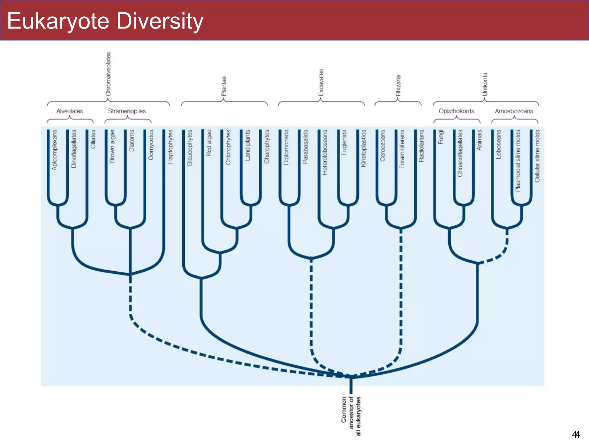
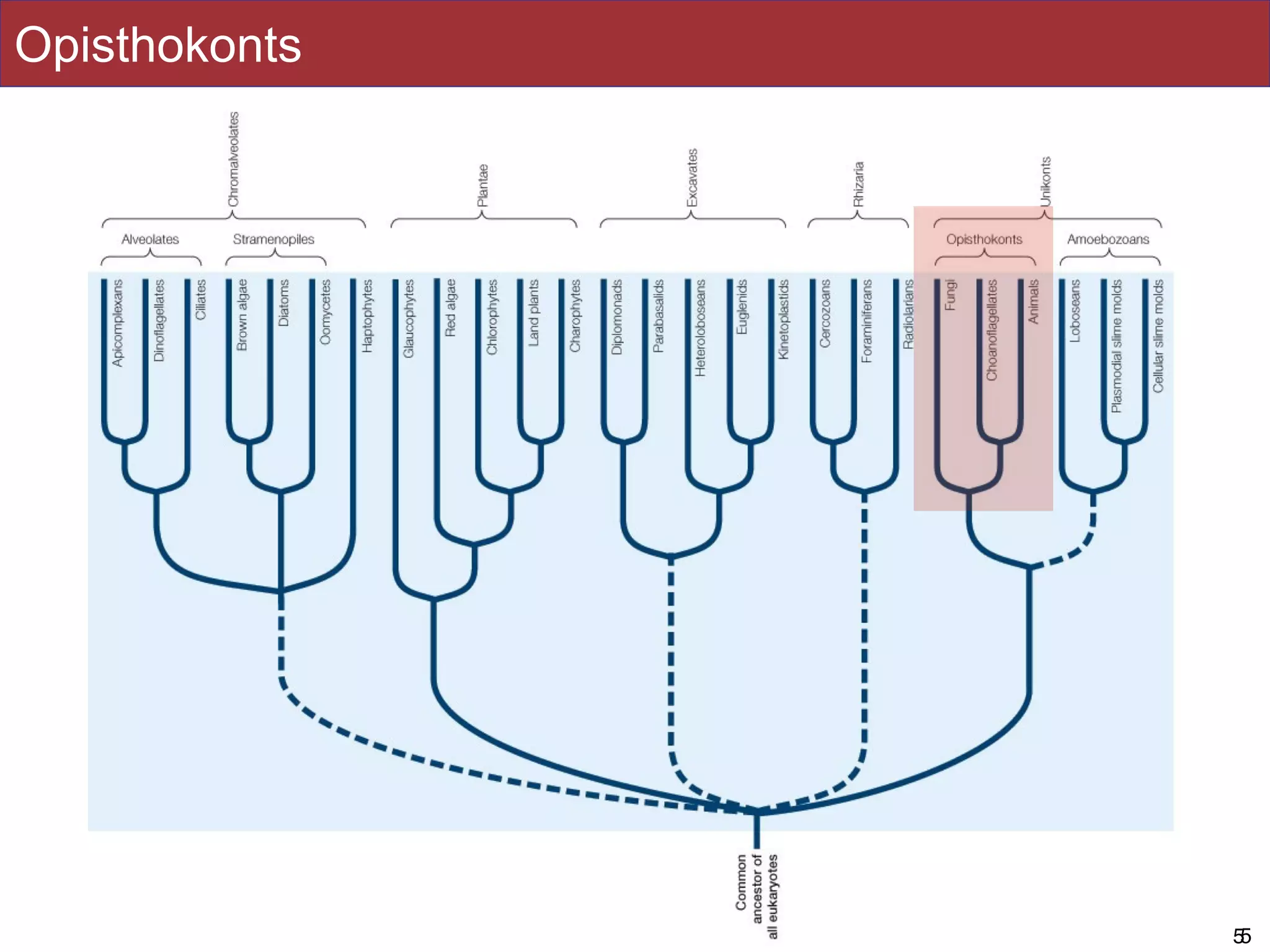

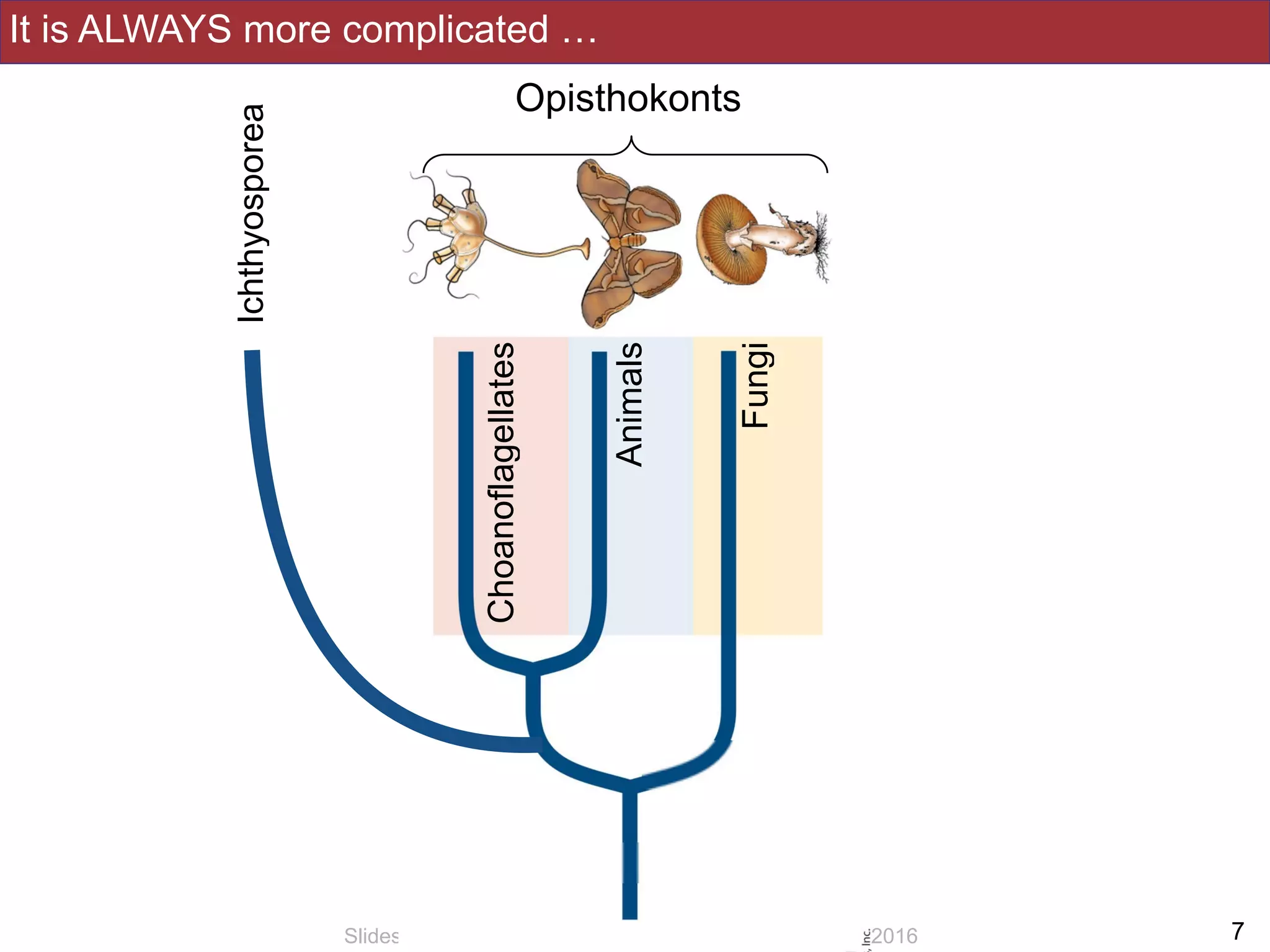
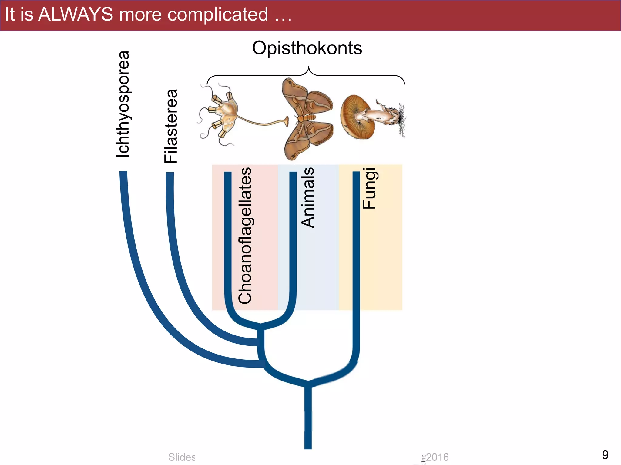
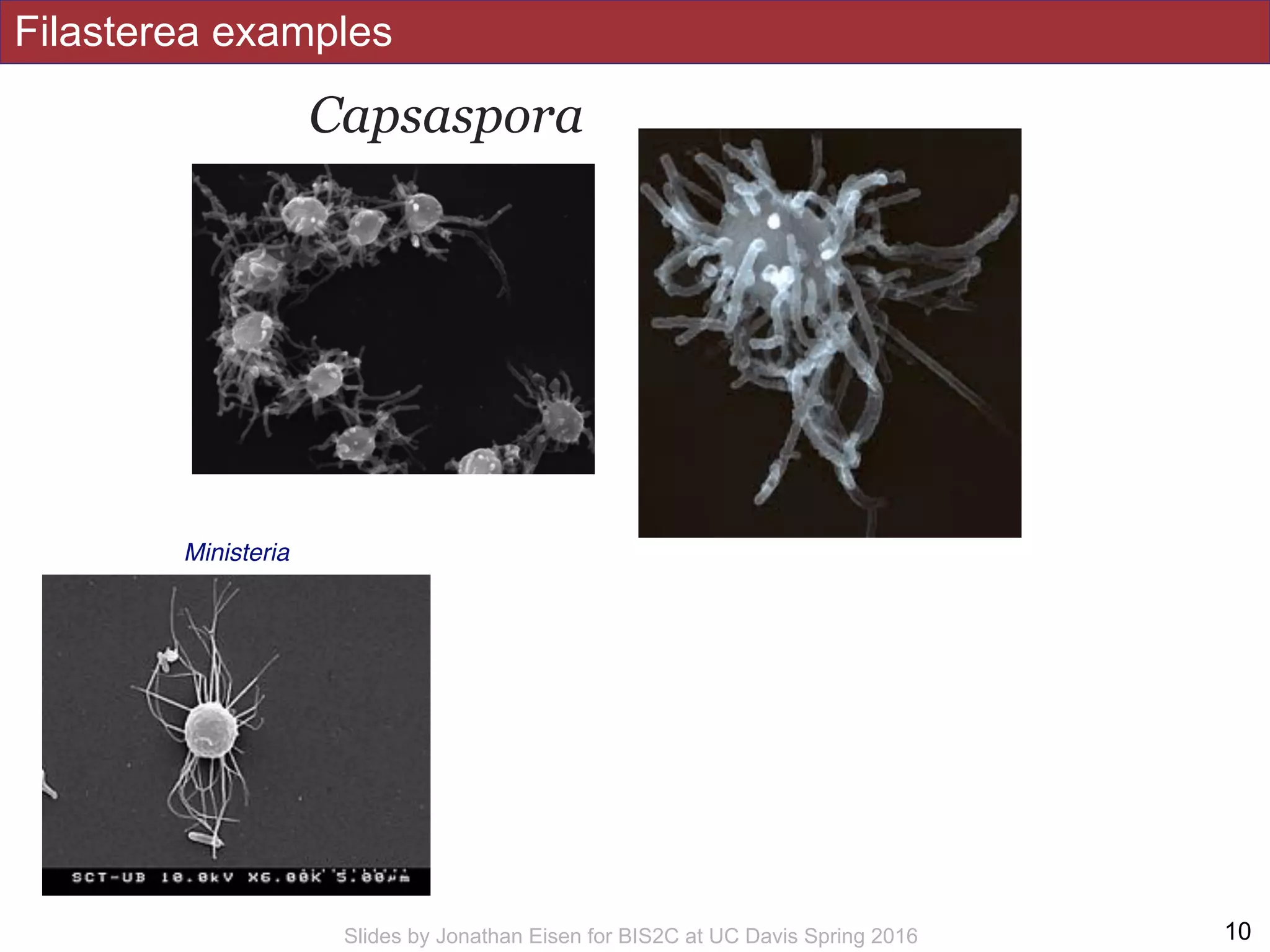
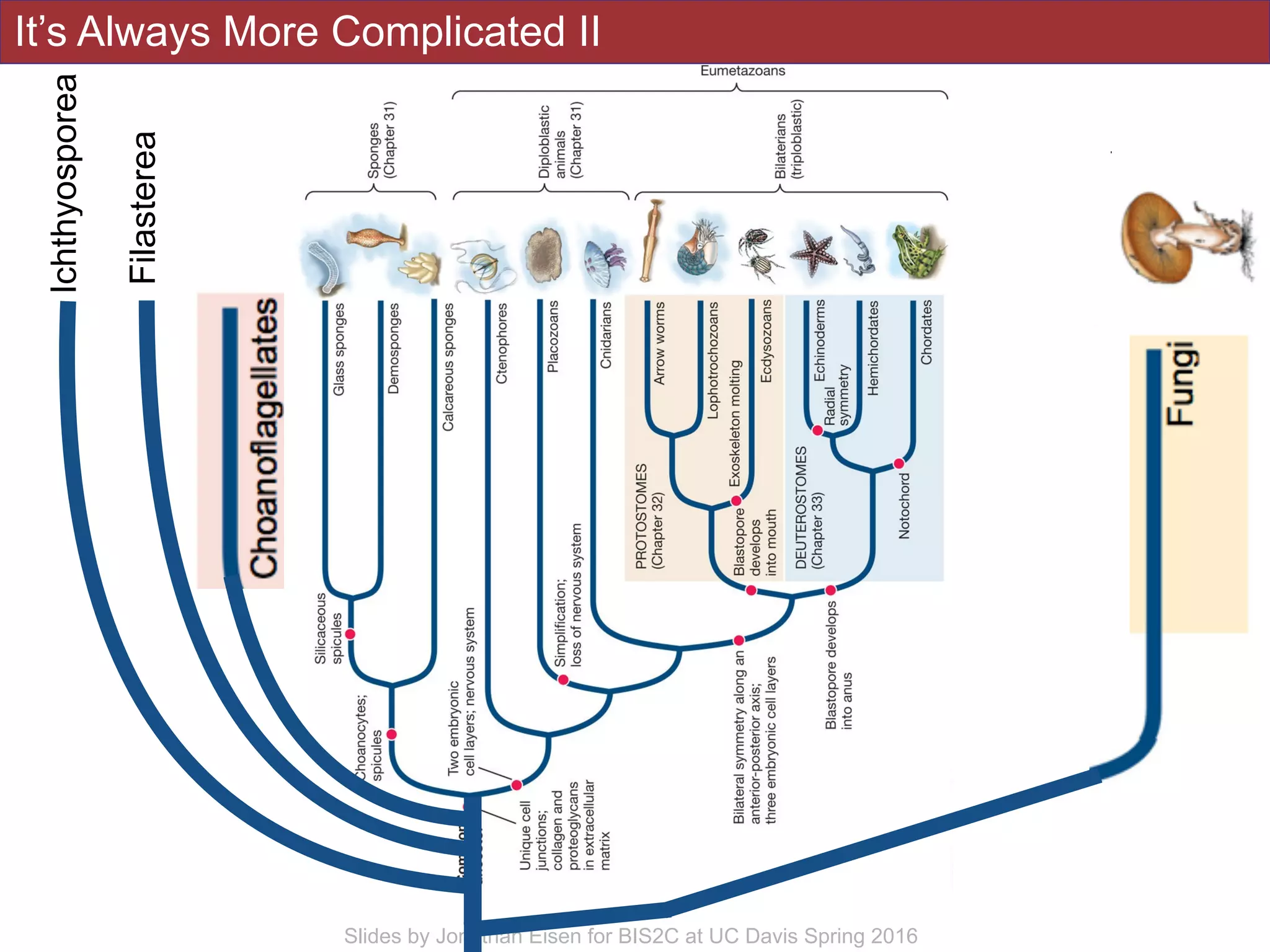
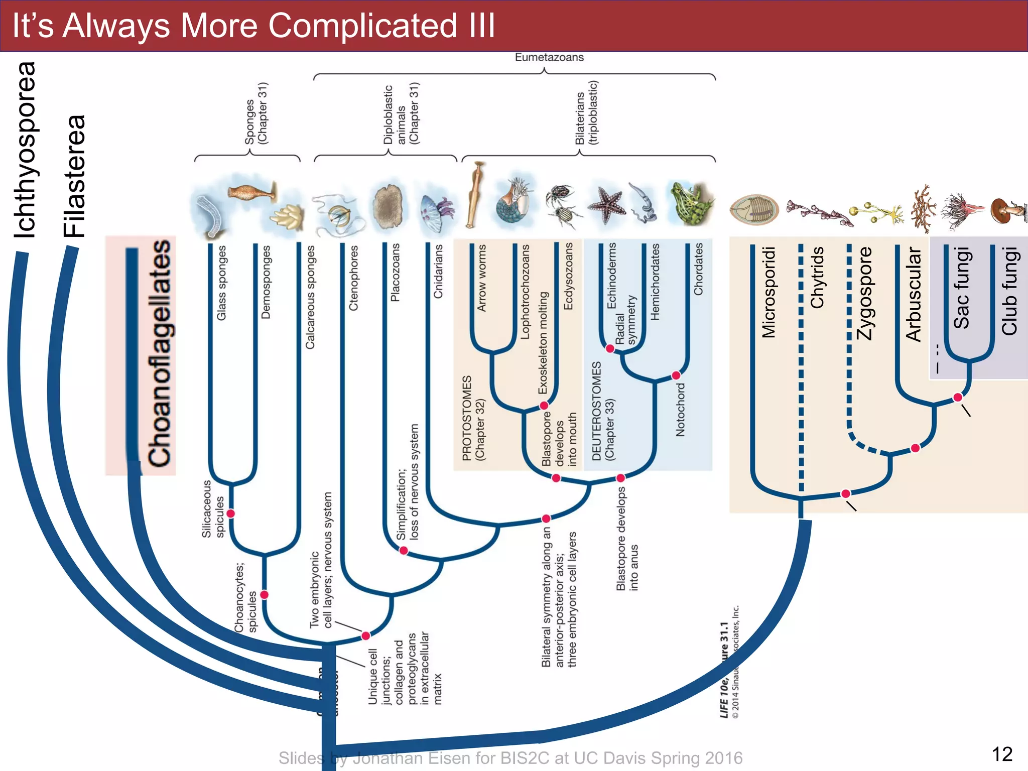
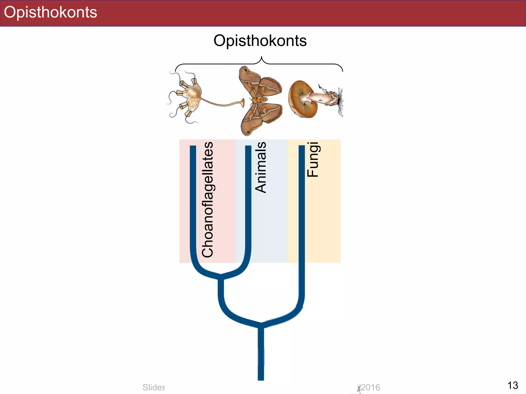
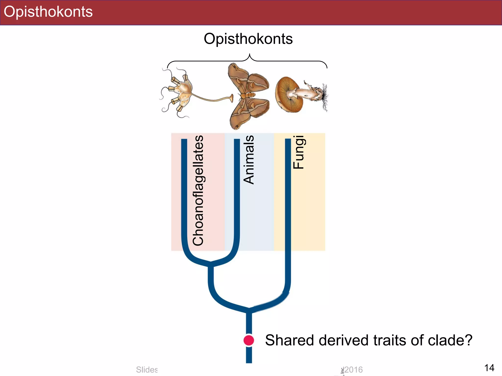
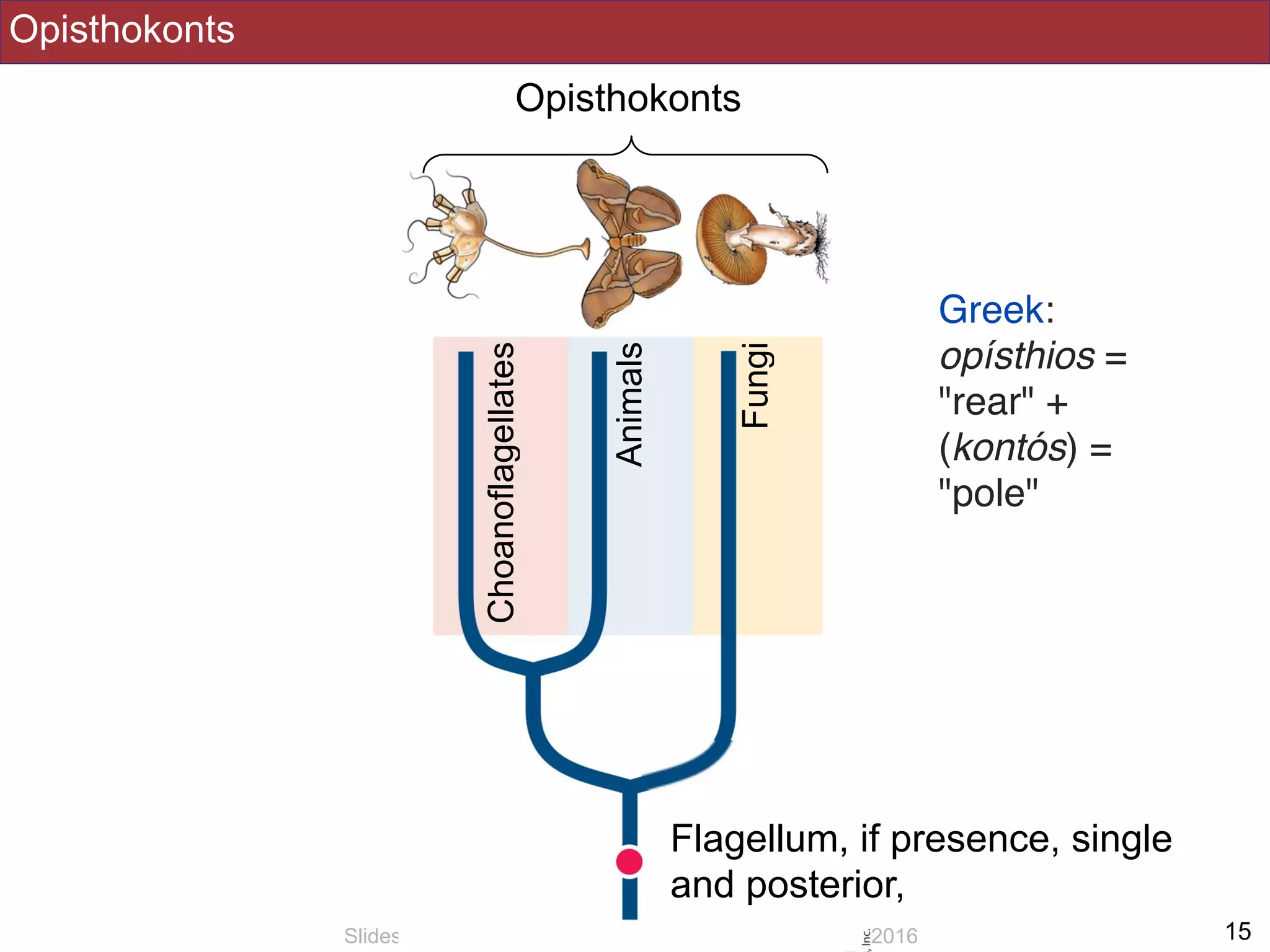
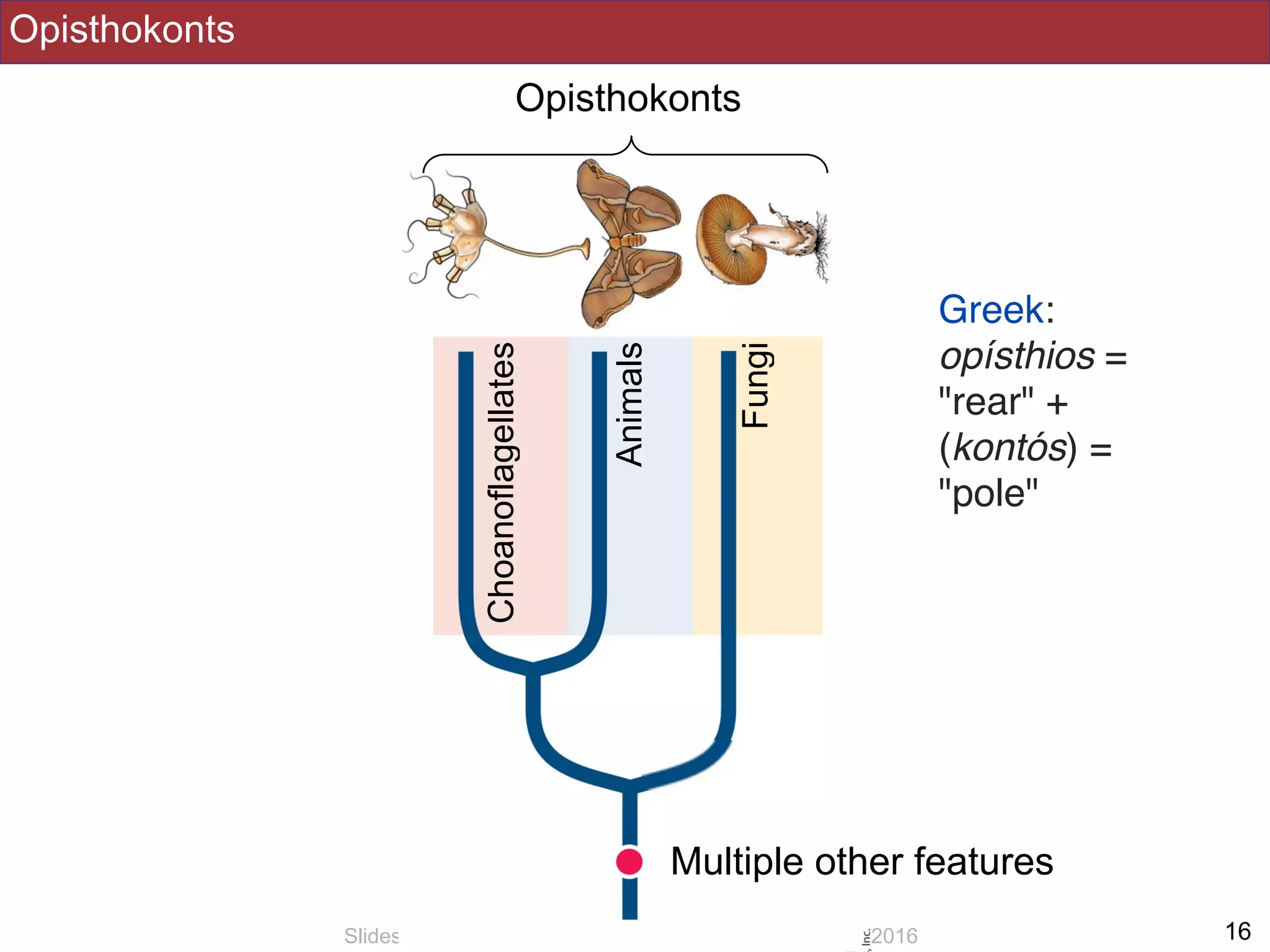
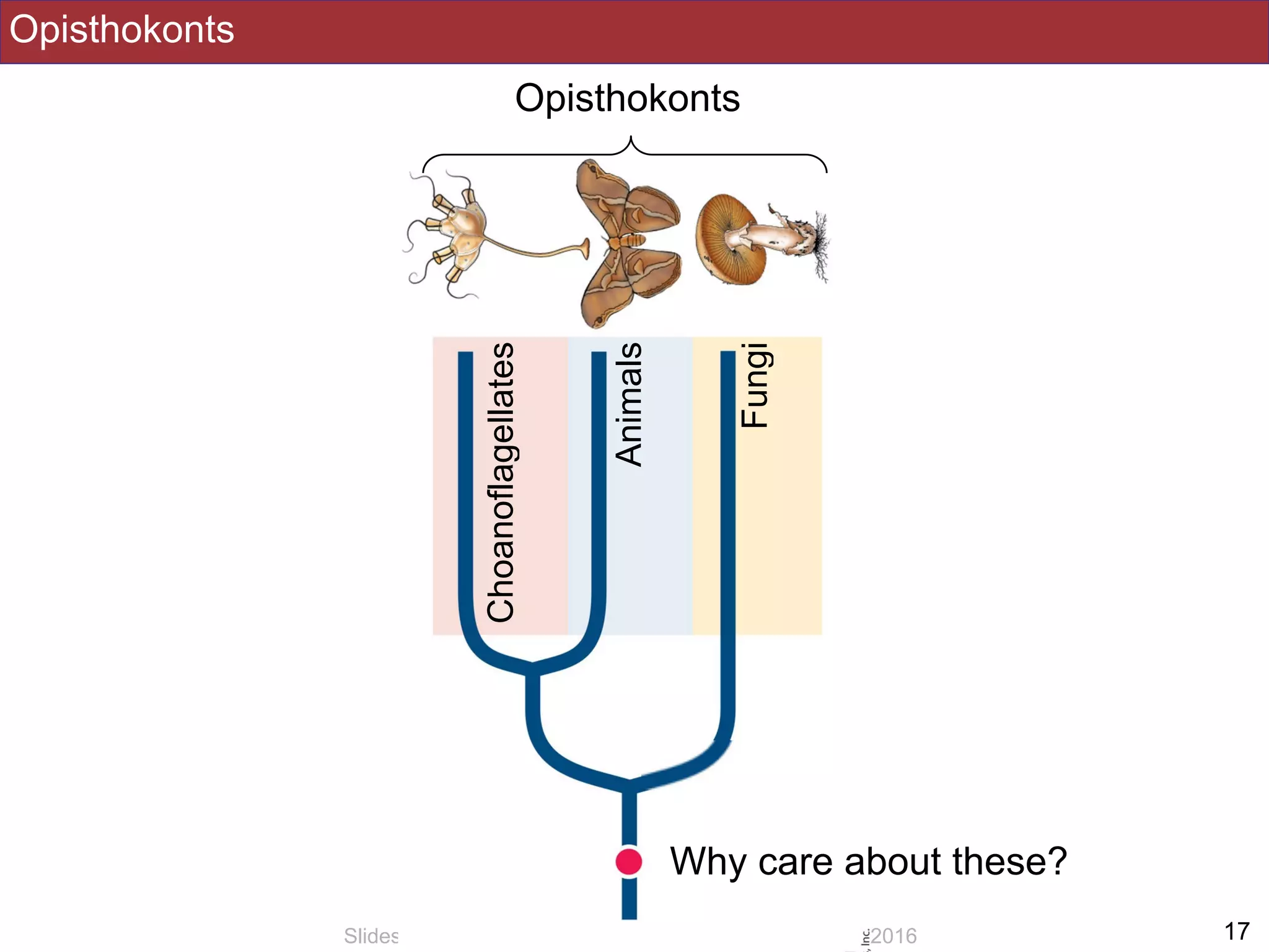
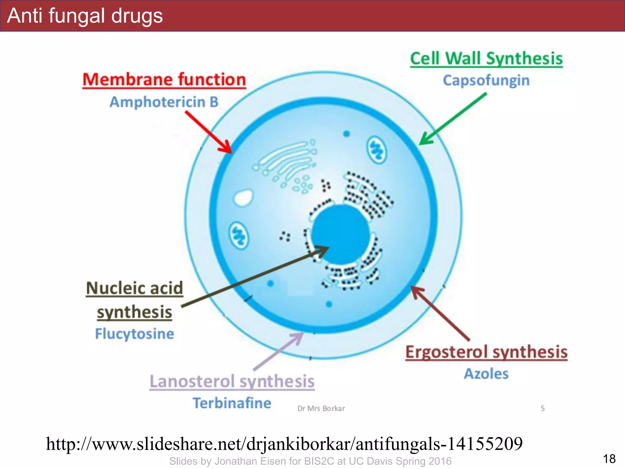
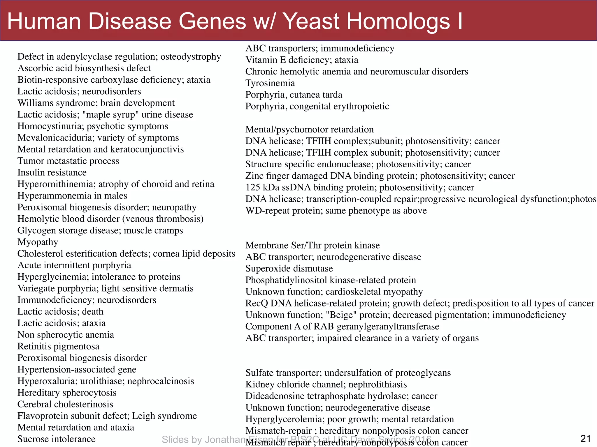

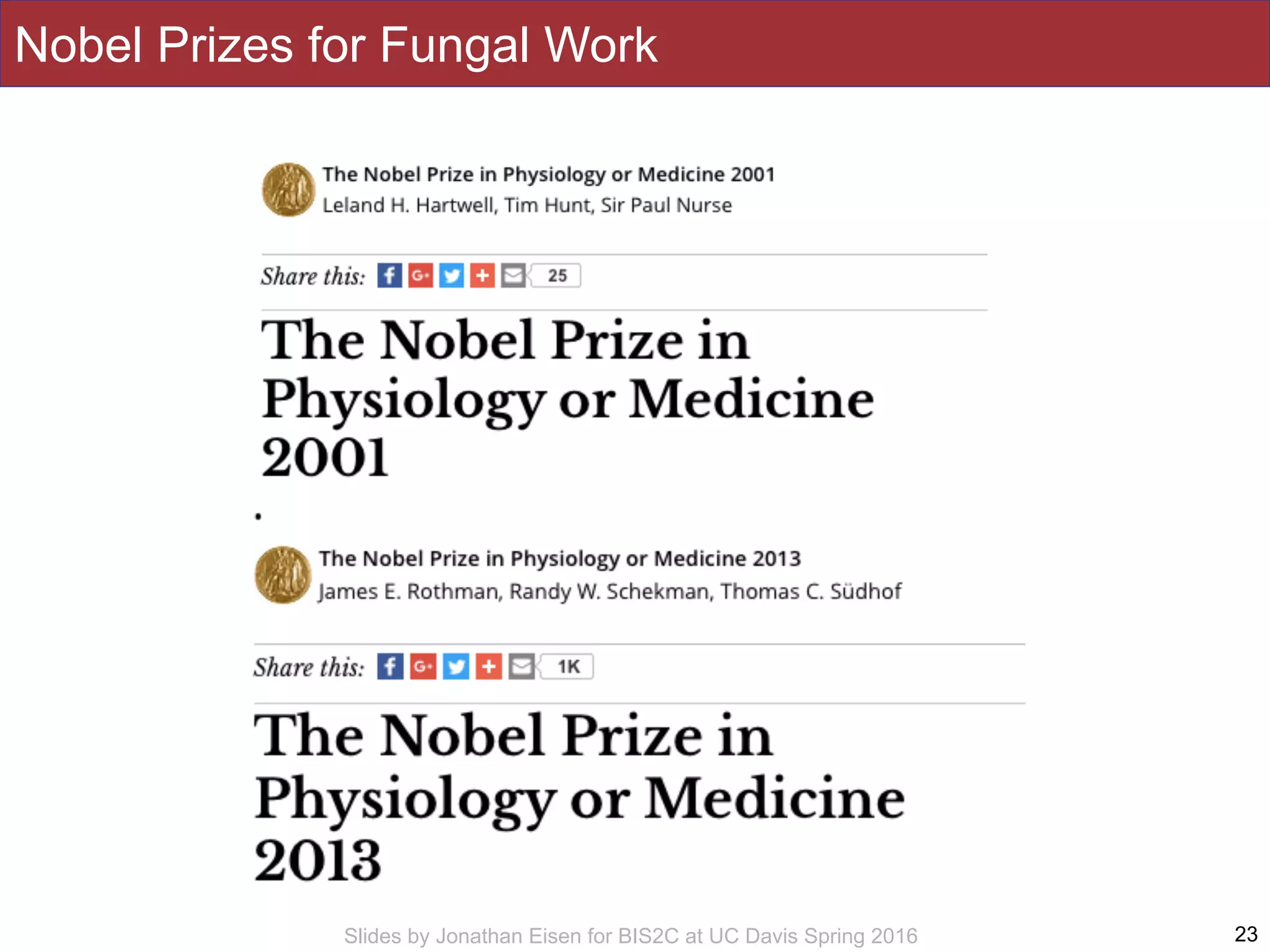

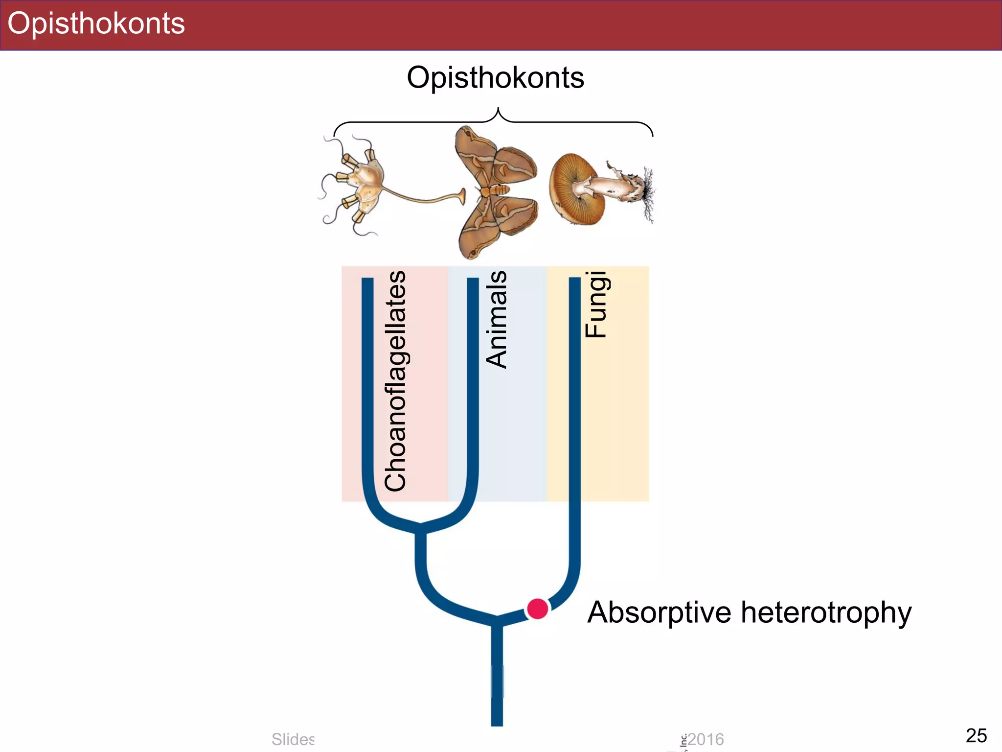

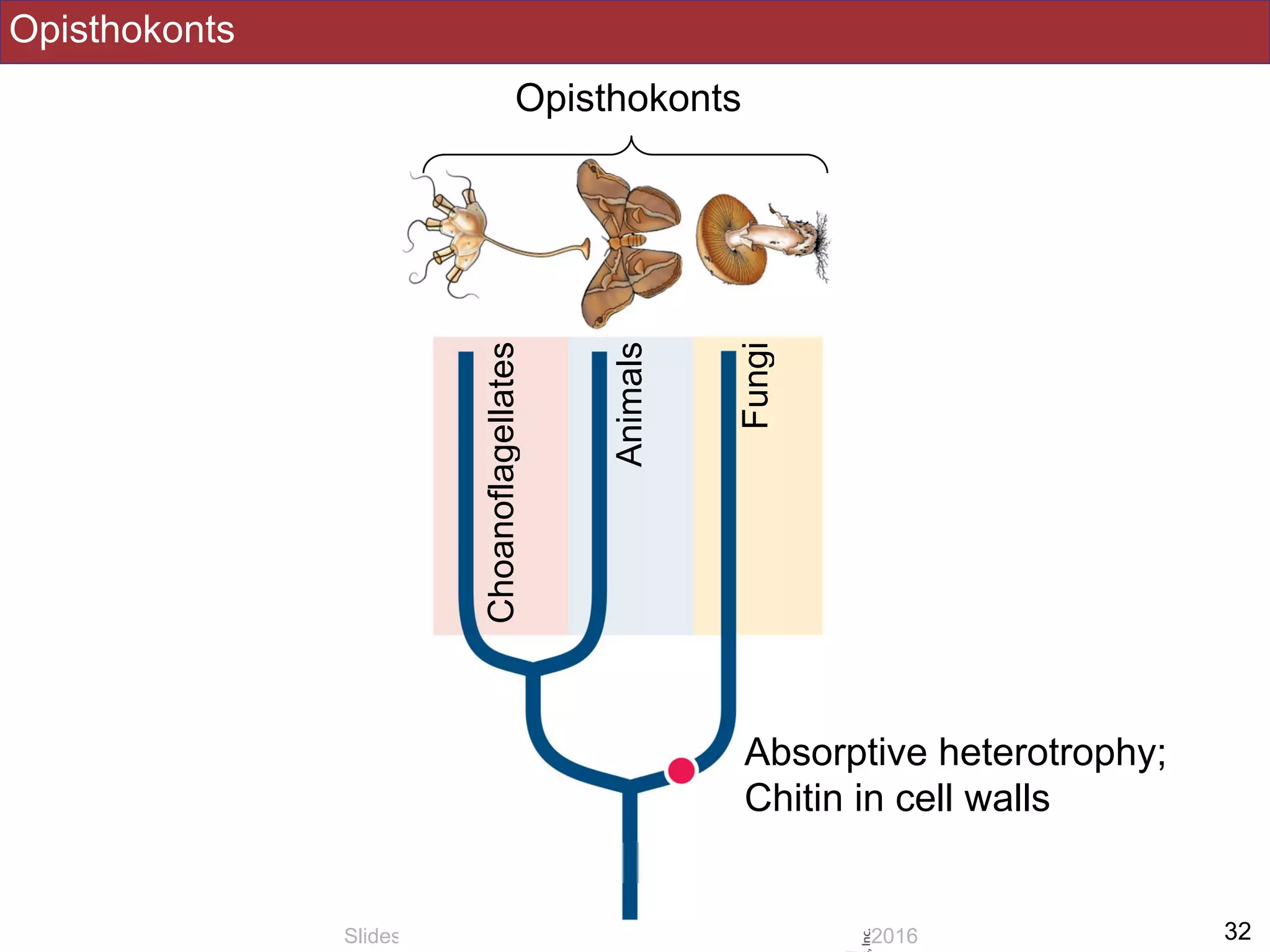
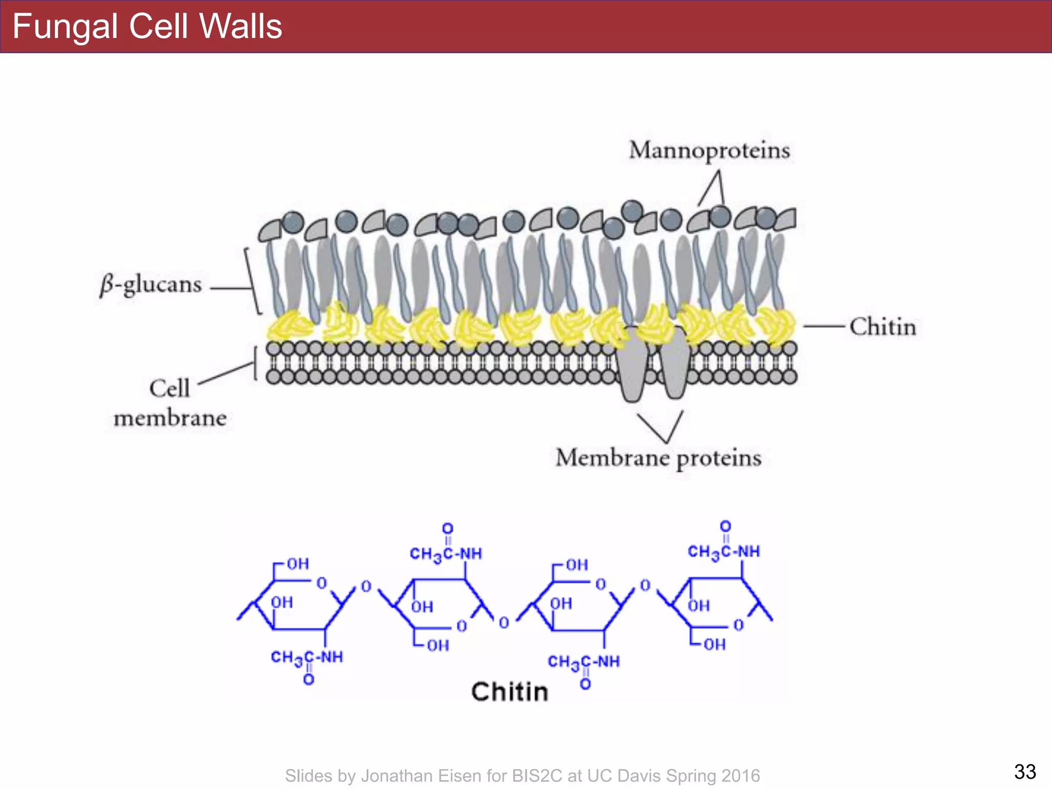
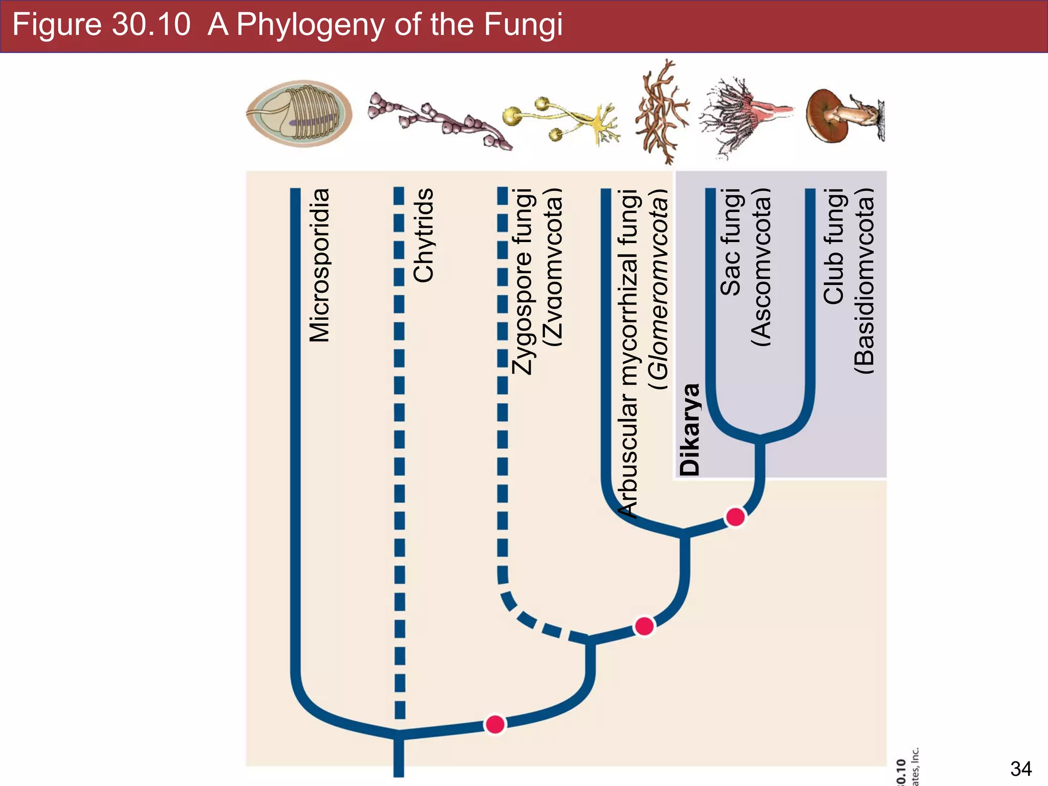
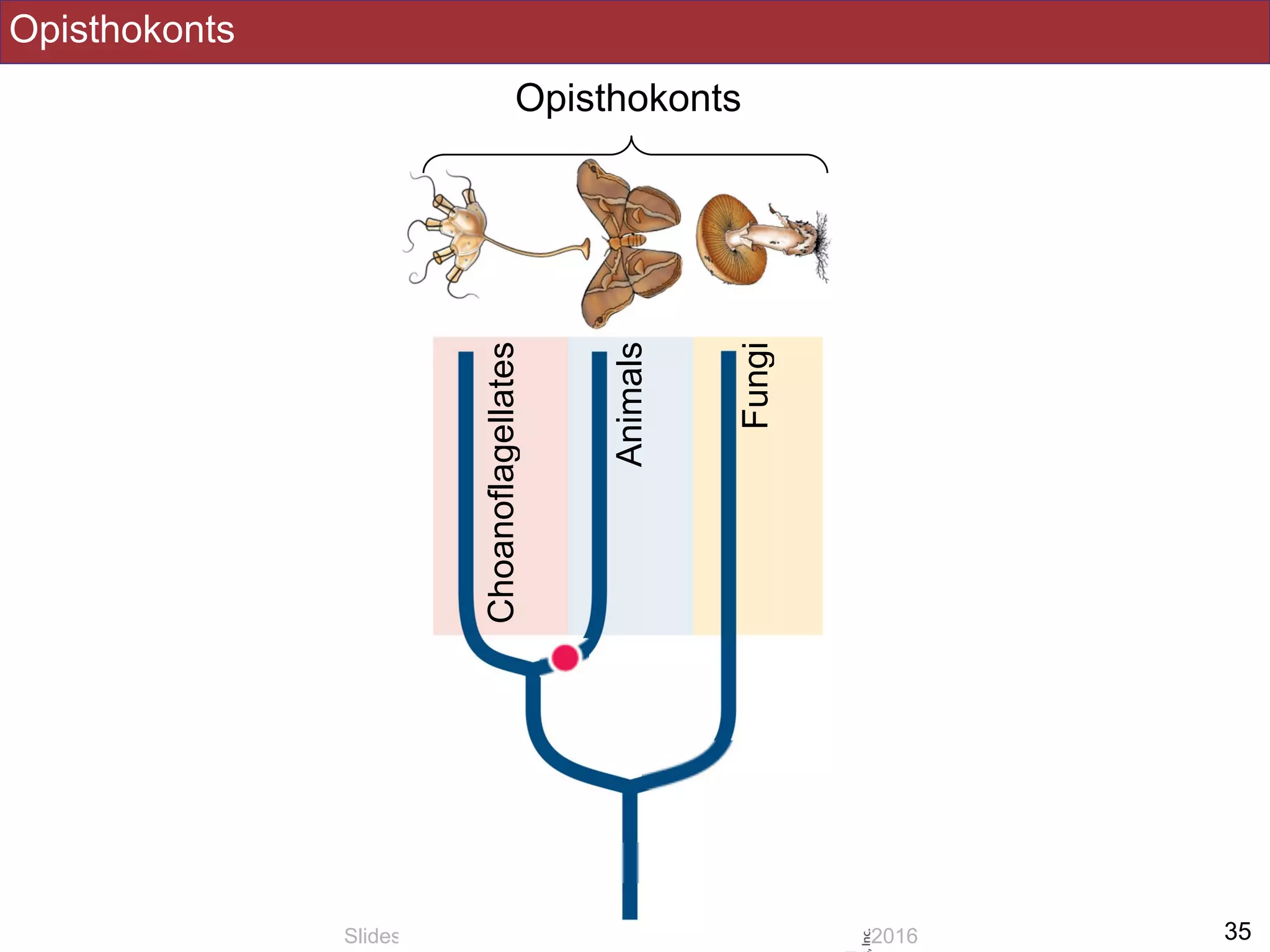
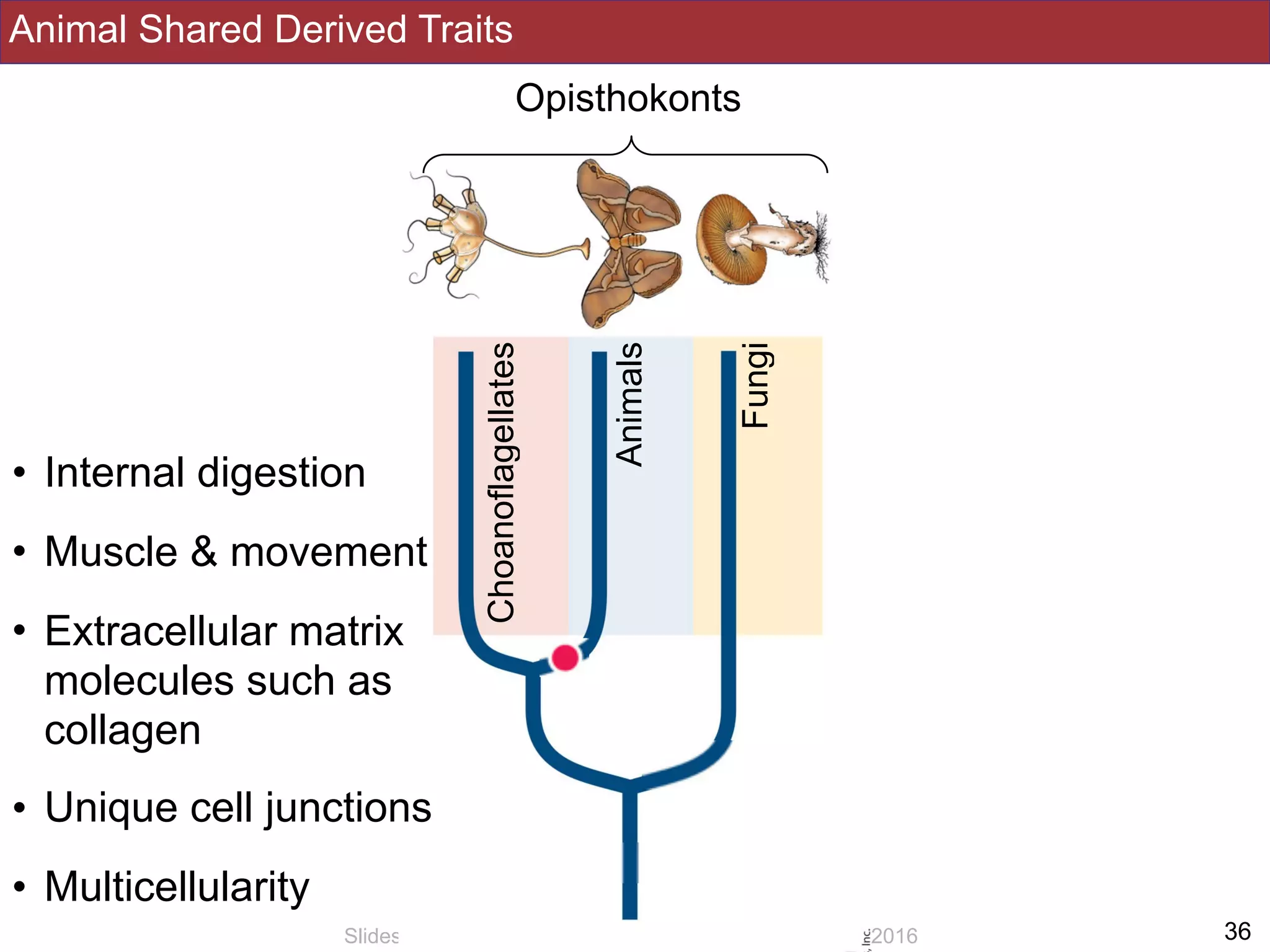
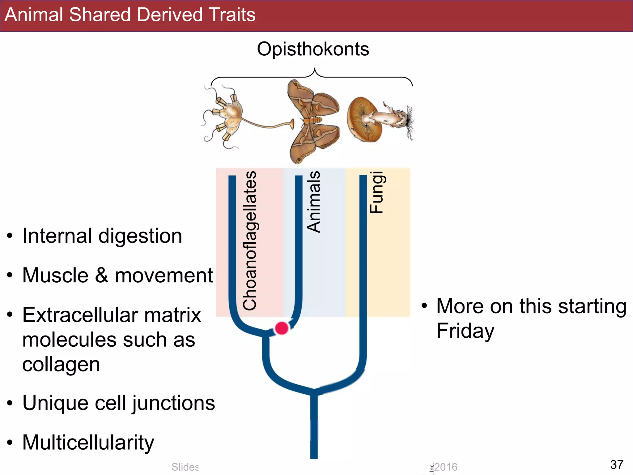
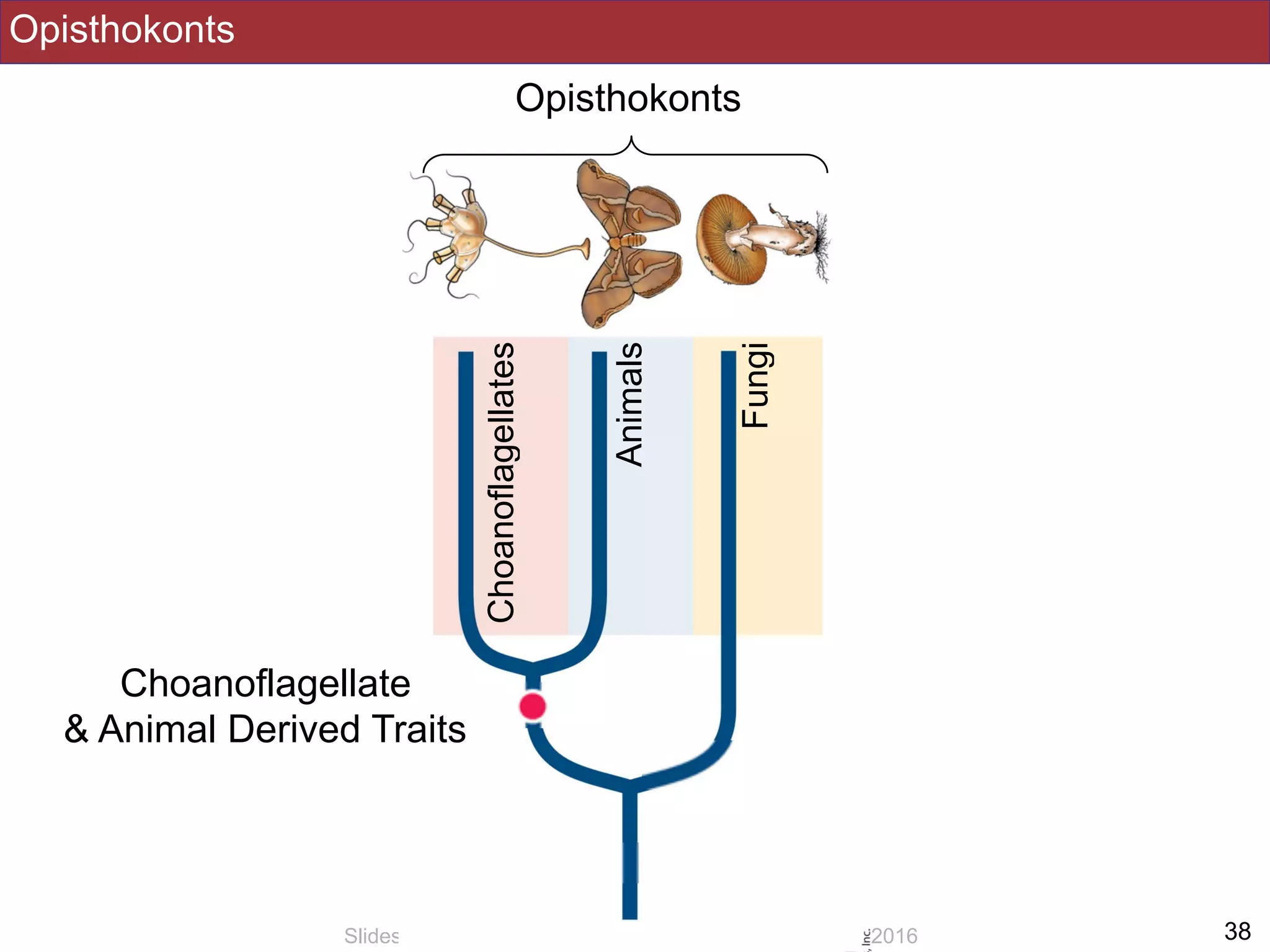
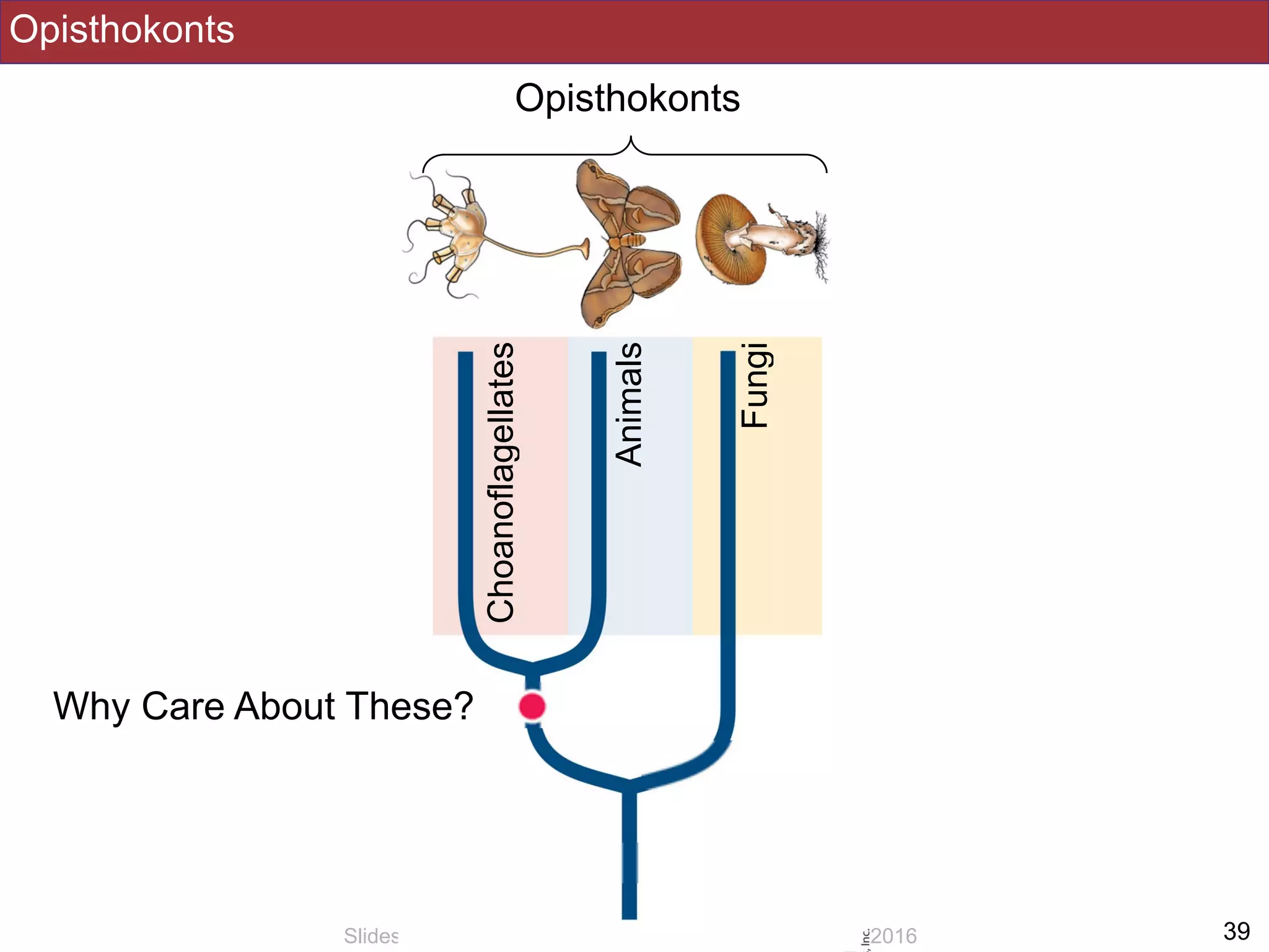

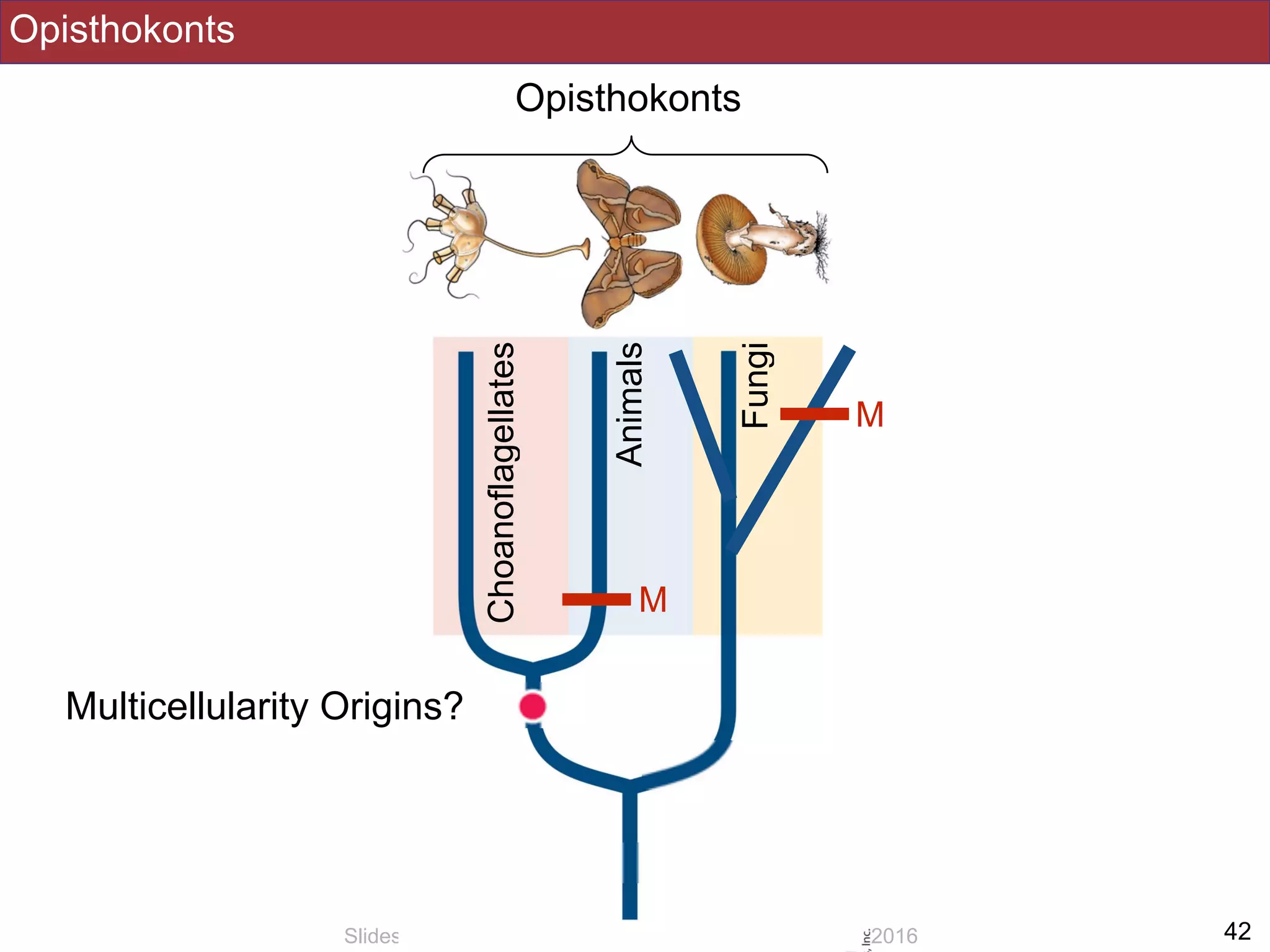
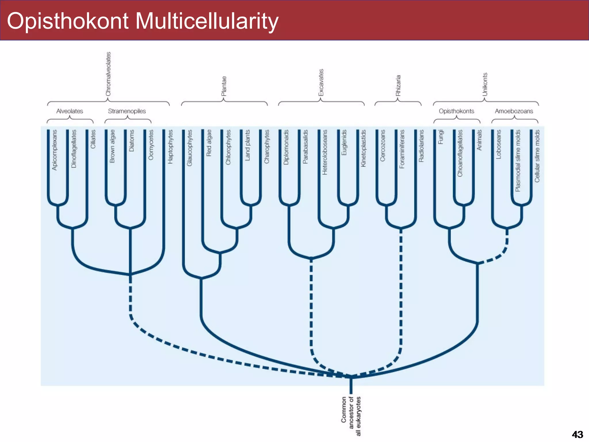


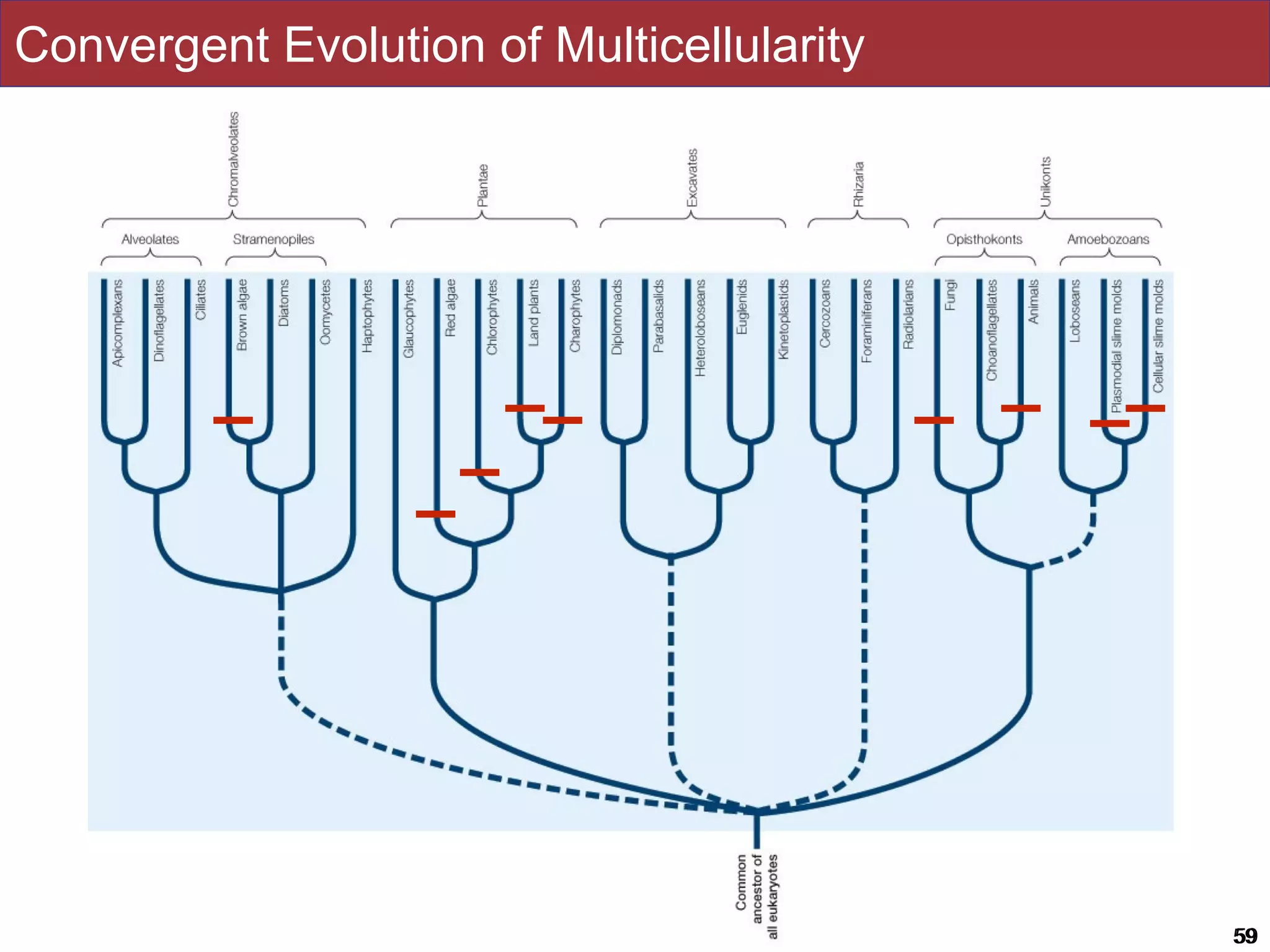
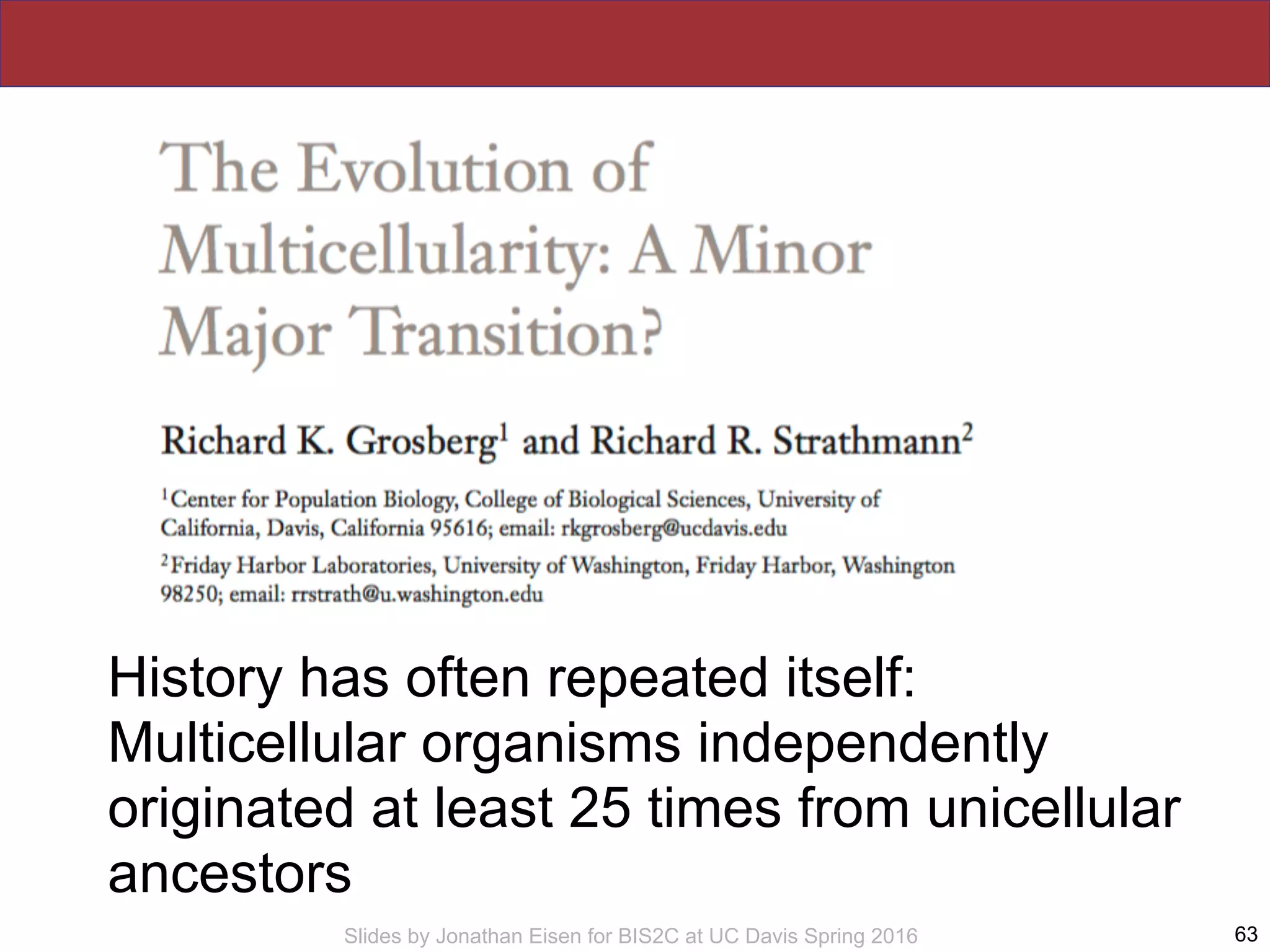
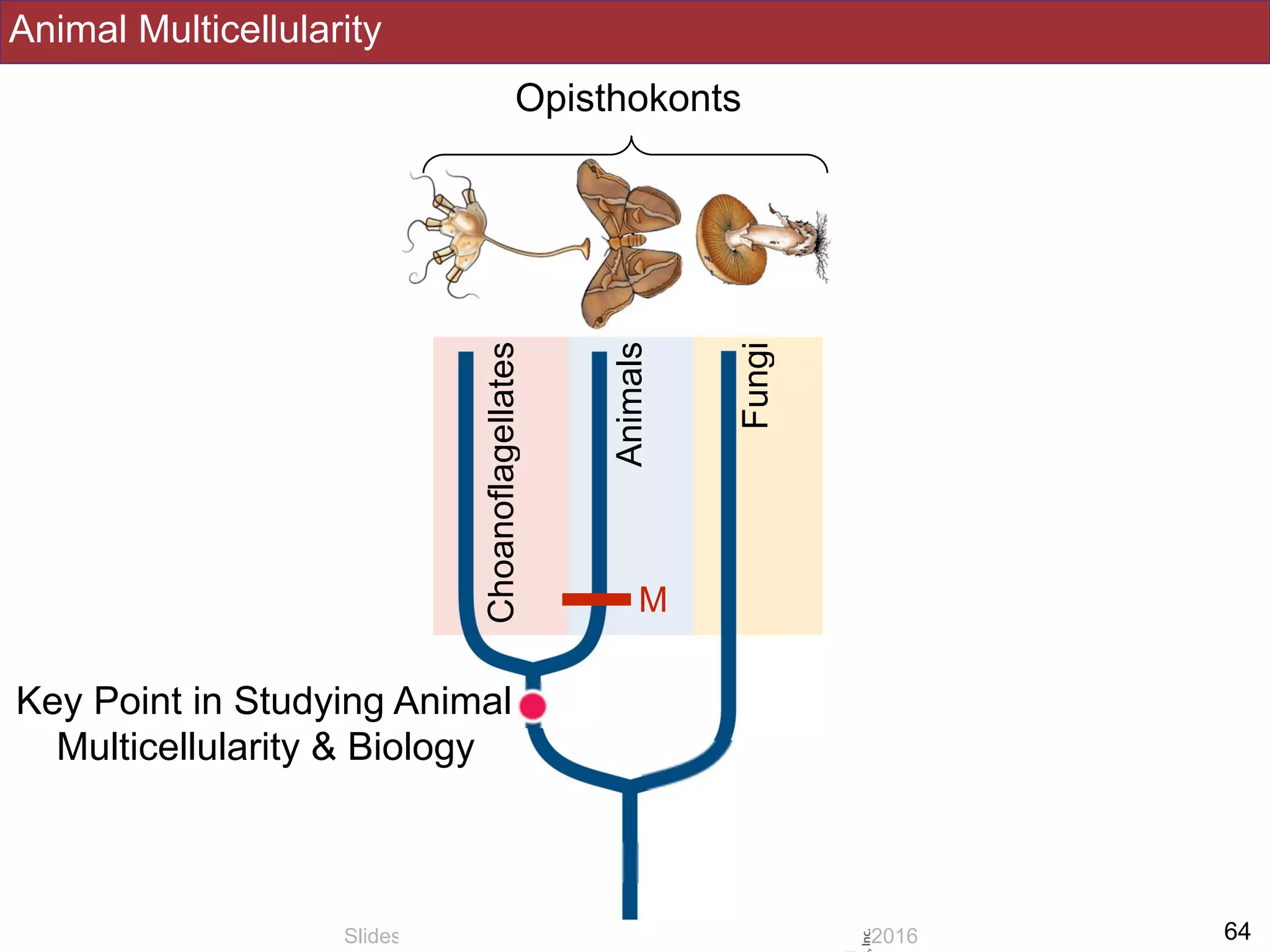
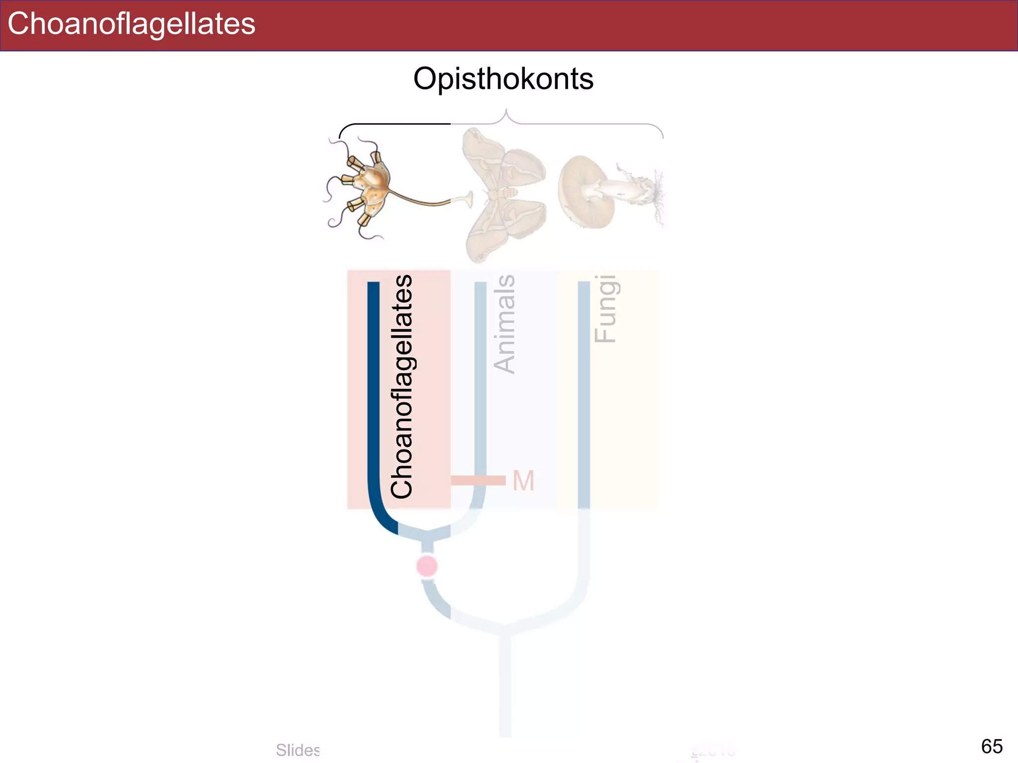
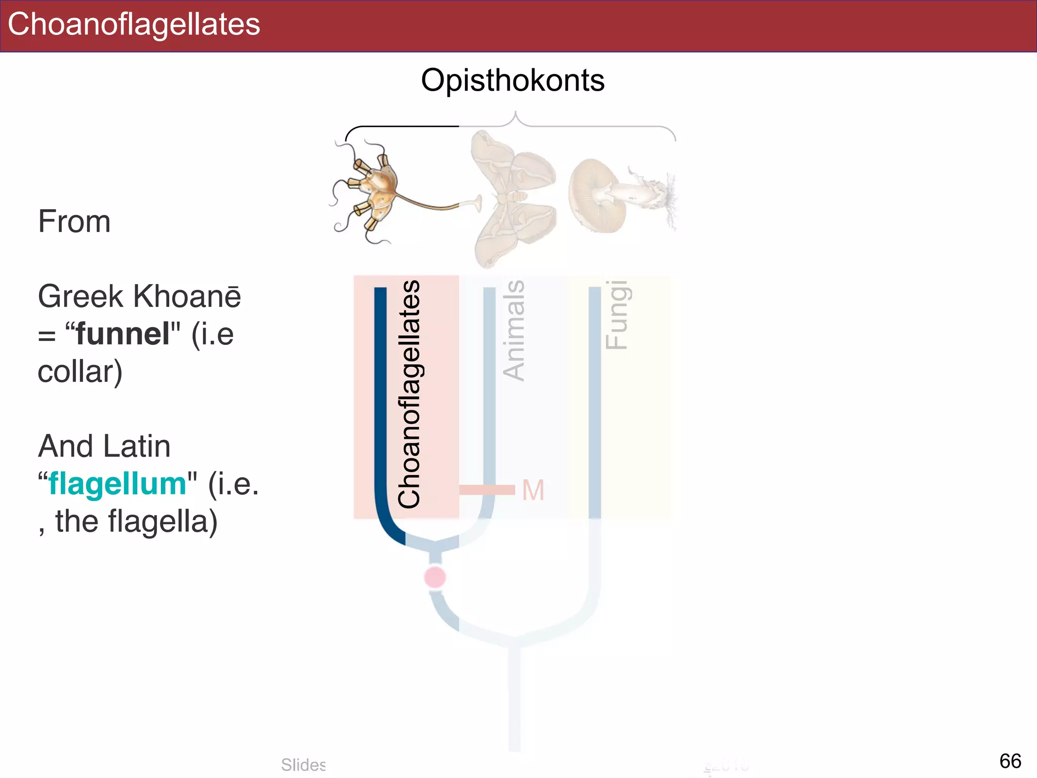


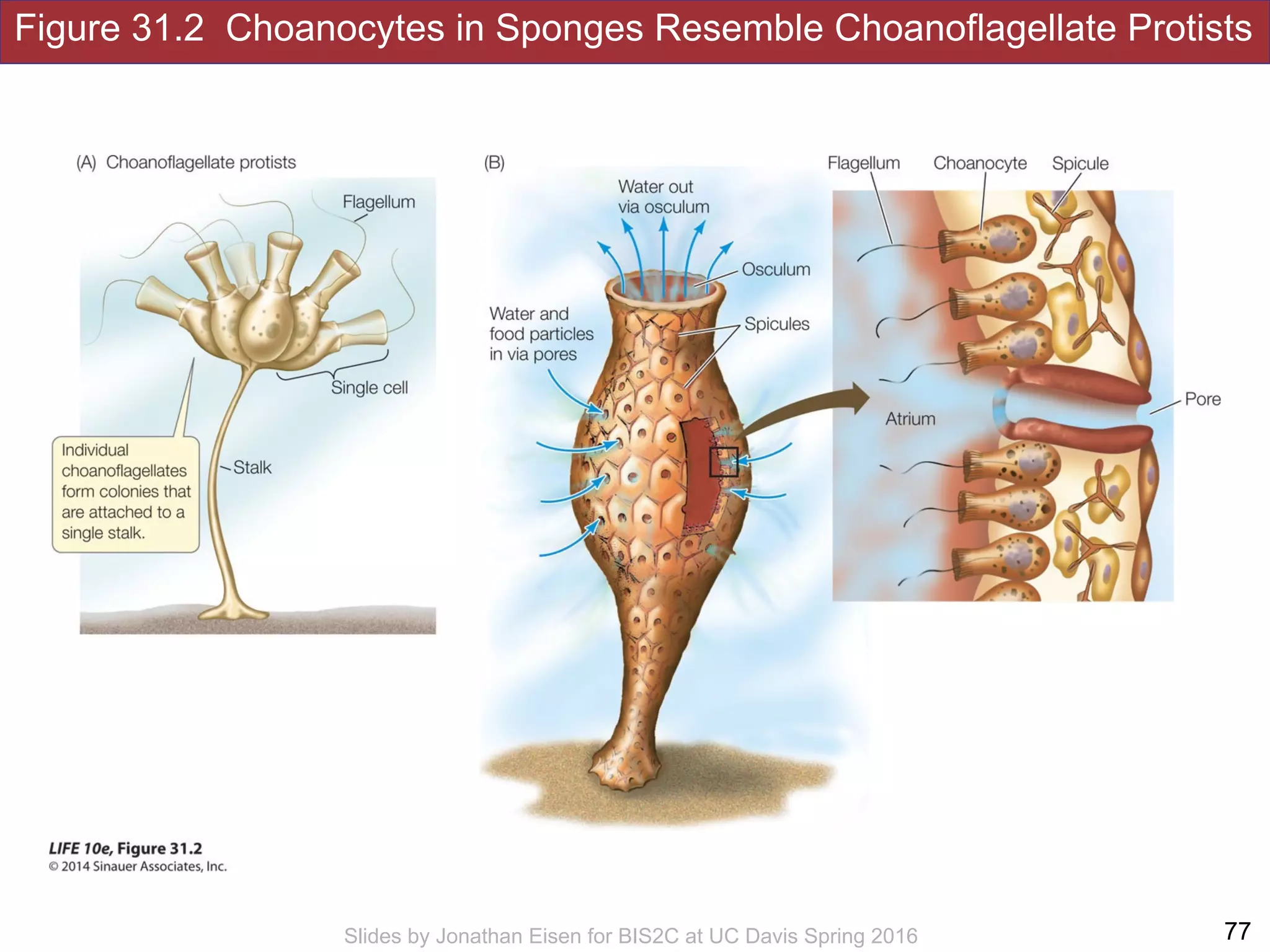
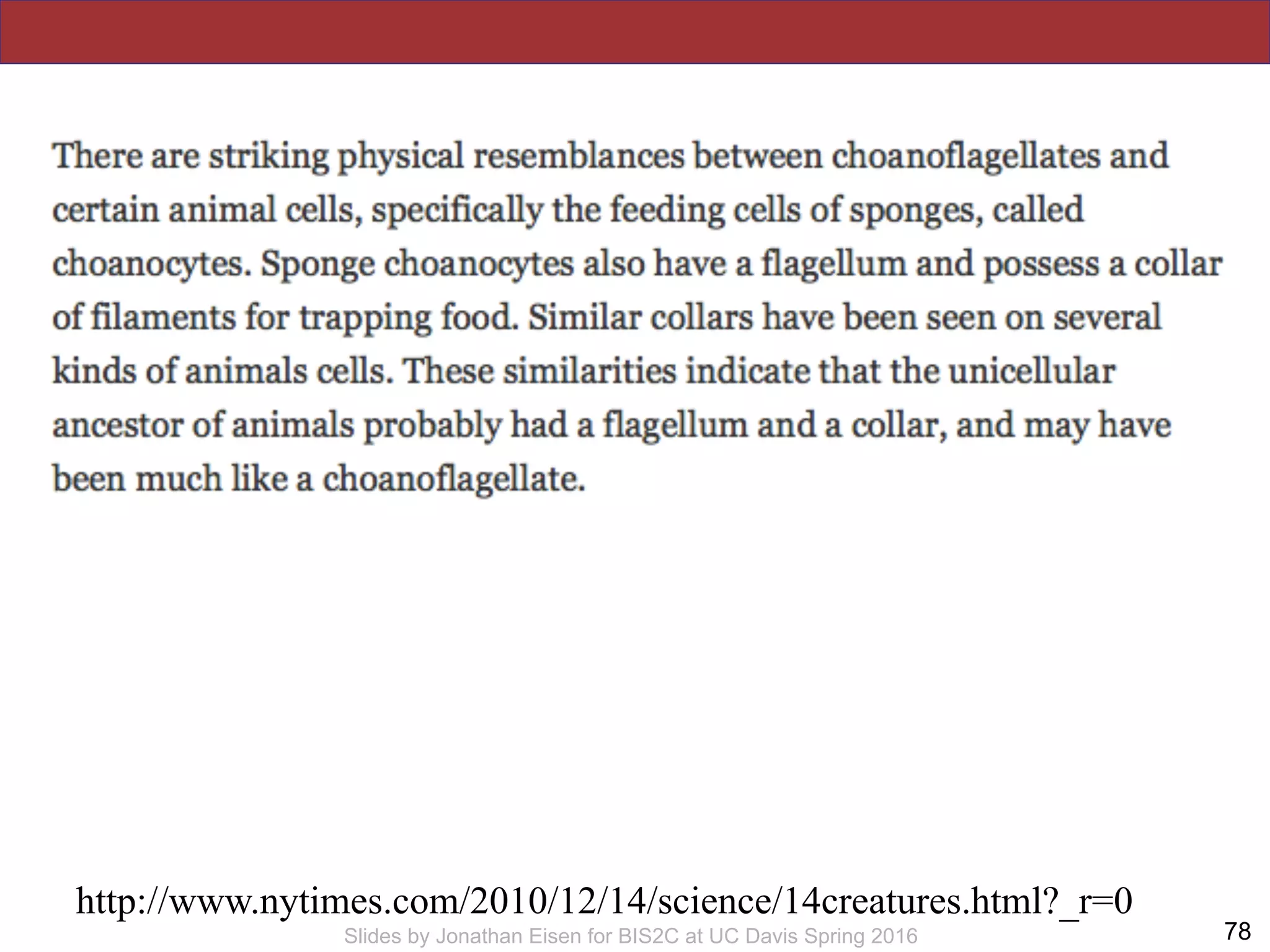
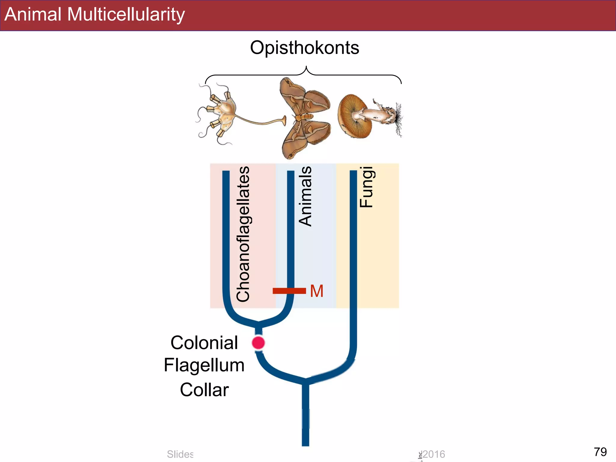


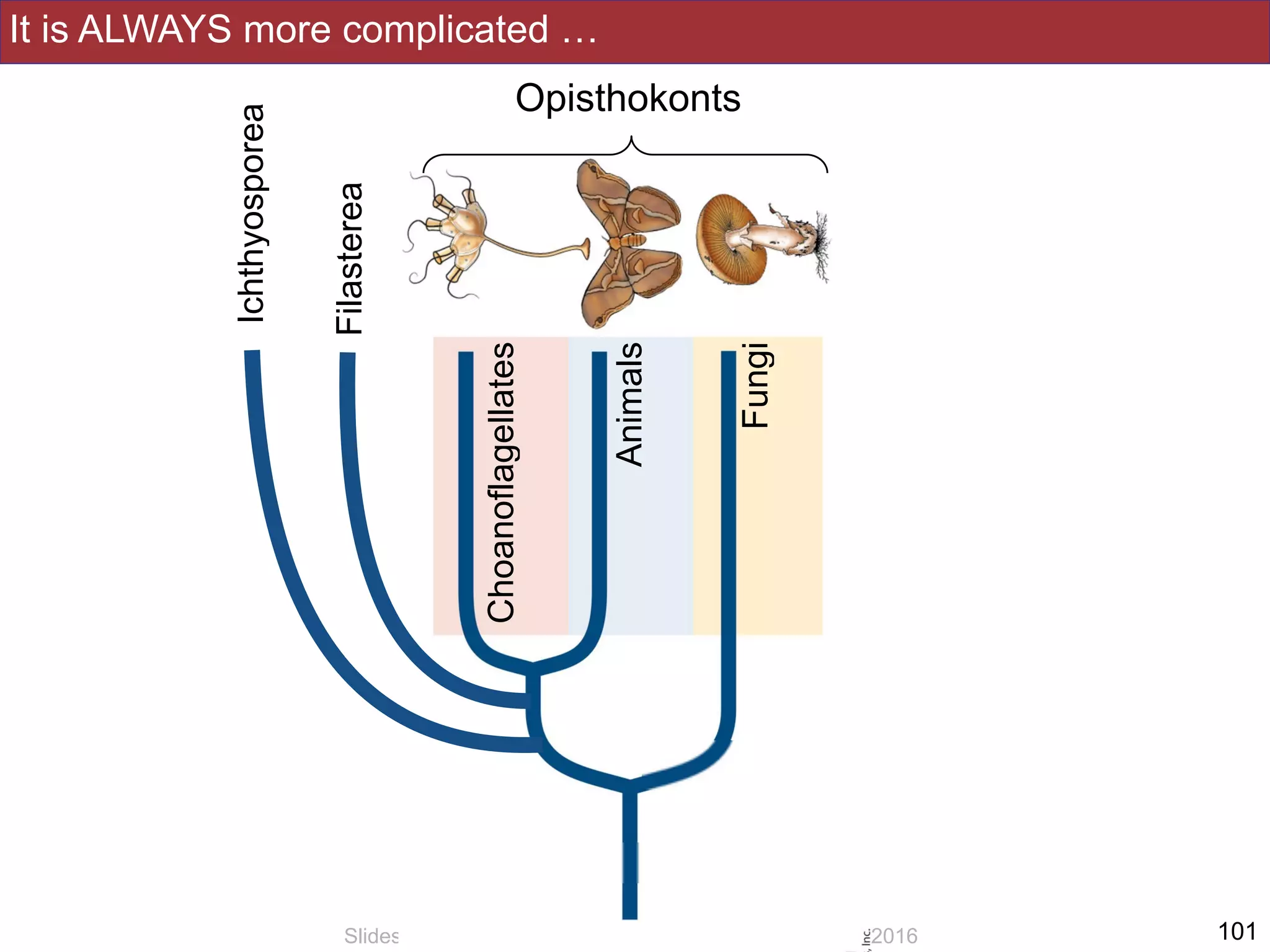


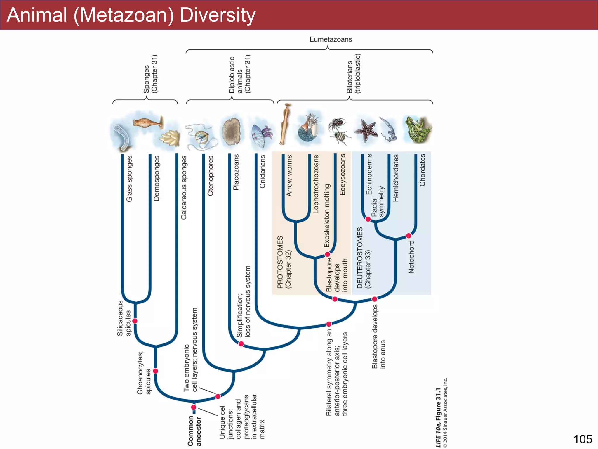
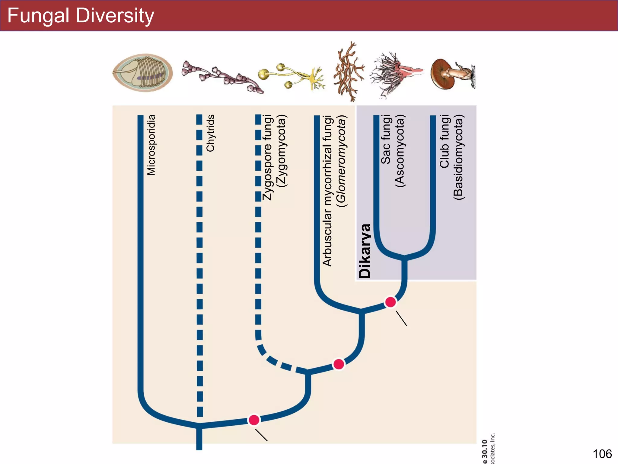

This document contains slides from a lecture on Opisthokonts. The lecture covers the key groups within the Opisthokonts, including fungi, animals, and choanoflagellates. It discusses shared derived traits of the opisthokont clade, as well as derived features of fungi, such as their absorptive heterotrophic nutrition. The slides also mention the relevance of studying opisthokonts and fungi to understanding human diseases and developing antifungal drugs.