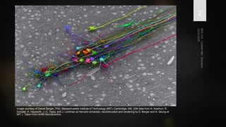
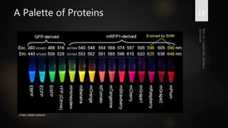
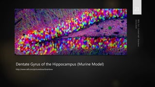
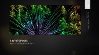
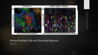
Brainbow is a technique that uses fluorescent proteins to map neural connections in the brain. It involves genetically engineering mice so that individual neurons express different combinations of colors. This allows their connections to be traced visually. The technique was improved over time by incorporating additional genetic tools. Brainbow has been applied to map connectomes in mice, fish, flies and more. It provides a way to visualize neural circuits and how they change over time or with conditions like disease.
This lecture covers key findings to the development of genomics as a field. This first part covers briefly Mendel to knowing that DNA is the genetic material by Hershey and Chase
This lecture covers some nice stories about the origins of the words "genome" and the derived word "genomics". the lecture also introduces viral, bacterial, and eukaryotic genomes.
Next generation Sequencing or massive parallel sequencing is a high throughput approach to sequence genetic material using the concept of massively parallel processing. It is also called second generation sequencing.This enables researchers a wide variety of applications & study biological systems.
The Emerging Global Collaboratory for Microbial Metagenomics ResearchersLarry Smarr
08.07.30
Invited Talk
Delivered From Calit2@UCSD
Monash University MURPA Lecture
Title: The Emerging Global Collaboratory for Microbial Metagenomics Researchers
Melbourne, Australia
Principles of DNA Structure, Replication and how it affects Plant breeding.pdfGedifewGebrie
A narrative review assignment on Principle of DNA structure, replication
and how it affects plant breeding
Submitted to: Assoc. Professor Tigist Abebe (PhD)
Bahir dar university
College of agriculture and environmental sciences
Department: Plant Sciences
Program: Plant Breeding and Genetics, Bahir Dar Ethiopia
Structure and genesis of mitochondrial and chloroplast, DNA replication , tra...Khalid Mukhtar
Presence of precise organelle DNA in mitochondria and chloroplasts became recognized over 3 years ago, proliferation of chloroplast DNA was first validated by the means of Chun et al, illustration of nuclear manipulation of the human mitochondrial genome chloroplast gene transcription managed transcription of cpDNA genes via the means of various factors from the nuclear basis, the number one elements affecting the transcription of cpDNA genes are NEP polymerase and non-intermediate subunits of PEP polymerase, where we explain the mechanism transporting barrel proteins from the outer mitochondrial membrane (OMM) through the TOM complex, and associated with chaperones TIM small cells within the IMS side and inserted into the OMM via sorting means and meeting equipment (SAM), we additionally annotate the chloroplast genome genes for some proteins required for the transcription and translation of encoded genes and, at the extreme, genes for photosynthesis, the locus of these repeats determines the site of unpaired reproduction the short (SSC) and extended unpaired reproductive site (LSC) in the chloroplast genome, leuco = white; plast = living) are colorless plastids that are identified in embryonic and germ cells.
In this paper, we briefly reviewed the numbers in life from a statistical genetic approach. The human genome comprises of 6 billion chemical bases of DNA. The DNA encodes 30,000 genes. It consists of two parts; the nuclear genome; which consists of 3,200,000,000 nucleotides of DNA, divided into 24 linear molecules, the shortest 50,000,000 nucleotides in length and the longest 260,000,000 nucleotides, each contained in a different chromosome and the mitochondrial genome; which contains approximately 16,600 base pairs encoding 37 genes. Most human cells have 46 chromosomes. However, the number of chromosomes in the nuclei of a person with Down syndrome is 47. The DNA of any two people on Earth is 99.6 percent identical, the 0.4 percent variation represents about 20 million base pairs. Almost all 98 percent of the human DNA is noncoding, while in bacteria, only 2% of the genetic material does not code for anything.
Visualizing Human Stem Cell Dynamics by Multicolor, Multiday High-Content Mic...InsideScientific
Visualizing the complex spatiotemporal dynamics of human stem cells as they proliferate and make cell fate decisions is key to improve our understanding of how to robustly engineer differentiated tissues for therapeutic applications.
In this webinar, Dr. Rafael Carazo Salas describes multicolor, multiday high-content microscopy pipelines that his group has recently developed to visualize the dynamical cell fate changes of human Pluripotent Stem Cells (hPSCs). In particular, he reviews the integrated experimental and computational approaches that his group has established, including novel “live” reporters of cell fate and multi-reporter hPSC lines generated by CRISPR/Cas9 allowing multiplexed monitoring of cell proliferation and fate dynamics, and exemplify the biological discoveries they are enabling.
Key Topics Include:
- Visualizing how human Pluripotent Stem Cells (hPSCs) proliferate and undergo early differentiation in vitro, by high content microscopy
- Learning about experimental and computational pipelines that enable cell fate monitoring at the collective and single-cell level
- Learning about novel “live” reporters of hPSC cell fate
Microbial Metagenomics Drives a New CyberinfrastructureLarry Smarr
06.03.03
Invited Talk
School of Biological Sciences
University of California, Irvine
Title: Microbial Metagenomics Drives a New Cyberinfrastructure
Irvine, CA
Apaf-1 and Apoptosome Activation in H. sapiensCorbett Hall
Mitochondrial stress triggers cytochrome C-mediated activation of Apaf-1, cleaving Caspase 9 and leading to Apoptosome formation in a second-order, "induced proximity" mechanism
Slide 1: Title Slide
Extrachromosomal Inheritance
Slide 2: Introduction to Extrachromosomal Inheritance
Definition: Extrachromosomal inheritance refers to the transmission of genetic material that is not found within the nucleus.
Key Components: Involves genes located in mitochondria, chloroplasts, and plasmids.
Slide 3: Mitochondrial Inheritance
Mitochondria: Organelles responsible for energy production.
Mitochondrial DNA (mtDNA): Circular DNA molecule found in mitochondria.
Inheritance Pattern: Maternally inherited, meaning it is passed from mothers to all their offspring.
Diseases: Examples include Leber’s hereditary optic neuropathy (LHON) and mitochondrial myopathy.
Slide 4: Chloroplast Inheritance
Chloroplasts: Organelles responsible for photosynthesis in plants.
Chloroplast DNA (cpDNA): Circular DNA molecule found in chloroplasts.
Inheritance Pattern: Often maternally inherited in most plants, but can vary in some species.
Examples: Variegation in plants, where leaf color patterns are determined by chloroplast DNA.
Slide 5: Plasmid Inheritance
Plasmids: Small, circular DNA molecules found in bacteria and some eukaryotes.
Features: Can carry antibiotic resistance genes and can be transferred between cells through processes like conjugation.
Significance: Important in biotechnology for gene cloning and genetic engineering.
Slide 6: Mechanisms of Extrachromosomal Inheritance
Non-Mendelian Patterns: Do not follow Mendel’s laws of inheritance.
Cytoplasmic Segregation: During cell division, organelles like mitochondria and chloroplasts are randomly distributed to daughter cells.
Heteroplasmy: Presence of more than one type of organellar genome within a cell, leading to variation in expression.
Slide 7: Examples of Extrachromosomal Inheritance
Four O’clock Plant (Mirabilis jalapa): Shows variegated leaves due to different cpDNA in leaf cells.
Petite Mutants in Yeast: Result from mutations in mitochondrial DNA affecting respiration.
Slide 8: Importance of Extrachromosomal Inheritance
Evolution: Provides insight into the evolution of eukaryotic cells.
Medicine: Understanding mitochondrial inheritance helps in diagnosing and treating mitochondrial diseases.
Agriculture: Chloroplast inheritance can be used in plant breeding and genetic modification.
Slide 9: Recent Research and Advances
Gene Editing: Techniques like CRISPR-Cas9 are being used to edit mitochondrial and chloroplast DNA.
Therapies: Development of mitochondrial replacement therapy (MRT) for preventing mitochondrial diseases.
Slide 10: Conclusion
Summary: Extrachromosomal inheritance involves the transmission of genetic material outside the nucleus and plays a crucial role in genetics, medicine, and biotechnology.
Future Directions: Continued research and technological advancements hold promise for new treatments and applications.
Slide 11: Questions and Discussion
Invite Audience: Open the floor for any questions or further discussion on the topic.
A brief information about the SCOP protein database used in bioinformatics.
The Structural Classification of Proteins (SCOP) database is a comprehensive and authoritative resource for the structural and evolutionary relationships of proteins. It provides a detailed and curated classification of protein structures, grouping them into families, superfamilies, and folds based on their structural and sequence similarities.
Professional air quality monitoring systems provide immediate, on-site data for analysis, compliance, and decision-making.
Monitor common gases, weather parameters, particulates.
Comparing Evolved Extractive Text Summary Scores of Bidirectional Encoder Rep...University of Maribor
Slides from:
11th International Conference on Electrical, Electronics and Computer Engineering (IcETRAN), Niš, 3-6 June 2024
Track: Artificial Intelligence
https://www.etran.rs/2024/en/home-english/
This pdf is about the Schizophrenia.
For more details visit on YouTube; @SELF-EXPLANATORY;
https://www.youtube.com/channel/UCAiarMZDNhe1A3Rnpr_WkzA/videos
Thanks...!
Seminar of U.V. Spectroscopy by SAMIR PANDASAMIR PANDA
Spectroscopy is a branch of science dealing the study of interaction of electromagnetic radiation with matter.
Ultraviolet-visible spectroscopy refers to absorption spectroscopy or reflect spectroscopy in the UV-VIS spectral region.
Ultraviolet-visible spectroscopy is an analytical method that can measure the amount of light received by the analyte.
Richard's entangled aventures in wonderlandRichard Gill
Since the loophole-free Bell experiments of 2020 and the Nobel prizes in physics of 2022, critics of Bell's work have retreated to the fortress of super-determinism. Now, super-determinism is a derogatory word - it just means "determinism". Palmer, Hance and Hossenfelder argue that quantum mechanics and determinism are not incompatible, using a sophisticated mathematical construction based on a subtle thinning of allowed states and measurements in quantum mechanics, such that what is left appears to make Bell's argument fail, without altering the empirical predictions of quantum mechanics. I think however that it is a smoke screen, and the slogan "lost in math" comes to my mind. I will discuss some other recent disproofs of Bell's theorem using the language of causality based on causal graphs. Causal thinking is also central to law and justice. I will mention surprising connections to my work on serial killer nurse cases, in particular the Dutch case of Lucia de Berk and the current UK case of Lucy Letby.
Introduction:
RNA interference (RNAi) or Post-Transcriptional Gene Silencing (PTGS) is an important biological process for modulating eukaryotic gene expression.
It is highly conserved process of posttranscriptional gene silencing by which double stranded RNA (dsRNA) causes sequence-specific degradation of mRNA sequences.
dsRNA-induced gene silencing (RNAi) is reported in a wide range of eukaryotes ranging from worms, insects, mammals and plants.
This process mediates resistance to both endogenous parasitic and exogenous pathogenic nucleic acids, and regulates the expression of protein-coding genes.
What are small ncRNAs?
micro RNA (miRNA)
short interfering RNA (siRNA)
Properties of small non-coding RNA:
Involved in silencing mRNA transcripts.
Called “small” because they are usually only about 21-24 nucleotides long.
Synthesized by first cutting up longer precursor sequences (like the 61nt one that Lee discovered).
Silence an mRNA by base pairing with some sequence on the mRNA.
Discovery of siRNA?
The first small RNA:
In 1993 Rosalind Lee (Victor Ambros lab) was studying a non- coding gene in C. elegans, lin-4, that was involved in silencing of another gene, lin-14, at the appropriate time in the
development of the worm C. elegans.
Two small transcripts of lin-4 (22nt and 61nt) were found to be complementary to a sequence in the 3' UTR of lin-14.
Because lin-4 encoded no protein, she deduced that it must be these transcripts that are causing the silencing by RNA-RNA interactions.
Types of RNAi ( non coding RNA)
MiRNA
Length (23-25 nt)
Trans acting
Binds with target MRNA in mismatch
Translation inhibition
Si RNA
Length 21 nt.
Cis acting
Bind with target Mrna in perfect complementary sequence
Piwi-RNA
Length ; 25 to 36 nt.
Expressed in Germ Cells
Regulates trnasposomes activity
MECHANISM OF RNAI:
First the double-stranded RNA teams up with a protein complex named Dicer, which cuts the long RNA into short pieces.
Then another protein complex called RISC (RNA-induced silencing complex) discards one of the two RNA strands.
The RISC-docked, single-stranded RNA then pairs with the homologous mRNA and destroys it.
THE RISC COMPLEX:
RISC is large(>500kD) RNA multi- protein Binding complex which triggers MRNA degradation in response to MRNA
Unwinding of double stranded Si RNA by ATP independent Helicase
Active component of RISC is Ago proteins( ENDONUCLEASE) which cleave target MRNA.
DICER: endonuclease (RNase Family III)
Argonaute: Central Component of the RNA-Induced Silencing Complex (RISC)
One strand of the dsRNA produced by Dicer is retained in the RISC complex in association with Argonaute
ARGONAUTE PROTEIN :
1.PAZ(PIWI/Argonaute/ Zwille)- Recognition of target MRNA
2.PIWI (p-element induced wimpy Testis)- breaks Phosphodiester bond of mRNA.)RNAse H activity.
MiRNA:
The Double-stranded RNAs are naturally produced in eukaryotic cells during development, and they have a key role in regulating gene expression .
THE IMPORTANCE OF MARTIAN ATMOSPHERE SAMPLE RETURN.Sérgio Sacani
The return of a sample of near-surface atmosphere from Mars would facilitate answers to several first-order science questions surrounding the formation and evolution of the planet. One of the important aspects of terrestrial planet formation in general is the role that primary atmospheres played in influencing the chemistry and structure of the planets and their antecedents. Studies of the martian atmosphere can be used to investigate the role of a primary atmosphere in its history. Atmosphere samples would also inform our understanding of the near-surface chemistry of the planet, and ultimately the prospects for life. High-precision isotopic analyses of constituent gases are needed to address these questions, requiring that the analyses are made on returned samples rather than in situ.
2. 2
Image courtesy of Daniel Berger, PhD, Massachusetts Institute of Technology (MIT), Cambridge, MA. (EM data from N. Kasthuri, R.
Schalek, K. Hayworth, J.-C. Tapia, and J. Lichtman at Harvard University; reconstruction and rendering by D. Berger and S. Seung at
MIT.) Taken from HHMI Biointeractive.
3. What is the Connectome?
HOW WE THINK WE THINK
3
4. Starting at the Synapse
Neurons connect with one another at
a synapse, or connection, between an
axon and a dendrite of two neurons
These connections carry electrical
potentials along a neural tract
All together, neural tracts establish
the reflexes, thinking patterns,
cognition, and personality of an
organism
4
http://www.drugabuse.gov/publications/teaching-packets/brain-
actions-cocaine-opiates-marijuana/section-i-introduction-to-brain/3-
neuronal-structure
5. Understanding Brain Function 5
https://upload.wikimedia.org/wikipedia/commons/th
umb/e/ec/PhrenologyPix.jpg/220px-
PhrenologyPix.jpg
http://classes.midlandstech.edu/carterp/Courses/bio110/chap08/Sli
de10.JPG
6. “
”
the size of the connectome would be…
an estimated 1011 neurons, with 1015
connections between them
(SPORNS ET AL. 246)
Mapping the Connectome Is Considerably More Data-Intensive than the Human
Genome Project, Based on the Number of Synapses
6
7. Reconstruction of Neocortex 7
(Kasthuri et al.) http://www.sciencedirect.com/science/article/pii/S0092867415008247
8. Shedding Light on the Synapse
THE USE OF GREEN FLUORESCENT PROTEIN AND ITS DERIVATIVES
8
9. “
”
Now it is such a bizarrely improbable coincidence that
anything so mind-bogglingly useful could have evolved
purely by chance that some thinkers have chosen to see it
as the final and clinching proof of the non-existence of
God.
DOUGLAS ADAMS, HITCHHIKERS GUIDE TO THE GALAXY
Cited by Martin Chalfie in both his textbook on GFP and his 2008 Nobel acceptance
speech
9
10. Green Fluorescent Protein
Reported in 1955 by Davenport and
Nicol in Aequorea victoria
In 1962, Shimomura et al. describe an
extract called “aequorin” with
fluorescent properties
Chalfie et al. began clonal expression
in early 1990s, eventually winning the
2008 Nobel Prize in Chemistry
10
(Kain 305)
11. Green Fluorescent Protein
Originally isolated from the
“squeezate” of Aequorea victoria
Fluoresces upon addition of calcium,
emitting a sharp peak at 509 nm
Mutations to the internal
chromophore can generate differently
colored fluorescence
11
RCSB ID: 4KW4 (GFP)
12. A Palette of Proteins
Wild-type GFP did not work well in
FRET studies, so alternative sources
were sought
Another natural source is the
molecule DsRed, found in many corals
of the Discosoma genus
Genetic mutants of GFP and DsRed
have provided molecular geneticists
with a large palette of easily-inserted
marker proteins
12
(Tsien, Nobel Lecture)
14. The Technical Challenge of a
Technicolor Approach
ADAPTING SITE-SPECIFIC RECOMBINASES TO MICE CONNECTOMICS
14
15. Cre/lox Recombinase System
Cre, derived from P1 bacteriophage, is
a topoisomerase-like enzyme
Can recognize lox sites and generate
excisions or inversions
The DNA in-between two lox sites can
be reorganized and transiently
expressed upon addition of Cre
15
RCSB ID: 3MGV (Cre recombinase)
30. Dividing Chick NPCs Time-Lapse 30
(Loulier et al.) Movie S1. Time-Lapse Imaging of Dividing Brainbow-Labeled Neural
Progenitors in the Chick Spinal Cord.
31. Chick Spinal Cord 31
(Loulier et al.) Movie S3. High-Resolution View of Combined
Integrative Cytbow and Nucbow Labels in an E17 Chicken Spinal Cord.
Editor's Notes
Figure 4 | Combinatorial XFP expression results from tandem copy
integration. a, With a Brainbow construct expressing three XFPs,
independent recombination of three transgene copies can, in principle,
generate ten distinct colour combinations. b, Oculomotor axons of Thy1-
Brainbow-1.0 line H (recombination with CreERT2). Boxes show sample
regions from different axons. c, Dentate gyrus of Thy1-Brainbow-1.0 line L
(recombination with CreERT2). d, A single FRT site inserted in Brainbow
constructs allows tandem transgene copy number reduction through Flpmediated
recombination. The PCR indicates the disappearance of transgene
repeats in Thy1-Brainbow-1.0 line H crossed with Flp-expressing mice
(inset). e, A Flp-recombined line derived from line H expresses XFPs in a
mutually exclusive manner. Scale bars, 10 mm.
FIGURE 5 | Four examples of Brainbow technologies at work. (c) Neurites of lamina and medulla neuron subtypes (ln, mn) in the adult Drosophila optic lobe are visualized by endogenous fluorescent protein signals using a Flybow-2.0B transgene, activated by hs-mFLP5 and NP4151-Gal4—an enhancer trap insertion into the Netrin B locus. The image represents a single optical section. Several neurons (arrowheads) are suitable for tracing in stacks. Photoreceptor axons are visualized by immunolabeling with mAb24B10 (blue). Scale bar, 20 μm. (d) Nuclei of epithelial cell clones in a 3rd instar larval wing disc of Drosophila are labeled by four fluorescent proteins using a Raeppli-NLS transgene, activated by tubulin-Gal4 and UAS-FLP. This approach facilitates the comprehensive analysis of clones in the entire tissue. (Reprinted with permission from Ref 10. Copyright 2014 The Company of Biologists Ltd.) Scale bar, 50 μm.
Fig. 3. Gal4 inducible expression with
UAS:Zebrabow transgenic fish. (A) Top, diagram of
the UAS:Zebrabow constructs; bottom, schematic of
UAS:Zebrabow expression. (B-D) Trigeminal sensory
neurons labeled with UAS:Zebrabow-V and a
somatosensory neuron-specific Gal4 driver, s1102t. In
the absence of Cre, only RFP is expressed (B). When
Cre is provided, six colors are observed in
heterozygotes (C)
Fig. 3. Gal4 inducible expression with
UAS:Zebrabow transgenic fish.
(G,H) Broad labeling can be achieved by co-injecting
Gal4 mRNA and Cre protein. Expression is strong
from embryonic stage (G, 24 hpf ) to larval stage (H, 3
dpf ). Scale bars: 50 μm.
Fig. 4. Axon labeling and tracing. The s1102t;UAS:Zebrabow-V
transgenic line labels somatosensory neurons and their axons.
(A,B) Central axons in the hindbrain at 5 dpf, viewed dorsally. Axonal
varicosities (presynaptic terminals) are visible in individual axons (B,
arrowheads). (C-F) Somatosensory neuron cell bodies are located in the
trigeminal ganglion (C, arrowhead). Each neuron forms an axonal arbor
that branches extensively in the skin. Four neurons (numbered) were
traced and are shown in E. The same image in the absence of color
information is more difficult to trace (D). Axonal morphology was imaged
every 2 hours from 28 to 44 hpf. Two time points are shown
FIGURE 5 | Four examples of Brainbow technologies at work. (a) Purkinje cells in the mouse cerebellum are visualized in seven colors (i–vii) using
Brainbow-3.1 and L7-Cre transgenes, as well as antibody amplification. (Reprinted with permission from Ref 16. Copyright 2013 Nature Publishing
Group) Scale bar, 20 μm. (b) Pyramidal neurons in the P28 cortex of a CAG-CreERTM mouse are labeled by combinations of co-electroporated MAGIC
Cytbow and Nucbow markers at E15. The image was acquired by two-photon microscopy. (Reprinted with permission from Ref 17. Copyright 2014
Elsevier Ltd.) Scale bar, 100 μm.
An E4 chick embryo neural tube section was observed 48 hr after electroporation withT2Cytbow, se-Cre, and Tol2 transposase expression vectors. A 200 μm thick volume of the section was imaged every 10 min with multichannel two-photon microscopy for a duration of 6.7 hr. The movie encompasses the time points shown in Figure 2. Streams of bipolar neural progenitors contacting the ventricular surface (left) and displaying typical radial interkinetic nuclear migration movements are observed. Progenitors belonging to the same stream share an identical color, indicative of its inheritance from a common ancestor. Several divisions produce cells that maintain their progenitor’s color (arrowheads, see close-ups in Figure 2). Scale bar represents 50 μm.
Zoomed view of the E17 spinal cord section shown in Figures 7E and S6, electroporated with T2Cytbow and T2Nucbow plasmids at E2. Several marker combinations are expressed, some of which shared by groups of nearby glial or neural cells.