Downloaded 19 times

![Berk SOYSAL 180206053
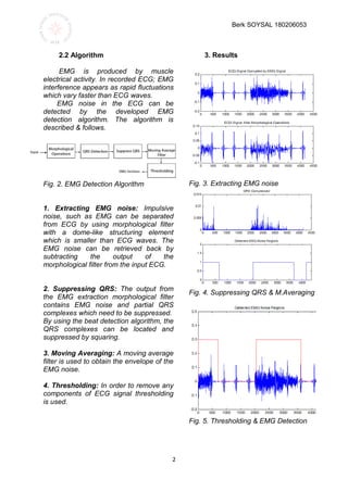
3
%% Berk Soysal EE434
close all;
% EMG signal construction
r = 0.03*randn(length(newdata),1);
t = 0.019*randn(length(newdata),1);
r(500:2000)=0;
t(1:2000)=0;
r(3000:4170)=0;
t(3500:4170)=0;
x=newdata+r+t; % ECG Signal Corrupted by
EMG Signal
subplot(4,1,1);plot(x); title(' ECG Signal
Corrupted by EMG Signal' );
SE=[0, 1, 0;1,1,1;0,1,0 ]; % Structuring Element
for Morphological Operations
x2 = imopen(x,SE); % Opening
x3 = imclose(x2,SE); % Closing
subplot(4,1,2);plot(x3); title(' ECG Signal After
Morphological Operations' );
% QRS Detector
x4 = diff(x3);
x4= diff(x4);
x4=x4.*x4;
subplot(4,1,3);plot(x4); title('QRS Compressed' );
for i=1:length(x4)
if ( x4(i) > 0.001 ) x5(i) =1;
else x5(i)=0;
end
end
%Moving Average Filter
movav=ones(200,1)/200;
x6=conv(movav,x5);
for i=1:length(x6)
if ( x6(i) > 0.001 ) x7(i) =0.3;
else x7(i)=0;
end
end
subplot(4,1,4); plot(x7); axis([0 length(x7) 0
0.5]);title('Detected EMG Noise Regions');
figure;plot(x); hold on
plot(x7,'r'); axis([0 length(x7) -0.2 0.5]);
title('Detected EMG Noise Regions');
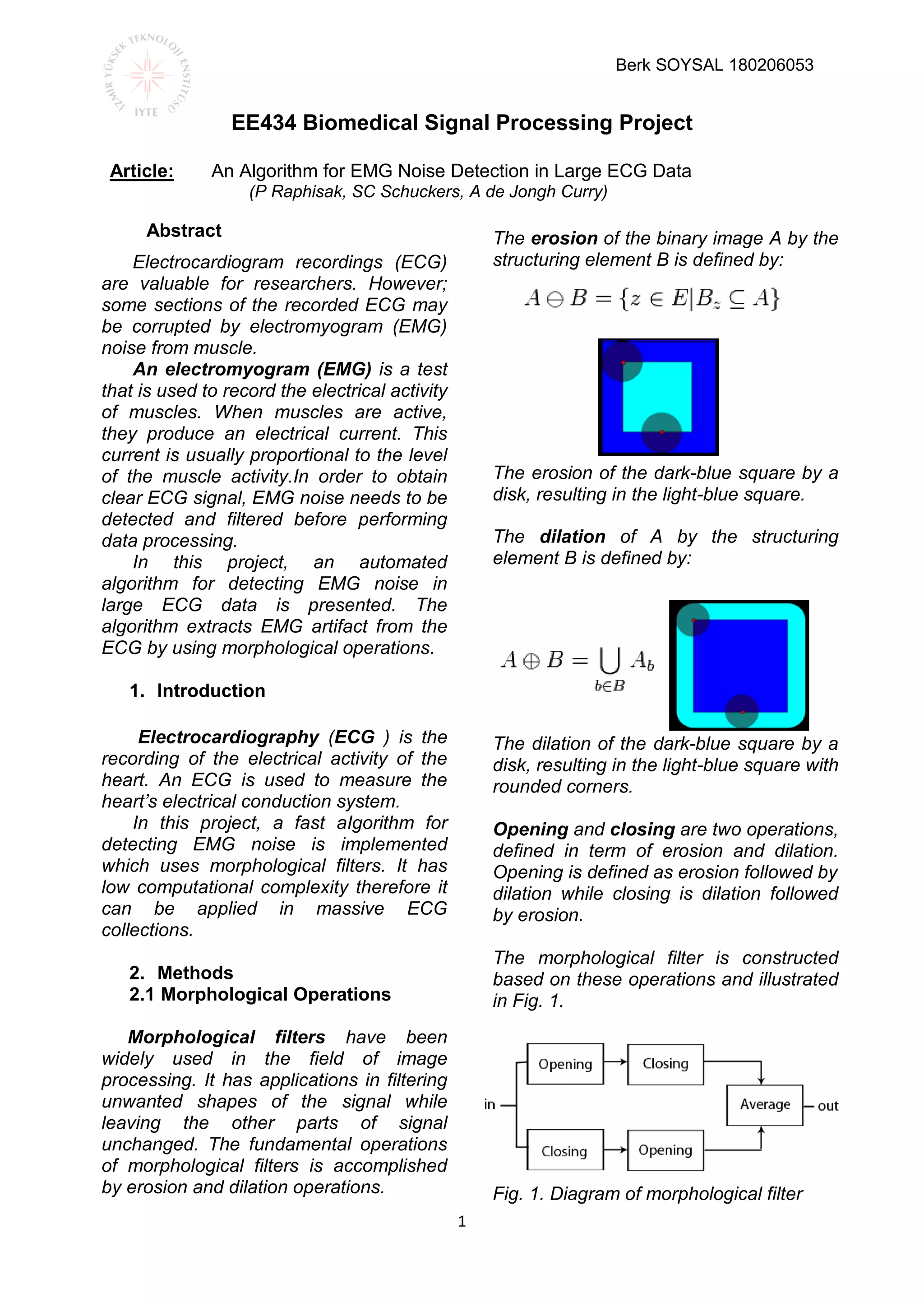
4.Conclusion
In this project, a fast EMG detection
algorithm was explained in detail. EMG is
extracted by two steps. First, a
morphological filtering was operated,
second EMG and QRS complexes were
extracted. Then, QRS complexes were
detected and suppressed. EMG was
detected by setting a threshold on its
moving average. Moving average size
and threshold value are 200 seconds and
0.001, respectively.
The algorithm achieved 100%
detection rate on the sample ECG signal.
Computational complexity of the
algorithm is very low therefore it would be
very convenient in medical cloud
applications.
5.References
[1] Sun Y, Ramires FJ, Weber KT. Fibrosis of
atria and great
vessels in response to angiotensin II or
aldosterone infusion.
Cardiovascular Research July 1997;35( 1): 138-
147.
[2] Raphisak P, de Jongh Curry A, Malkin RA,
Schuckers
SC. Heart rate variability in rats with aldosterone-
induced
chronic heart failure. IEEE Engineering Medicine
and Biology Society Annual Conference
Proceedings September
Impulsive noise suppression and
background normalimtion of electrocardiogram
signals using morphological operators. EEE
Transactions on Biomedical Engineering Febuary
1989;2(2):262-273.
[3] Chu CH, Delp EJ.
1990; 167-170.
[4] Wang D, He DC. A fast implementation of I-d
grayscale
morphological filters. IEEE Transactions on
Circuits and
Systems TI September 1994;41(9):634436.
[5] MacDonald R, Jenkins J, Anbaecher R,
Throne R. A
software trigger for intracardiac waveform
detection with
automatic threshold adjustment. Computers in
Cardiology
2003;1:228-231.](https://image.slidesharecdn.com/biomedicalprojectreport-160726074909/85/Biomedical-project-report-detecting-emg-noise-3-320.jpg)

The document presents an automated algorithm for detecting electromyogram (EMG) noise in electrocardiogram (ECG) data using morphological operations. The algorithm efficiently filters out EMG noise to ensure clear ECG signals, utilizing techniques such as moving averaging and thresholding, and achieves a 100% detection rate with low computational complexity. This approach is suitable for application in large medical data collections and cloud-based medical systems.


![CTEV [ clubfoot] DR ARUN LAL ,DR MOHAMED ASHRAF travancore medical college k...](https://cdn.slidesharecdn.com/ss_thumbnails/ctevclubfootdrarunlaldrmohamedashraftravancoremedicalcollegekollamkeralaindia-260208063247-18fc466c-thumbnail.jpg?width=640&height=640&fit=bounds)
