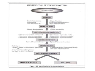
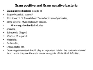
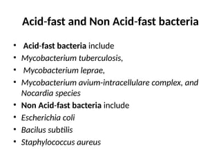
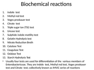
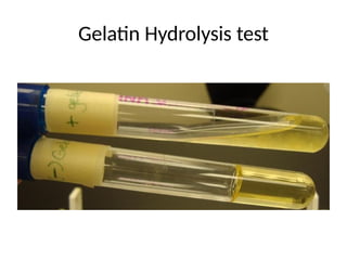
Biochemical reactions
• Gram positive bacteria include all
• Staphylococci (S. aureus)
• Streptococci (St faecalis) and Cornybacterium diphtheriae,
• some Listeria, Mycobacterium species.
• Gram negative family includes
• Shigella,
• Salmonella (S typhi)
• Proteus (P. vugaris)
• Klebsiella,
• Escherichia,
• Enterobacter etc.
• Gram negative enteric bacilli play an important role in the contamination of
food. Hence they are the main causative agents of intestinal infection.
Gram positive and Gram negative bacteria
Acid-fast and Non Acid-fast bacteria
• Acid-fast bacteria include
• Mycobacterium tuberculosis,
• Mycobacterium leprae,
• Mycobacterium avium-intracellulare complex, and
Nocardia species
• Non Acid-fast bacteria include
• Escherichia coli
• Bacilus subtilis
• Staphylococcus aureus
1. Indole test
2. Methyl red test
3. Voges proskauer test
4. Citrate test
5. Triple sugar ion (TSI) test
6. Urease test
7. Sulphide indole motility test
8. Gelatin Hydrolysis test
9. Nitrate Reduction Broth
10. Catalase Test
11. Coagulase Test
12. Oxidase Test
13. Starch Hydrolysis Test
• Usually four tests are used for differentiation of the various members of
Enterobactericeae. They are Indole test, Methyl red test, Voges proskauer
test and Citrate test; collectively known as IMViC series of reactions
Biochemical reactions
Indole test
• Indole tests looks for the presence or absence of
tryptophanase enzyme production of the bacteria. If
the enzyme is present, it will degrade the aminoacid
tryptophan in the media and will produce Indole,
ammonia and pyruvic acid.
• This is tested in a peptone water culture, after 48 to
96 hrs of incuation at 37C. 0.5ml of this culture will
react with Kovac's reagent to produce a cherry red
complex, which indicates a positive indole test.
Methyl Red Test
• This test detects the ability of microorganism to ferment
glucose and to produce acidic end products. Enteric
organism produces pyruvic acid from glucose metabolism.
• Some enteric will then use the mixed acid pathway
to metabolize pyruvic acid to other acidic products such
as lactic acid, acetic acid and formic acids.
• This will reduce the pH of the media. Methyl red is a pH
indicator which is red at the acidic pH (below 4.4) and
yellow at alkaline pH (above 7).
• The formation of red color after the addition of Methyl red
reagent indicates the accumulation of acidic end products
in the medium and is an indicative of positive test
Voges Proskauer Test
• This test determines the ability of microorganism to
ferment glucose. The end products of glucose
metabolism,pyruvic acid, is further metabolized by using
Butylene glucol pathway to produce neutral end such as
acetoin and 2,3 butanediol.
• When Barrit's reagent A ( 40% KOH) and Barrit's reagent B
(5% solution of alpha naphthol) is added to culture broth,
it will detect the presence of acetoin, the precursor in the
2,3- butanediol synthesis.
• Acetoin in the presence of Oxygen and Barrit's reagent
is oxidized to diacetyl, where alpha naphthol act as a
catalyst. Diacetyl then reacts with guanidine components
of peptone to produce a cherry red colour
Citrate Utilization Test
• This test determines the ability of microorganism to utilize Citrate.
Koser’s citrate medium uses citrate as sole source of carbon. The ability
to use the citrate can be determined by the production of turbidity.
Enteric gram negative bacteria gives positive to this test.
• Some bacteria have the capability to convert the salts of organic acids,
for example, Sodium citrate to alkaline carbonates in presence of citrate
lyase and oxaloacetate decarboxylase to release carbondioxide and
gives turbidity. Sodium citrate is one of the important metabolite of
Kreb's cycle.
• The other products of the reaction are acetate, Lactic acid, formic acid
etc. The carbondioxide reacts with sodium and water to form sodium
carbonate
Triple sugar ion (TSI) test
• The triple sugar- ion agar test is designed to differentiate among the
different groups or genera of the Enterobacteriaceae, which are all gram
negative bacilli capable of fermenting glucose with the production of acid,
and to distinguish them from other gram negative intestinal bacilli which
produce hydrogen sulfide.
• TSI Agar contains three fermentative sugars, lactose and sucrose in 1%
concentrations and glucose in a concentration of 0.1%. Due to the
building of acid during fermentation, the pH falls. The acid base
indicator Phenol red is incorporated for detecting carbohydrate
fermentation that is indicated by the change in colour of the medium
from orange red to yellow in the presence of acids.
• Carbohydrate fermentation is detected by the presence of gas and a visible
colour change (from red to yellow) of the pH indicator, phenol red. The
production of hydrogen sulfide is indicated by the presence of a
precipitate that blackens the medium in the butt of the tube.
Urease test
• This is done in Christenton’s Urease medium.
• Urea is a nitrogen containing compound that is produced during the
decarboxylation process of the amino acid arginine in the urea
cycle. Certain bacteria produce the enzyme urease during its
metabolism process and that will break down the urea in the
medium to ammonia and carbon dioxide to produce purple-pink
colour. The test is to e examined for colour only after four hours after
incubation.
The urease test is useful in identifying the genera Proteus vulgaris,
Providentia, and Morganella, which liberate this enzyme.
Gelatin Hydrolysis test
• Gelatin, a protein derived from the animal protein collagen.
• Some microorganisms possess an enzyme called gelatinase, which
breaks down gelatin into amino acids. Gelatin deeps contain the substrate
gelatin, which is a protein produced by the hydrolysis of collagen.
Organisms which hydrolyze gelatin will cause the gelatin to liquefy.
• The gelatin hydrolysis tests for an organism's ability to break down the
protein gelatin which is derived from collagen. Gelatin causes the media
to thicken, especially at cooler (below 28oC) temperatures.
• If the organism can release gelatinase enzymes the gelatin is broken down
or liquefied. The media is checked over a period of about a week after
inoculation and incubation at room temperature, for gelatinase activity.
The tube is placed on ice for a few minutes and if the media fails to
solidify it is considered a positive test. The gelatinase reaction may be
slow or incomplete
Gelatin Hydrolysis test
Catalase
Test
• The enzyme catalase is present in most cytochrome containing aerobic and
facultative anaerobic bacteria. Catalase has one of the highest turnover numbers
of all enzymes such that one molecule of catalase can convert millions of
molecules of hydrogen peroxide to water and oxygen in a second.
• Catalase production and activity can be detected by adding the substrate H2O2 to
an appropriately incubated (18- to 24-hour) tryptic soy agar slant culture.
Organisms which produce the enzyme break down the hydrogen peroxide, and the
resulting O2 production produces bubbles in the reagent drop, indicating a positive
test. Organisms lacking the cytochrome system also lack the catalase enzyme and
are unable to break down hydrogen peroxide, into O2 and water and are catalase
negative.
Coagulase Test
• Coagulases are enzymes that clot blood plasma by a mechanism
that is similar to normal clotting.
• The coagulase test identifies whether an organism produces this
exoenzyme. This enzyme clots the plasma component of blood. The
only significant disease-causing bacteria of humans that produce
coagulase are Staphylococcus aureus.
• Thus this enzyme is a good indicator of the pathogenic potential of
S. aureus.
• In the test, the sample is added to rabbit plasma and held at 37° C
for a specified period of time.
• Formation of clot within 4 hours is indicated as a positive result and
indicative of a virulent Staphylococcus aureus strain. The absence of
coagulation after 24 hours of incubation is a negative result.
•
Oxidase Test
The test depends on the ability of certain bacteria to produce
indophenol blue from the oxidation of dimethyl-p-
phenylenediamine and α-naphthol.
To the culture, add dimethyl-p- phenylenediamine and α-
naphthol. If blue colour is appered indictaes positive oxidase
test.
Pseudomonas aeruginosa is an oxidase positive
organism.
Starch Hydrolysis Test
• Starch agar is an example of differential medium which tests the ability of an
organism to produce certain alpha- amylase and oligo-1, 6-glucosidase that
hydrolyze starch. Starch molecules are too large to enter into the bacterial
cells, so some bacteria will secrete exoenzymes that will degrade starch into
subunits that can be then easily utilized by the organism.
• Starch agar is a simple nutritive medium with starch added. Since no colour
change occurs in the medium when organisms hydrolyze starch, iodine
solution is added to the plate after incubation. Iodine turns blue, purple, or
black (the colour depends on the concentration of the iodine used) in the
presence of starch. A clearing (No colour) around the bacterial growth shows
that the organism has hydrolyzed starch.
biochemical reactions converted (1).pptx

biochemical reactions converted (1).pptx

  • 1.
  • 3.
    • Gram positivebacteria include all • Staphylococci (S. aureus) • Streptococci (St faecalis) and Cornybacterium diphtheriae, • some Listeria, Mycobacterium species. • Gram negative family includes • Shigella, • Salmonella (S typhi) • Proteus (P. vugaris) • Klebsiella, • Escherichia, • Enterobacter etc. • Gram negative enteric bacilli play an important role in the contamination of food. Hence they are the main causative agents of intestinal infection. Gram positive and Gram negative bacteria
  • 4.
    Acid-fast and NonAcid-fast bacteria • Acid-fast bacteria include • Mycobacterium tuberculosis, • Mycobacterium leprae, • Mycobacterium avium-intracellulare complex, and Nocardia species • Non Acid-fast bacteria include • Escherichia coli • Bacilus subtilis • Staphylococcus aureus
  • 6.
    1. Indole test 2.Methyl red test 3. Voges proskauer test 4. Citrate test 5. Triple sugar ion (TSI) test 6. Urease test 7. Sulphide indole motility test 8. Gelatin Hydrolysis test 9. Nitrate Reduction Broth 10. Catalase Test 11. Coagulase Test 12. Oxidase Test 13. Starch Hydrolysis Test • Usually four tests are used for differentiation of the various members of Enterobactericeae. They are Indole test, Methyl red test, Voges proskauer test and Citrate test; collectively known as IMViC series of reactions Biochemical reactions
  • 7.
    Indole test • Indoletests looks for the presence or absence of tryptophanase enzyme production of the bacteria. If the enzyme is present, it will degrade the aminoacid tryptophan in the media and will produce Indole, ammonia and pyruvic acid. • This is tested in a peptone water culture, after 48 to 96 hrs of incuation at 37C. 0.5ml of this culture will react with Kovac's reagent to produce a cherry red complex, which indicates a positive indole test.
  • 8.
    Methyl Red Test •This test detects the ability of microorganism to ferment glucose and to produce acidic end products. Enteric organism produces pyruvic acid from glucose metabolism. • Some enteric will then use the mixed acid pathway to metabolize pyruvic acid to other acidic products such as lactic acid, acetic acid and formic acids. • This will reduce the pH of the media. Methyl red is a pH indicator which is red at the acidic pH (below 4.4) and yellow at alkaline pH (above 7). • The formation of red color after the addition of Methyl red reagent indicates the accumulation of acidic end products in the medium and is an indicative of positive test
  • 9.
    Voges Proskauer Test •This test determines the ability of microorganism to ferment glucose. The end products of glucose metabolism,pyruvic acid, is further metabolized by using Butylene glucol pathway to produce neutral end such as acetoin and 2,3 butanediol. • When Barrit's reagent A ( 40% KOH) and Barrit's reagent B (5% solution of alpha naphthol) is added to culture broth, it will detect the presence of acetoin, the precursor in the 2,3- butanediol synthesis. • Acetoin in the presence of Oxygen and Barrit's reagent is oxidized to diacetyl, where alpha naphthol act as a catalyst. Diacetyl then reacts with guanidine components of peptone to produce a cherry red colour
  • 10.
    Citrate Utilization Test •This test determines the ability of microorganism to utilize Citrate. Koser’s citrate medium uses citrate as sole source of carbon. The ability to use the citrate can be determined by the production of turbidity. Enteric gram negative bacteria gives positive to this test. • Some bacteria have the capability to convert the salts of organic acids, for example, Sodium citrate to alkaline carbonates in presence of citrate lyase and oxaloacetate decarboxylase to release carbondioxide and gives turbidity. Sodium citrate is one of the important metabolite of Kreb's cycle. • The other products of the reaction are acetate, Lactic acid, formic acid etc. The carbondioxide reacts with sodium and water to form sodium carbonate
  • 11.
    Triple sugar ion(TSI) test • The triple sugar- ion agar test is designed to differentiate among the different groups or genera of the Enterobacteriaceae, which are all gram negative bacilli capable of fermenting glucose with the production of acid, and to distinguish them from other gram negative intestinal bacilli which produce hydrogen sulfide. • TSI Agar contains three fermentative sugars, lactose and sucrose in 1% concentrations and glucose in a concentration of 0.1%. Due to the building of acid during fermentation, the pH falls. The acid base indicator Phenol red is incorporated for detecting carbohydrate fermentation that is indicated by the change in colour of the medium from orange red to yellow in the presence of acids. • Carbohydrate fermentation is detected by the presence of gas and a visible colour change (from red to yellow) of the pH indicator, phenol red. The production of hydrogen sulfide is indicated by the presence of a precipitate that blackens the medium in the butt of the tube.
  • 12.
    Urease test • Thisis done in Christenton’s Urease medium. • Urea is a nitrogen containing compound that is produced during the decarboxylation process of the amino acid arginine in the urea cycle. Certain bacteria produce the enzyme urease during its metabolism process and that will break down the urea in the medium to ammonia and carbon dioxide to produce purple-pink colour. The test is to e examined for colour only after four hours after incubation. The urease test is useful in identifying the genera Proteus vulgaris, Providentia, and Morganella, which liberate this enzyme.
  • 16.
    Gelatin Hydrolysis test •Gelatin, a protein derived from the animal protein collagen. • Some microorganisms possess an enzyme called gelatinase, which breaks down gelatin into amino acids. Gelatin deeps contain the substrate gelatin, which is a protein produced by the hydrolysis of collagen. Organisms which hydrolyze gelatin will cause the gelatin to liquefy. • The gelatin hydrolysis tests for an organism's ability to break down the protein gelatin which is derived from collagen. Gelatin causes the media to thicken, especially at cooler (below 28oC) temperatures. • If the organism can release gelatinase enzymes the gelatin is broken down or liquefied. The media is checked over a period of about a week after inoculation and incubation at room temperature, for gelatinase activity. The tube is placed on ice for a few minutes and if the media fails to solidify it is considered a positive test. The gelatinase reaction may be slow or incomplete
  • 17.
  • 18.
    Catalase Test • The enzymecatalase is present in most cytochrome containing aerobic and facultative anaerobic bacteria. Catalase has one of the highest turnover numbers of all enzymes such that one molecule of catalase can convert millions of molecules of hydrogen peroxide to water and oxygen in a second. • Catalase production and activity can be detected by adding the substrate H2O2 to an appropriately incubated (18- to 24-hour) tryptic soy agar slant culture. Organisms which produce the enzyme break down the hydrogen peroxide, and the resulting O2 production produces bubbles in the reagent drop, indicating a positive test. Organisms lacking the cytochrome system also lack the catalase enzyme and are unable to break down hydrogen peroxide, into O2 and water and are catalase negative.
  • 20.
    Coagulase Test • Coagulasesare enzymes that clot blood plasma by a mechanism that is similar to normal clotting. • The coagulase test identifies whether an organism produces this exoenzyme. This enzyme clots the plasma component of blood. The only significant disease-causing bacteria of humans that produce coagulase are Staphylococcus aureus. • Thus this enzyme is a good indicator of the pathogenic potential of S. aureus. • In the test, the sample is added to rabbit plasma and held at 37° C for a specified period of time. • Formation of clot within 4 hours is indicated as a positive result and indicative of a virulent Staphylococcus aureus strain. The absence of coagulation after 24 hours of incubation is a negative result. •
  • 21.
    Oxidase Test The testdepends on the ability of certain bacteria to produce indophenol blue from the oxidation of dimethyl-p- phenylenediamine and α-naphthol. To the culture, add dimethyl-p- phenylenediamine and α- naphthol. If blue colour is appered indictaes positive oxidase test. Pseudomonas aeruginosa is an oxidase positive organism.
  • 22.
    Starch Hydrolysis Test •Starch agar is an example of differential medium which tests the ability of an organism to produce certain alpha- amylase and oligo-1, 6-glucosidase that hydrolyze starch. Starch molecules are too large to enter into the bacterial cells, so some bacteria will secrete exoenzymes that will degrade starch into subunits that can be then easily utilized by the organism. • Starch agar is a simple nutritive medium with starch added. Since no colour change occurs in the medium when organisms hydrolyze starch, iodine solution is added to the plate after incubation. Iodine turns blue, purple, or black (the colour depends on the concentration of the iodine used) in the presence of starch. A clearing (No colour) around the bacterial growth shows that the organism has hydrolyzed starch.